
C57BL/6-Ccr8tm1(CCR8)Bcgen Ccl1tm1(CCL1) Bcgen/Bcgen • 121836
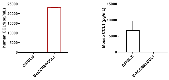
Strain specific CCL1 expression analysis in homozygous B-hCCR8/hCCL1 mice by ELISA. Serum were collected from wild-type mice and homozygous B-hCCR8/hCCL1 mice that stimulated with 7.5ug anti-mCD3ε antibody in vivo (n=2 or 3), and analyzed by ELISA with species-specific CCL1 ELISA kit. Mouse CCL1 was detectable in wild-type mice. Human CCL1 was exclusively detectable in homozygous B-hCCR8/hCCL1 mice but not in wild-type mice.
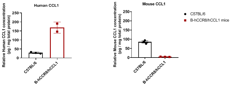
Detection of CCL1 in tumor microenvironment of C57BL/6 and B-hCCR8/hCCL1 mice bearing MC38 cells by ELISA. Murine colon cancer MC38 cells were subcutaneously implanted into C57BL/6 and B-hCCR8/hCCL1 mice (n=2 or 3). Tumor tissues were harvested when tumor volume ~ 600mm3 and analyzed by ELISA. Human CCL1 was detectable in homozygous B-hCCR8/hCCL1 mice, the content is about 150pg per mg total protein.
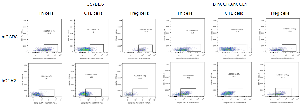
CCR8 (also named CD198) protein expression analysis in B-hCCR8/hCCL1 mice by FACS. Murine colon cancer MC38 cells were subcutaneously implanted into homozygous B-hCCR8/hCCL1 mice. TILs were analysis in tumor volume at approximately 600 mm3. Human CCR8 were detectable in Th cells and Treg cells of homozygous B-hCCR8/hCCL1 mice. However, according to data, human CCR8 were also detectable in CTL cells of wild type C57BL/6 mice and humanized mice. It could be the antibodies have more unspecific binding with CTLs.
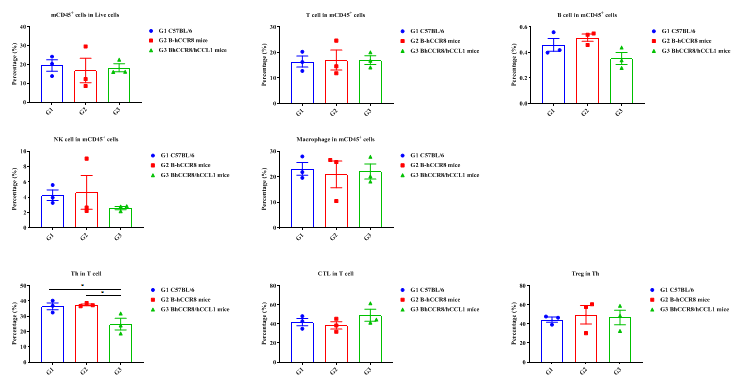
Tumor-infiltrating lymphocytes analysis in different mice. The percentage of Treg cells (% in Th cells) in B-hCCR8/hCCL1 mice has no significant difference compared with the C57BL/6 mice and B-hCCR8 mice.

Antitumor activity of anti-human CCR8 antibody in B-hCCR8/hCCL1 mice. (A) Anti-human CCR8 antibody inhibited MC38 tumor growth in B-hCCR8/hCCL1 mice. Murine colon cancer MC38 cells (5×10⁵) were subcutaneously implanted into homozygous B-hCCR8/hCCL1 mice (female, 7-week-old, n=6). Mice were grouped when tumor volume reached approximately 100-150 mm3, at which time they were intraperitoneally injected with anti-human CCR8 antibody indicated in panel. (B) Body weight changes during treatment. As shown in panel A, anti-human CCR8antibody was efficacious in controlling tumor growth in B-hCCR8/hCCL1 mice, demonstrating that the B-hCCR8/hCCL1 mice provide a powerful preclinical model for in vivo evaluation of anti-human CCR8 antibodies. Values are expressed as mean ± SEM.
The overage of this tumor model is 41.7%.

MC38 tumor growth curves from individual mice. Murine colon cancer MC38 cells (5×10⁵) were subcutaneously implanted into homozygous B-hCCR8/hCCL1 mice (female, 7-week-old, n=6). Mice were grouped when tumor volume reached approximately 100-150 mm3, at which time they were intraperitoneally injected with anti-human CCR8 antibody indicated in panel. Results indicate that anti-human CCR8 antibody was efficacious in controlling tumor growth in B-hCCR8/hCCL1 mice, demonstrating that the B-hCCR8/hCCL1 mice provide a powerful preclinical model for in vivo evaluation of anti-human CCR8 antibodies.

Analysis of tumor infiltrates lymphocytes by FACS. Tumor cells were harvested at the endpoint of experiment (n=6). Flow cytometry analysis of the lymphocytes were performed to assess cell number and proportion changes. The proportions of CD3+ T cells, CD4+ T cells, Treg cells, and hCCR8+ Treg cells The anti-CCR8 antibody treatment group showed no significant changes in compared to the control group. Values are expressed as mean ± SEM. Significance was determined by one-way ANOVA test. *P < 0.05, **P < 0.01, ***P < 0.001.