
C.B6-Cd3etm1(CD3E)Bcgen Cd3dtm1(CD3D)Bcgen Cd3gtm1(CD3G)Bcgen/Bcgen • 111719
| Product name | B-hCD3EDG mice(C) |
|---|---|
| Catalog number | 111719 |
| Strain name | C.B6-Cd3etm1(CD3E)Bcgen Cd3dtm1(CD3D)Bcgen Cd3gtm1(CD3G)Bcgen/Bcgen |
| Strain background | C57BL/6; BALB/cCrSlcNifdc |
| NCBI gene ID | 915,916,917 (Human) |
| Aliases | T3D; IMD19; CD3DELTA; CD3-DELTA; T3E; TCRE; IMD18; CD3epsilon; T3G; IMD17; CD3GAMMA; CD3-GAMMA |
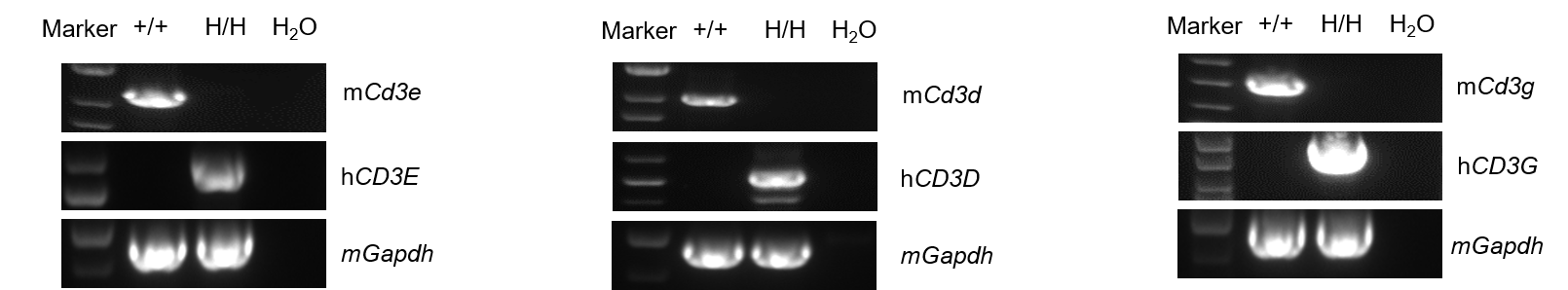
Strain specific analysis of CD3E, D, G mRNA expression in wild-type BALB/cCrSlcNifdc mice and B-hCD3EDG mice(C) by RT-PCR. Thymus RNA were isolated from wild-type BALB/cCrSlcNifdc mice (+/+) and homozygous B-hCD3EDG mice(C) (H/H), then cDNA libraries were synthesized by reverse transcription, followed by PCR with mouse or human CD3E, D, G primers. Mouse Cd3e, d, g mRNA was only detectable in wild-type mice. Human CD3E, D, G mRNA was exclusively detectable in homozygous B-hCD3EDG mice(C), but not in wild-type mice.
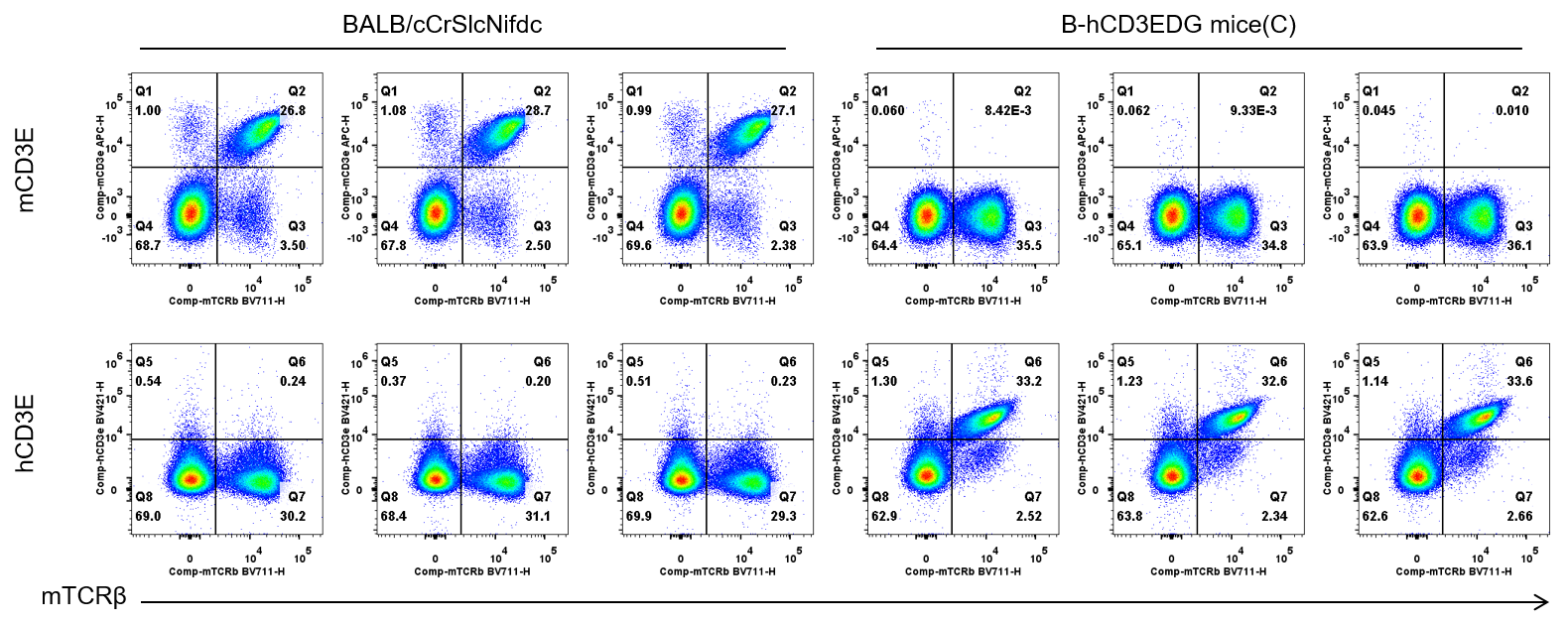
Strain specific CD3E expression analysis in wild-type BALB/cCrSlcNifdc mice and homozygous humanized B-hCD3EDG mice(C) by flow cytometry. Splenocytes were collected from wild-type BALB/cCrSlcNifdc mice and homozygous B-hCD3EDG mice(C) (female, n=3, 6-week-old). Protein expression was analyzed with anti-mouse CD3e antibody (Biolegend, 100312) and anti-human CD3e antibody (BD Horizon™ , 562426) by flow cytometry. Mouse CD3E was only detectable in wild-type BALB/cCrSlcNifdc mice. Human CD3E was exclusively detectable in homozygous B-hCD3EDG mice(C), but not in wild-type BALB/cCrSlcNifdc mice.
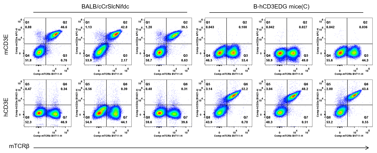
Strain specific CD3E expression analysis in wild-type BALB/cCrSlcNifdc mice and homozygous humanized B-hCD3EDG mice(C) by flow cytometry. Blood cells were collected from wild-type BALB/cCrSlcNifdc mice and homozygous B-hCD3EDG mice(C) (female, n=3, 6-week-old). Protein expression was analyzed with anti-mouse CD3e antibody (Biolegend, 100312) and anti-human CD3e antibody (BD Horizon™ , 562426) by flow cytometry. Mouse CD3E was only detectable in wild-type BALB/cCrSlcNifdc mice. Human CD3E was exclusively detectable in homozygous B-hCD3EDG mice(C), but not in wild-type BALB/cCrSlcNifdc mice.

Frequency of leukocyte subpopulations in spleen by flow cytometry. Splenocytes were isolated from wild-type BALB/cCrSIcNifdc mice and homozygous B-hCD3EDG mice(C) (female, 6-week-old, n=3). A. Flow cytometry analysis of the splenocytes was performed to assess the frequency of leukocyte subpopulations. B. Frequency of T cell subpopulations. Frequencies of T cells, B cells, NK cells, DCs, neutrophils, monocytes, macrophages, CD4+ T cells, CD8+ T cells and Tregs in B-hCD3EDG mice(C) were similar to those in BALB/cCrSIcNifdc. The frequency of leukocyte subpopulations in thymus, lymph node and blood of B-hCD3EDG mice(C) were also comparable to wild-type BALB/cCrSIcNifdc mice (Data not shown). Values are expressed as mean ± SEM.
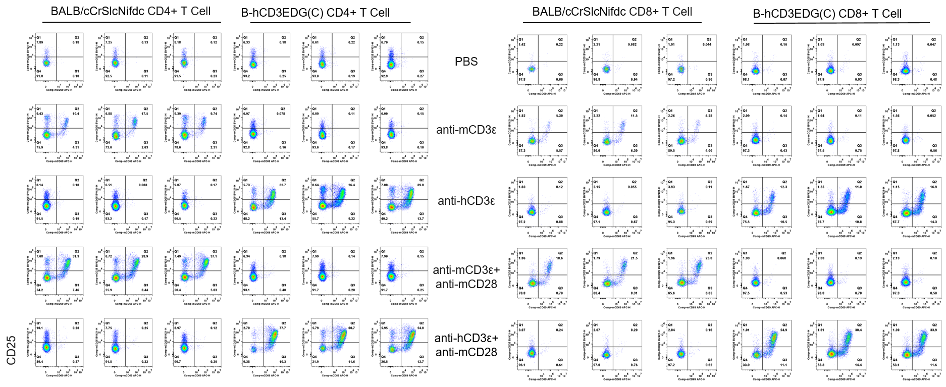
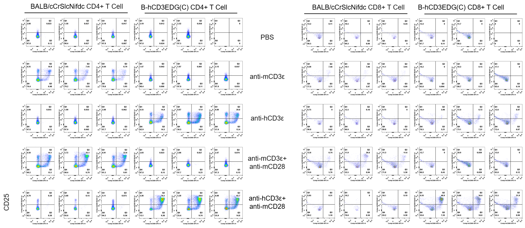
CD25 and CD69 expression analysis in wild-type BALB/cCrSlcNifdc mice and homozygous humanized B-hCD3EDG mice(C) by flow cytometry. T cells were isolated from splenocytes of BALB/cCrSlcNifdc and B-hCD3EDG mice(C) (n=3, 6-week-old), and were incubated in the presence of anti-mCD3ε antibody (2ug/ml,BioXcell,BE0001-2) , anti-hCD3ε antibody (2ug/ml,BioXcell,BE0001-1) and anti-mCD28 antibody (5ug/ml,BioXcell,BE0015-1) for 24h. T cell proliferation was tested by flow cytometry. T cell activation in B-hCD3EDG mice(C) was significantly up-regulated by anti-hCD3ε antibody or anti-hCD3ε antibody and anti-mCD28 antibody.
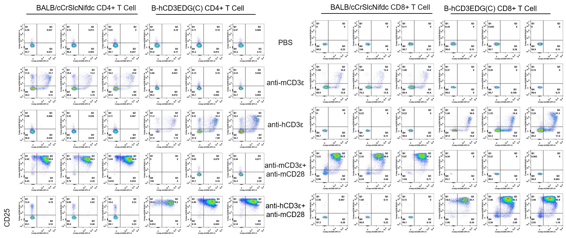
CD25 and CD69 expression analysis in wild-type BALB/cCrSlcNifdc mice and homozygous humanized B-hCD3EDG mice(C) by flow cytometry. T cells were isolated from splenocytes of BALB/cCrSlcNifdc and B-hCD3EDG mice(C) (n=3, 6-week-old), and were incubated in the presence of anti-mCD3ε antibody (2ug/ml,BioXcell,BE0001-2) , anti-hCD3ε antibody (2ug/ml,BioXcell,BE0001-1) and anti-mCD28 antibody (5ug/ml,BioXcell,BE0015-1) for 48h. T cell proliferation was tested by flow cytometry. T cell activation in B-hCD3EDG mice(C) was significantly up-regulated by anti-hCD3ε antibody or anti-hCD3ε antibody and anti-mCD28 antibody.
CD25 and CD69 expression analysis in wild-type BALB/cCrSlcNifdc mice and homozygous humanized B-hCD3EDG mice(C) by flow cytometry. T cells were isolated from splenocytes of BALB/cCrSlcNifdc and B-hCD3EDG mice(C) (n=3, 6-week-old), and were incubated in the presence of anti-mCD3ε antibody(2ug/ml,BioXcell,BE0001-2) , anti-hCD3ε antibody (2ug/ml,BioXcell,BE0001-1) and anti-mCD28 antibody (5ug/ml,BioXcell,BE0015-1) for 72h. T cell proliferation was tested by flow cytometry. T cell activation in B-hCD3EDG mice(C) was significantly up-regulated by anti-hCD3ε antibody or anti-hCD3ε antibody and anti-mCD28 antibody.
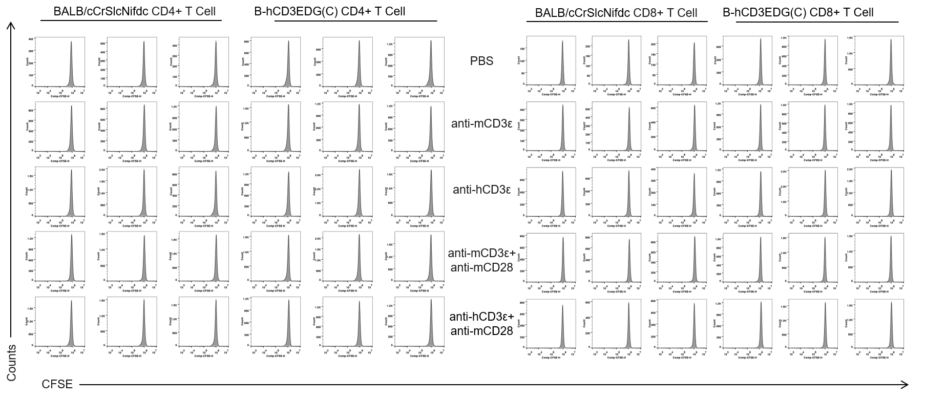
The proliferation of CD4+T cells and CD8+T cells was analyzed by flow cytometry. T cells were isolated from splenocytes of BALB/cCrSlcNifdc and B-hCD3EDG mice(C) (n=3, 6-week-old), and were incubated in the presence of anti-mCD3ε antibody(2ug/ml,BioXcell,BE0001-2) , anti-hCD3ε antibody (2ug/ml,BioXcell,BE0001-1) and anti-mCD28 antibody (5ug/ml,BioXcell,BE0015-1) for 24h. T cell proliferation was tested by flow cytometry. T cell activation in B-hCD3EDG mice(C) was significantly up-regulated by anti-hCD3ε antibody and anti-mCD28 antibody.
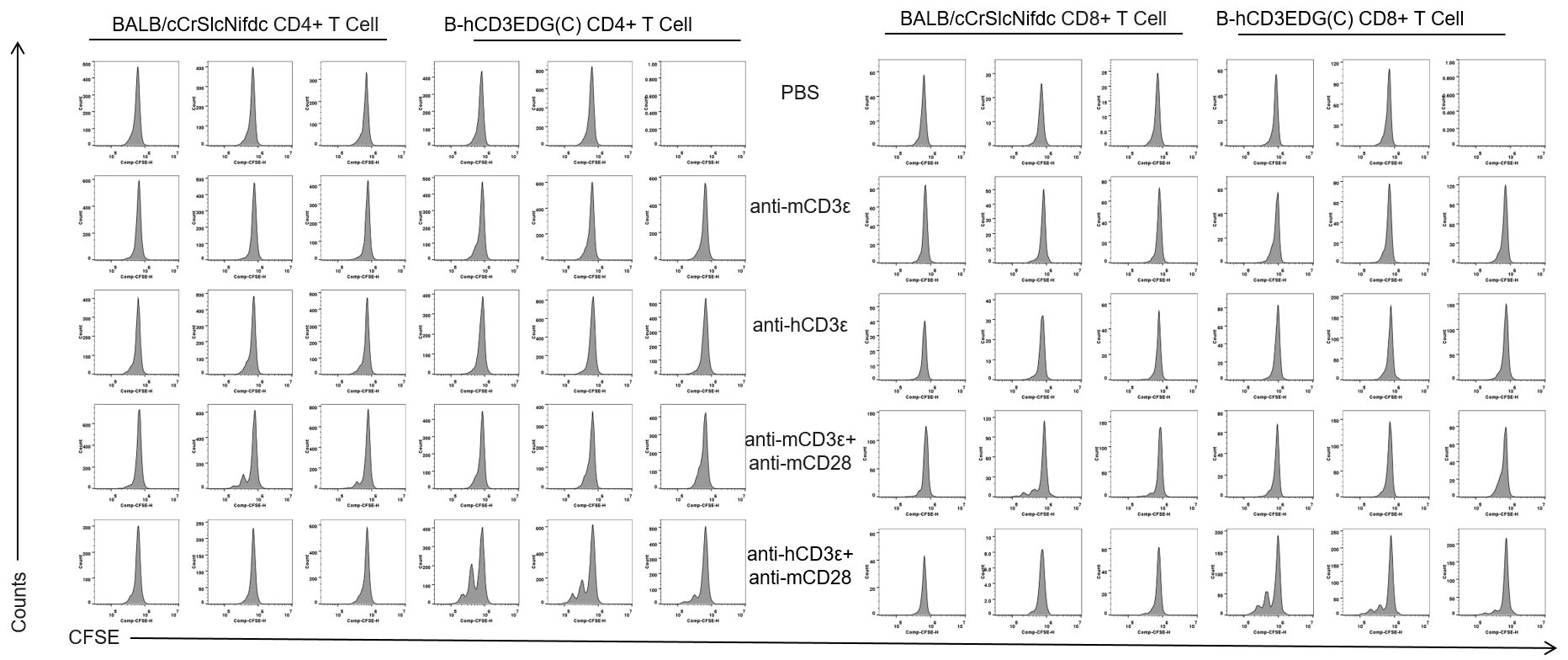
The proliferation of CD4+T cells and CD8+T cells was analyzed by flow cytometry. T cells were isolated from splenocytes of BALB/cCrSlcNifdc and B-hCD3EDG mice(C) (n=3, 6-week-old), and were incubated in the presence of anti-mCD3ε antibody(2ug/ml,BioXcell,BE0001-2) , anti-hCD3ε antibody (2ug/ml,BioXcell,BE0001-1) and anti-mCD28 antibody (5ug/ml,BioXcell,BE0015-1) for 48h. T cell proliferation was tested by flow cytometry. T cell activation in B-hCD3EDG mice(C) was significantly up-regulated by anti-hCD3ε antibody and anti-mCD28 antibody.
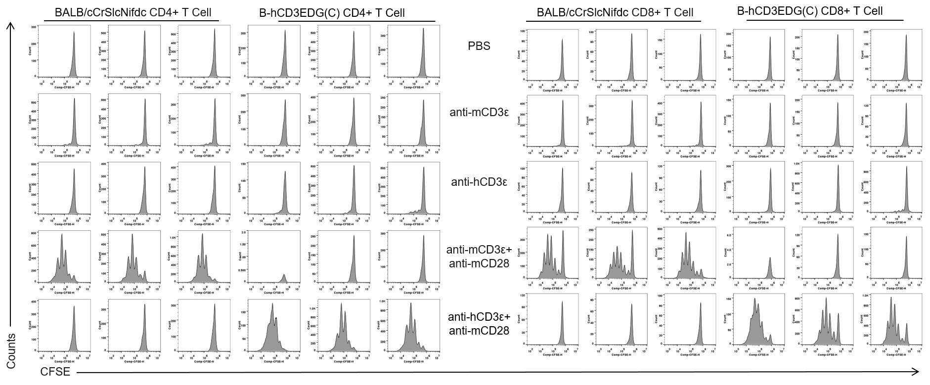
The proliferation of CD4+T cells and CD8+T cells was analyzed by flow cytometry. T cells were isolated from splenocytes of BALB/cCrSlcNifdc and B-hCD3EDG mice(C) (n=3, 6-week-old), and were incubated in the presence of anti-mCD3ε antibody(2ug/ml,BioXcell,BE0001-2) , anti-hCD3ε antibody (2ug/ml,BioXcell,BE0001-1) and anti-mCD28 antibody (5ug/ml,BioXcell,BE0015-1) for 72h. T cell proliferation was tested by flow cytometry. T cell activation in B-hCD3EDG mice(C) was significantly up-regulated by anti-hCD3ε antibody and anti-mCD28 antibody.
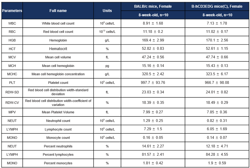
Complete blood count (CBC) of B-hCD3EDG mice(C). Values are expressed as mean ± SD.
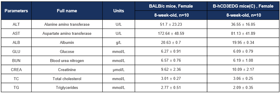
Biochemical test of B-hCD3EDG mice(C). Values are expressed as mean ± SD.