| Strain Name |
C57BL/6-Pdcd1tm1(PDCD1)Bcgen Cd274tm1(CD274)BcgenLag3tm3(LAG3)Bcgen/Bcgen
|
Common Name | B-hPD-1/hPD-L1/hLAG3 plus mice |
| Background | C57BL/6 | Catalog number |
130572 |
|
Related Genes |
PD-1 (Programmed death-1)
CD274 (CD274 antigen) LAG3 (lymphocyte-activation gene 3) |
||
|
NCBI Gene ID |
18566,60533,16768 | ||
PD-1 (Programmed death-1) is mainly expressed on the surface of T cells and primary B cells. The two PD-1 ligands, PD-L1 and PD-L2, are widely expressed on antigen-presenting cells (APCs). PD-L1 expression is favorable for tumorigenesis and growth, for induction of anti-tumor T cell apoptosis, and for escaping responses by the immune system. Inhibition of PD-1 binding to its ligand can result in tumor cells that are exposed to the killing version of the immune system, and thus is a target for cancer treatments. PD-L1 (Programmed cell death ligand-1), also known as B7-H1 and CD274, is mainly expressed in antigen-presenting cells (APCs) and activated T cells, and is one of the two ligands of PD-1. The interaction between PD1 and PD-L1 plays an important role in the negative regulation of the immune response. PD-L1 is highly expressed in a variety of solid tumors. PD-1 and PD-L1 interactions can reduce T cell activation and promote tumor immune escape. The PD-1/PD-L1 signaling pathway can be blocked and antitumor immune response can be restored by using by anti-PD-1 or anti-PD-L1 antibodies to block the binding of PD1 to PD-L1. LAG3 (Lymphocyte activation gene 3, CD223) is a lymphocyte activation gene and a member of the Ig family. It is mainly expressed in activated T cells, NK cells, B cells and plasmacytoid DCs. LAG3 is a negative regulator of immunity and mainly binds to MHC class Ⅱ molecules to regulate the function of dendritic cells. The expression of LAG3 is associated with the negative immunoregulatory function of specific T cells. Inhibition of LAG3 function enhances the antitumor effect of specific CD8+ T cells, therefore LAG3 is a potential target for tumor immunotherapy.
Protein expression analysis

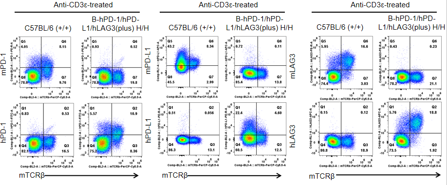
Strain specific PD-1, PD-L1 and LAG3 expression analysis in homozygous B-hPD-1/hPD-L1/LAG3(plus) mice by flow cytometry.
Splenocytes were collected from WT and homozygous B-hPD-1/hPD-L1/hLAG3 (H/H) mice stimulated with anti-CD3ε in vivo, and analyzed by flow cytometry with species-specific anti-PD-1, anti-PD-L1 and anti-LAG3 antibody. Mouse PD-1, PD-L1 and LAG3 were detectable in WT mice. Human PD-1, PD-L1 and LAG3 were exclusively detectable in homozygous B-hPD-1/hPD-L1/hLAG3 but not WT mice.
Combination therapy of pembrolizumab and relatlimab

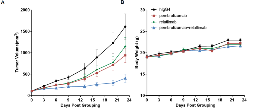
Antitumor activity of pembrolizumab and relatlimab in B-hPD-1/hPD-L1/hLAG3(plus) mice.
(A) human PD-1 antibody combined with anti-human LAG3 antibody inhibited MC38 tumor growth in B-hPD-1/hPD-L1/hLAG3(plus) mice. Murine colon cancer MC38 cells were subcutaneously implanted into homozygous B-hPD-1/hPD-L1/hLAG3(plus) mice (female, 7-8 week-old, n=8). Mice were grouped when tumor volume reached approximately 100 mm3, at which time they were treated with pembrolizumab and relatlimab with doses and schedules indicated in panel A. (B) Body weight changes during treatment. As shown in panel A, combination of pembrolizumab and relatlimab shows more inhibitory effects than individual groups, demonstrating that the B-hPD-1/hPD-L1/hLAG3(plus) mice provide a powerful preclinical model for in vivo evaluating combination therapy efficacy of hPD-1 antibodies and hLAG3 antibodies. Values are expressed as mean ± SEM. (All antibodies were made in house).


