B-hSIRPA/hCD47, Prkdc KO mice
| Strain Name |
C57BL/6-Sirpatm1(SIRPA)Bcgen Cd47tm1(CD47)Bcgen Prkdctm1Bcgen/Bcgen
|
Common Name | B-hSIRPA/hCD47, Prkdc KO mice |
| Background | C57BL/6 | Catalog number | 130565 |
|
Aliases |
BIT, CD172A, MFR, MYD-1, P84, PTPNS1, SHPS1, SIRP, IAP, MER6, OA3, DNA-PKC, DNA-PKcs, DNAPK, DNAPKc, DNPK1, HYRC, HYRC1, IMD26, XRCC7, p350, CD132, CIDX | ||
|
NCBI Gene ID |
19261,16423,19090 | ||
Protein expression analysis
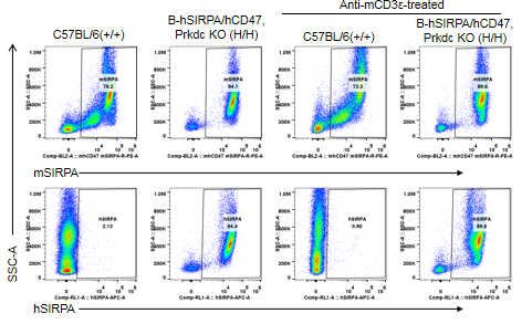
Species specific SIRPα expression analysis in B-hSIRPA/hCD47,Prkdc KO mice by flow cytometry. Splenocytes isolated from C57BL/6 (+/+) mice and homozygous B-hSIRPA/hCD47,Prkdc KO mice (H/H) stimulated with or without anti-mCD3ε in vivo and were analyzed by flow cytometry with anti-SIRPα antibodies. Human SIRPα was exclusively detectable in homozygous B-hSIRPA/hCD47,Prkdc KO mice but not in C57BL/6 mice. Mouse SIRPα was detectable in C57BL/6 and homozygous B-hSIRPA/hCD47,Prkdc KO mice, indicating that this anti-mouse SIRPα antibody was cross-reacting with human SIRPα.
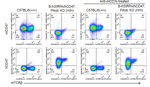
Species specific CD47 expression analysis in B-hSIRPA/hCD47,Prkdc KO mice by flow cytometry. Splenocytes were isolated from C57BL/6 (+/+) mice and homozygous B-hSIRPA/hCD47,Prkdc KO mice (H/H) stimulated with or without anti-mCD3ε in vivo and were analyzed by flow cytometry with anti-CD47 antibodies. Mouse CD47 was only detectable in C57BL/6 mice. Human CD47 was exclusively detectable in homozygous B-hSIRPA/hCD47,Prkdc KO mice but not in C57BL/6 mice. Human CD47 was not detectable on T cells in B-hSIRPA/hCD47,Prkdc KO mice stimulated with or without anti-mCD3ε antibody.
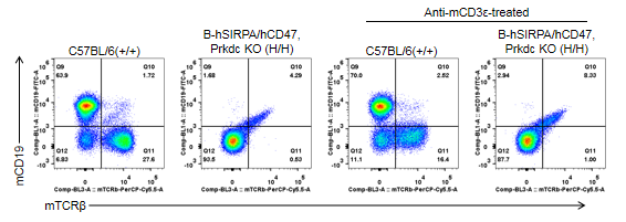
Analysis of leukocyte subpopulations in spleen by FACS
Splenocytes were isolated from C57BL/6 and B-hSIRPA/hCD47,Prkdc KO mice (n=2, 5-week-old). Flow cytometry analysis of the splenocytes was performed to assess T cells and B cells. T, B cells were undetectable in B-hSIRPA/hCD47,Prkdc KO mice stimulated with or without anti-mCD3ε antibody.
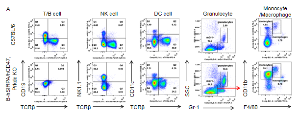
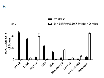
Analysis of spleen leukocyte subpopulations by FACS
Splenocytes were isolated from female C57BL/6 and B-hSIRPA/hCD47,Prkdc KO mice (n=3, 8-week-old). Flow cytometry analysis of the splenocytes was performed to assess leukocyte subpopulations. A. Representative FACS plots. Single live cells were gated for CD45+ population and used for further analysis as indicated here. B. Results of FACS analysis. Percent of NK cells, granulocytes, macrophages and monocytes in homozygous B-hSIRPA/hCD47,Prkdc KO mice were increased compared to those in the C57BL/6 mice, while B cells and all T cells were undetectable in B-hSIRPA/hCD47,Prkdc KO mice. Results indicated that knockout of Prkdc gene prevents the development of B cells and T cells in spleen.
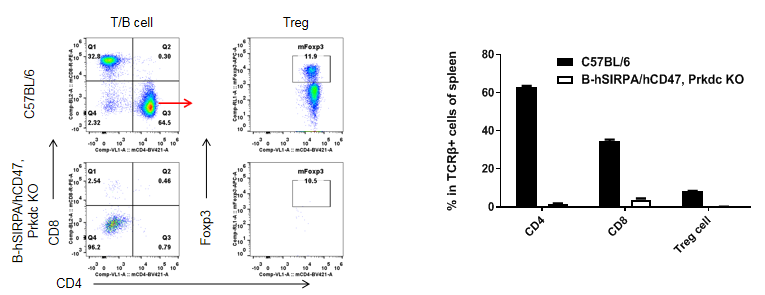
Analysis of leukocyte subpopulations in spleen by FACS
Splenocytes were isolated from C57BL/6 and B-hSIRPA/hCD47,Prkdc KO mice (n=3, 8-week-old). Flow cytometry analysis of the splenocytes was performed to assess leukocyte subpopulations. A. Representative FACS plots. Single live CD45+ cells were gated for CD3+ T cell population and used for further analysis as indicated here. B. Results of FACS analysis. CD4+ T cells, CD8+ T cells and Tregs were undetectable in homozygous B-hSIRPA/hCD47,Prkdc KO. Results indicated that knockout of Prkdc gene prevented differentiation of T cells in spleen.
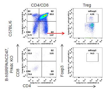
Analysis of leukocyte subpopulations in spleen by FACS
Thymocytes were isolated from C57BL/6 and B-hSIRPA/hCD47,Prkdc KO mice (n=3, 8-week-old). Flow cytometry analysis of the thymocytes was performed to assess leukocyte subpopulations. A. Representative FACS plots. Single live CD45+ cells were gated for CD3+ T cell population and used for further analysis as indicated here. B. Results of FACS analysis. All subtypes of T cells (CD4-CD8-DN cells, CD4+, CD8+, CD4+CD8+DP T cells and Tregs) were undetectable in thymus.


