B-luc Daudi
|
Common name |
B-luc Daudi |
Catalog number | 310677 |
| Aliases |
NA |
Disease | Burkitt's lymphoma |
|
Organism |
Human |
Strain | NA |
| Tissue types |
Peripheral blood |
Tissue |
Peripheral blood |
Description
Targeting strategy
Gene targeting strategy for B-luc Daudi. The luciferase cDNA sequence with CAG promoter was inserted in the AAVS1 locus exon1 of wild-type Daudi .
Phenotypic Analysis

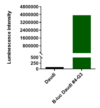
Luminescence signal intensity of B-luc Daudi cells. Luminescence intensity was measured using the Bright-GloTM luciferase Assay (Promega, Cat E2610). B-luc Daudi cells have a strong luminescence signal. The 4-G3 clone of B-luc Daudi cells can be used in vivo experiments.

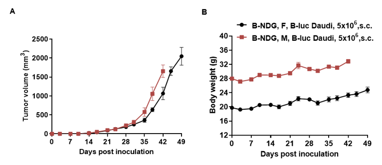
Subcutaneous xenograft tumor growth of B-luc Daudi cells. B-luc Daudi cells (5x10⁶) were subcutaneously implanted into the B-NDG mice (female and male, 7-week-old, n=8).Tumor size and mice body weight were measured twice a week. (A) Tumor average volume ± SEM, (B) Mice body weight (Mean± SEM). Volume was expressed in mm3 using the formula: V=0.5a X b2, where a and b were the long and short diameters of the tumor, respectively. As shown in panel A, B-luc Daudi were able to establish tumor in vivo and can be used for efficacy study.

B-luc Daudi cells were inoculated into the tail vein of B-NDG mice, and imaging was performed on days 0, 3, 7, 10, 14, 17, 21, 24, and 28 for detection and tumor imaging signal intensity analysis
Animal weight monitoring after B-NDG mice were inoculated with B-luc Daudi cells.
Results: B-luc Daudi cells were able to establish tumor model steadily and track tumor volume effectively .


