Mouse Immunodeficiency System CDX Model
Xenotransplantation of human-derived tumor cell lines (CDX) refers to the tumor model established by inoculating human tumor cells into immunodeficient mice (such as severely immunodeficient B-NDG mice) after in vitro subculture. The commonly used inoculation sites are subcutaneous, orthotopic or intravenous. This tumor model has the advantages of good modeling stability and high success rate, and is widely used in tumor cell proliferation research and in the screening of anti-tumor drugs in vivo.
Using our self-developed immunodeficient B-NDG mice, Biocytogen establishes a variety of solid tumor models for anti-tumor drug screening, including lung cancer, rectal adenocarcinoma, colon cancer, breast cancer, pancreatic cancer, bladder cancer, liver cancer and a variety of hematological tumor models such as acute lymphoid leukemia and acute myeloid leukemia. Using these models, we provide high-quality pharmacodynamic (PD) services.
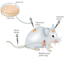
Xenograft model [1]
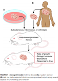
Xenograft Model [2]
Case 1: Melanoma Model
Human melanoma A2058 cells were inoculated subcutaneously in B-NDG mice to successfully establish a melanoma CDX model.
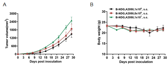
A. Tumor growth curve, B. Body weight change of tumor-bearing mice
Case 2: Human B-cell lymphoma model
The B cell lymphoma model was successfully established by injecting Raji cells into B-NDG, NOD-scid and BALB/C nude mice via the tail vein.
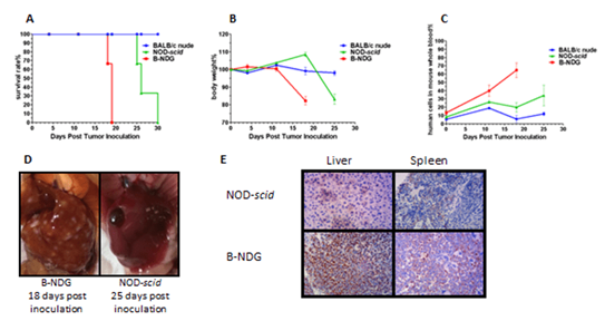
(A) Kaplan-Merier survival curves, (B) relative body weight change, (C) q-PCR quantification of peripheral blood human cell ratios in mice, (D) liver tumors, (E) immunohistochemical staining of liver and spleen
Case 3: Hepatoma Model
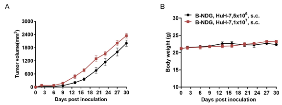
A. Tumor growth curve, B. Body weight change of tumor-bearing mice
Case 4: Colon Cancer Model
Human colorectal adenocarcinoma HT-29 cells were inoculated subcutaneously in B-NDG mice to successfully establish a colorectal adenocarcinoma model.
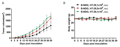
A. Tumor growth curve, B. Body weight change of tumor-bearing mice
Available CDX models
|
Bone marrow |
KG-1, K-562, NCI-H929 |
|
Brain |
U-87 MG, LN-229 |
|
Breast |
MDA-MB-231, DU4475, BT-474, MCF-7 |
|
Colorectal |
HT-29, LOVO, HCT-8, LS 174T, HCT 116, COLO 205 |
|
Kidney |
5637, G401 |
|
Liver |
Hep G2, PLC/PRF/5, Hep 382.1-7, HuH-7 |
|
Lung |
NCI-H520, A549, NCI-H1975, HCC827, NCI-H460 |
|
Ovary |
SK-OV-3 |
|
Pancreas |
PANC-1, MIA PaCa-2, BxPC-3 |
|
Prostate |
Du 145, LNCaP clone FGC |
|
SKIN |
A-431, A375, A2058 |
|
Stomach |
NCI-N87, SNU-5, NUGC-4, Hs 746T |
|
Peripheral blood |
|
|
Lymphoma |
THP-1, Raji, NAMALWA, SU-DHL-1, Daudi |
CDX Models Using Luciferase-Expressing Cell Lines
|
Cell LineName |
Disease |
|
B-luc-GFP Raji |
Burkitt's lymphoma |
|
B-luc-GFP HT-29 |
Colorectal adenocarcinoma |
|
B-luc-GFP NCI-H1975 |
Lung carcinoma, non-small cell (NSCLC) |
|
B-luc K-562 |
Chronic myelogenous leukemia (CML) |
|
B-luc MIA PaCa-2 |
Pancreatic carcinoma |
|
B-luc B16-F10 |
Melanoma |
|
B-luc EL4 |
Lymphoma |
|
B-luc Hep 3B plus |
Hepatocellular carcinoma (HCC) |
|
B-luc Daudi |
Lymphoma |
|
B-Luc A20 |
Reticulum cell sarcoma |
REFERENCES:
【1】Dranoff, G. Experimental mouse tumour models: what can be learnt about human cancer immunology? Nat Rev Immunol 12, 61-66 (2011).
【2】Kohnken, R., Porcu, P. & Mishra, A. Overview of the Use of Murine Models in Leukemia and Lymphoma Research. Front Oncol 7, 22 (2017).


