B-hCD3EDG/hEGFR mice

C57BL/6-Cd3etm1(CD3E)Bcgen Cd3dtm1(CD3D)BcgenCd3gtm1(CD3G)Bcgen Egfrtm2(EGFR)Bcgen/Bcgen • 113562

B-hCD3EDG/hEGFR mice

Product nameB-hCD3EDG/hEGFR mice
Catalog number113562
Strain nameC57BL/6-Cd3etm1(CD3E)Bcgen Cd3dtm1(CD3D)BcgenCd3gtm1(CD3G)Bcgen Egfrtm2(EGFR)Bcgen/Bcgen
Strain backgroundC57BL/6
NCBI gene ID916,915,917,1956 (Human)
AliasesIMD18, T3E, TCRE; CD3-DELTA, IMD19, T3D; CD3-GAMMA, IMD17, T3G; ERBB, ERBB1, HER1, NISBD2, PIG61, mENA

이 페이지에서

  • Description
  • Phenotypic analysis

포스터

모두 보기

    출판물

      Description
      • The CD3 complex is an important marker on the surface of T cells. CD3 has four peptide chains, namely, the γ chain, the δ chain, the ε chain, and the ζ chain. The transmembrane region of CD3 molecules is connected to the transmembrane region of TCRα and β chains through salt Bridges to form the TCR-CD3 complex, which jointly participates in the recognition of antigens by T cells. In this process, TCR recognizes and binds to antigenic peptides presented by MHC molecules, resulting in the phosphorylation of the immune receptor tyrosine activation motifs ITAM in the CD3 cytoplasmic region, which can then recruit other tyrosine protein kinases (such as ZAP-70), thereby causing activation of downstream pathways such as MAPK, NF-κB and other signaling pathways. Under the action of multiple transcription factors, it can cause T cell proliferation, migration, cytokine production and effector function.
      • EGFR is expressed in various tissues. Upon binding of ligands like EGF to the EGFR receptor, EGFR dimers are formed. The activation of EGFR dimers stimulates intracellular tyrosine kinases, resulting in phosphorylation that triggers downstream signaling cascades, leading to cancer cell proliferation and the proliferation of new blood vessels within cancer cells. The mechanism of action of EGFR-targeting drugs primarily involves blocking or inhibiting the activity of EGFR, thereby preventing the growth and spread of cancer cells.
      Protein expression analysis in spleen T cells

      CD3E expression analysis in wild-type C57BL/6 mice and homozygous humanized B-hCD3EDG/hEGFR mice by flow cytometry. Spleen T cells were collected from wild-type C57BL/6 mice (+/+) (Female, 6-week-old, n=1) and homozygous B-hCD3EDG/hEGFR mice (H/H; H/H; H/H; H/H) (Female, 6-week-old, n=1). Protein expression was analyzed with anti-human CD3E antibody (BD Horizon™, 562426) and anti-mouse CD3E antibody (Biolegend, 100312) by flow cytometry. Human CD3E was exclusively detectable in homozygous B-hCD3EDG/hEGFR mice, but not in wild-type C57BL/6 mice.

      Protein expression analysis in blood T cells

      CD3E expression analysis in wild-type C57BL/6 mice and homozygous humanized B-hCD3EDG/hEGFR mice by flow cytometry. Blood T cells were collected from wild-type C57BL/6 mice (+/+) (Female, 6-week-old, n=1) and homozygous B-hCD3EDG/hEGFR mice (H/H; H/H; H/H; H/H) (Female, 6-week-old, n=1). Protein expression was analyzed with anti-human CD3E antibody (BD Horizon™, 562426) and anti-mouse CD3E antibody (Biolegend, 100312) by flow cytometry. Human CD3E was exclusively detectable in homozygous B-hCD3EDG/hEGFR mice, but not in wild-type C57BL/6 mice.

      mRNA expression analysis

      Strain specific analysis of EGFR mRNA expression in wild-type C57BL/6 mice and B-hCD3EDG/hEGFR mice by RT-PCR. Liver RNA were isolated from wild-type C57BL/6 mice (+/+) and homozygous B-hCD3EDG/hEGFR mice (H/H; H/H; H/H; H/H), then cDNA libraries were synthesized by reverse transcription, followed by PCR with mouse or human EGFR primers. Mouse Egfr mRNA was only detectable in wild-type mice. Human EGFR mRNA was exclusively detectable in homozygous B-hCD3EDG/hEGFR mice but not in wild-type mice.

      Protein expression analysis

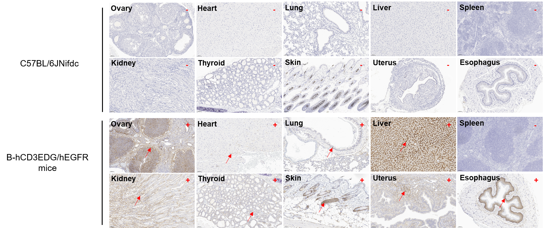
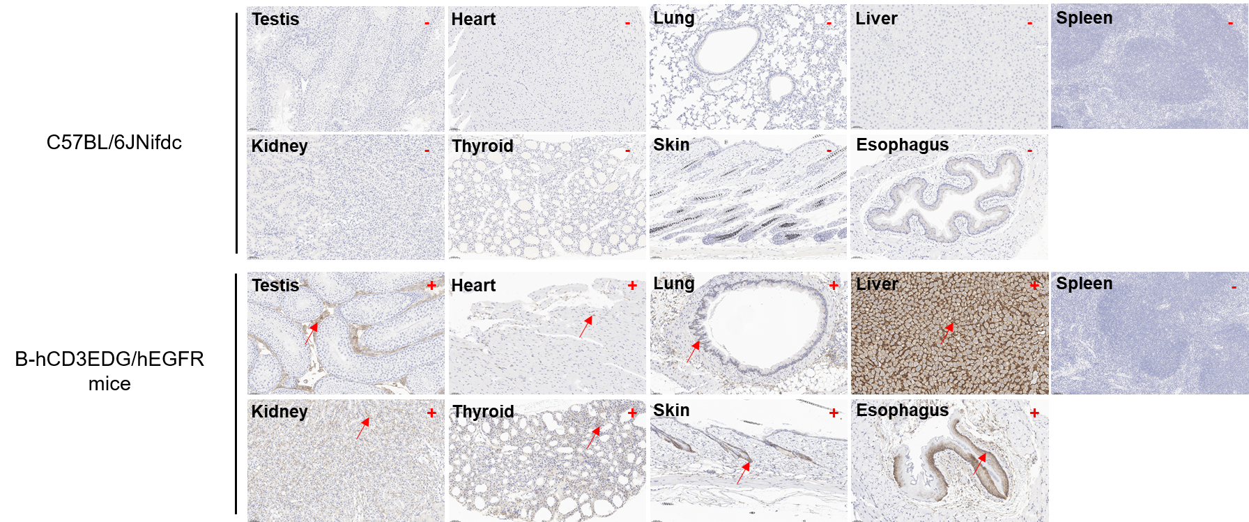
      Immunohistochemical (IHC) analysis of EGFR expression in homozygous B-hCD3EDG/hEGFR mice. The ovary, testis, heart, lung, liver, spleen, skin, thyroid, esophagus, kidney and uterus were collected from C57BL/6 mice (+/+) (G1: female, 5-week-old, n=2; G3: male, 5-week-old , n=2) and homozygous B-hCD3EDG/hEGFR mice (H/H; H/H; H/H; H/H) (G2: female, 5-week-old, n=2; G4: male, 5-week-old , n=2) , analyzed by IHC with anti-EGFR (abcam, ab32198). EGFR was detectable in both C57BL/6 mice and homozygous B-hCD3EDG/hEGFR mice because of antibody crossover. The arrow indicates tissue cells with positive EGFR staining (brown). “+” indicates that the tissue is positive, and “-” indicates that the tissue is negative. "unknown" means it can not be determined.