Targeting strategy
Gene targeting strategy for B-hCD3EDG/hTFR1 mice.
The exons 4-19 of mouse Tfr1 gene that encode extracellular domain are replaced by human counterparts in B-hCD3EDG/hTFR1 mice. The genomic region of mouse Tfr1 gene that encodes cytoplasmic portion is retained. The promoter, 5’UTR and 3’UTR region of the mouse gene are also retained. The chimeric TFR1 expression is driven by endogenous mouse Tfr1 promoter, while mouse Tfr1 gene transcription and translation will be disrupted.
In B-hCD3EDG/hTFR1 mice, chimeric human CD3EDG was expressed, while mouse Cd3edg were knocked out.
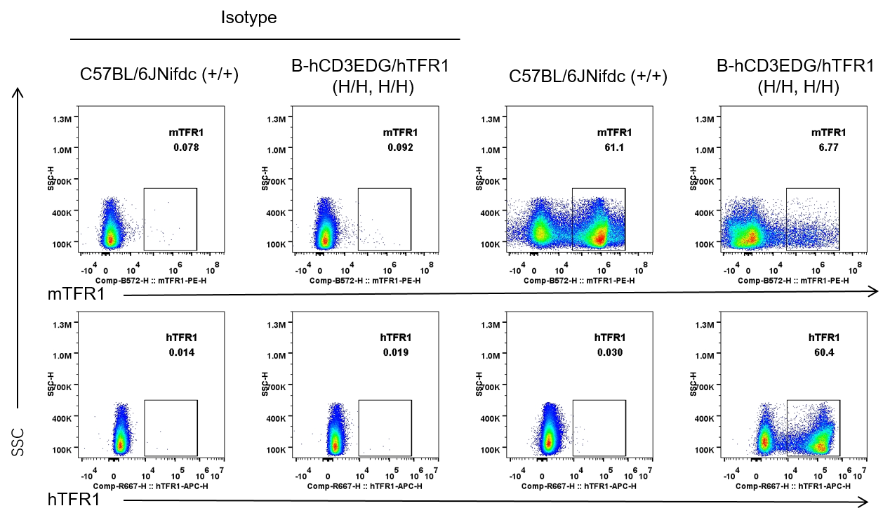
Protein expression analysis in bone marrow erythrocytes
Strain specific TFR1 expression analysis in wild-type C57BL/6JNifdc mice and homozygous humanized B-hCD3EDG/hTFR1 mice by flow cytometry. Bone marrow erythrocytes were collected from wild-type C57BL/6JNifdc mice (+/+) and homozygous B-hCD3EDG/hTFR1 mice (H/H, H/H). Protein expression was analyzed with anti-mouse TFR1 antibody (Biolegend, 113808) and anti-human TFR1 antibody (Biolegend, 334107) by flow cytometry. Mouse TFR1 was only detectable in wild-type C57BL/6JNifdc mice. Human TFR1 was exclusively detectable in homozygous B-hCD3EDG/hTFR1 mice, but not in wild-type C57BL/6JNifdc mice.
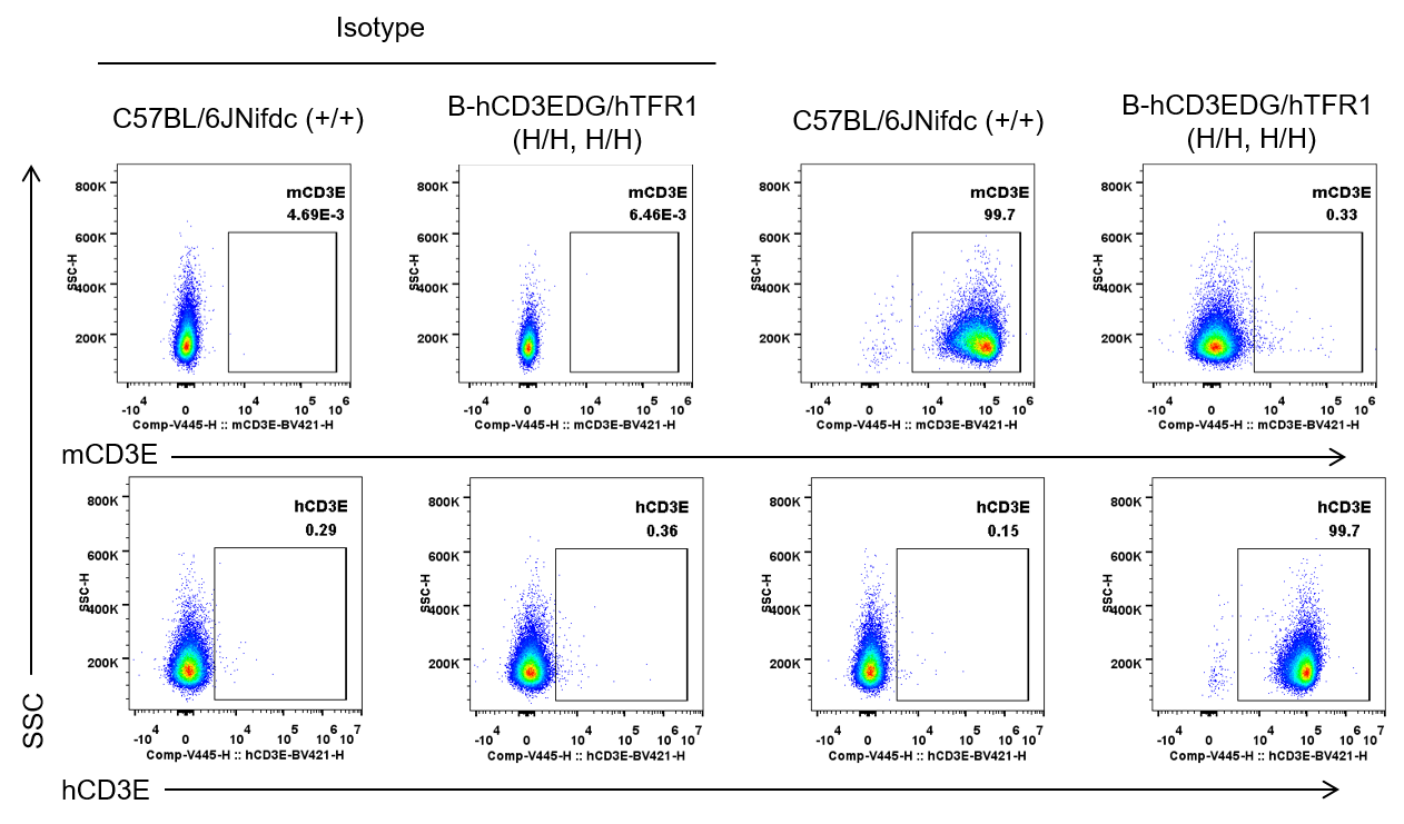
Protein expression analysis in T cells
Strain specific CD3E expression analysis in wild-type C57BL/6JNifdc mice and homozygous humanized B-hCD3EDG/hTFR1 mice by flow cytometry. Splenocytes were collected from wild-type C57BL/6JNifdc mice (+/+) and homozygous B-hCD3EDG/hTFR1 mice (H/H, H/H). Protein expression was analyzed with anti-mouse CD3E antibody (Biolegend, 100341) and anti-human CD3E antibody (BD Horizon™, 562426) by flow cytometry. Mouse CD3E was only detectable in wild-type C57BL/6JNifdc mice. Human CD3E was exclusively detectable in homozygous B-hCD3EDG/hTFR1 mice.
Protein expression analysis in B cells
Strain specific CD3E expression analysis in wild-type C57BL/6JNifdc mice and homozygous humanized B-hCD3EDG/hTFR1 mice by flow cytometry. Splenocytes were collected from wild-type C57BL/6JNifdc mice (+/+) and homozygous B-hCD3EDG/hTFR1 mice (H/H, H/H). Protein expression was analyzed with anti-mouse CD3E antibody (Biolegend, 100341) and anti-human CD3E antibody (BD Horizon™, 562426) by flow cytometry. CD3E cannot be detected in B cells of wild-type C57BL/6JNifdc mice and homozygous B-hCD3EDG/hTFR1 mice.
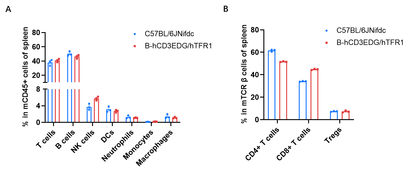
Frequency of leukocyte subpopulations in spleen
Frequency of leukocyte subpopulations in spleen by flow cytometry. Splenocytes were isolated from wild-type C57BL/6JNifdc mice (male, n=3, 9-week-old) and homozygous B-hCD3EDG/hTFR1 mice (male, n=3, 9-week-old). A. Flow cytometry analysis of the splenocytes was performed to assess the frequency of leukocyte subpopulations. B. Frequency of T cell subpopulations. Percentages of T cells, B cells, NK cells, dendritic cells, Neutrophils, monocytes, macrophages, CD4+ T cells, CD8+ T cells and Tregs in B-hCD3EDG/hTFR1 mice were similar to those in C57BL/6JNifdc mice. Values are expressed as mean ± SEM. Significance was determined by two-way ANOVA test. *P < 0.05, **P < 0.01, ***p < 0.001.
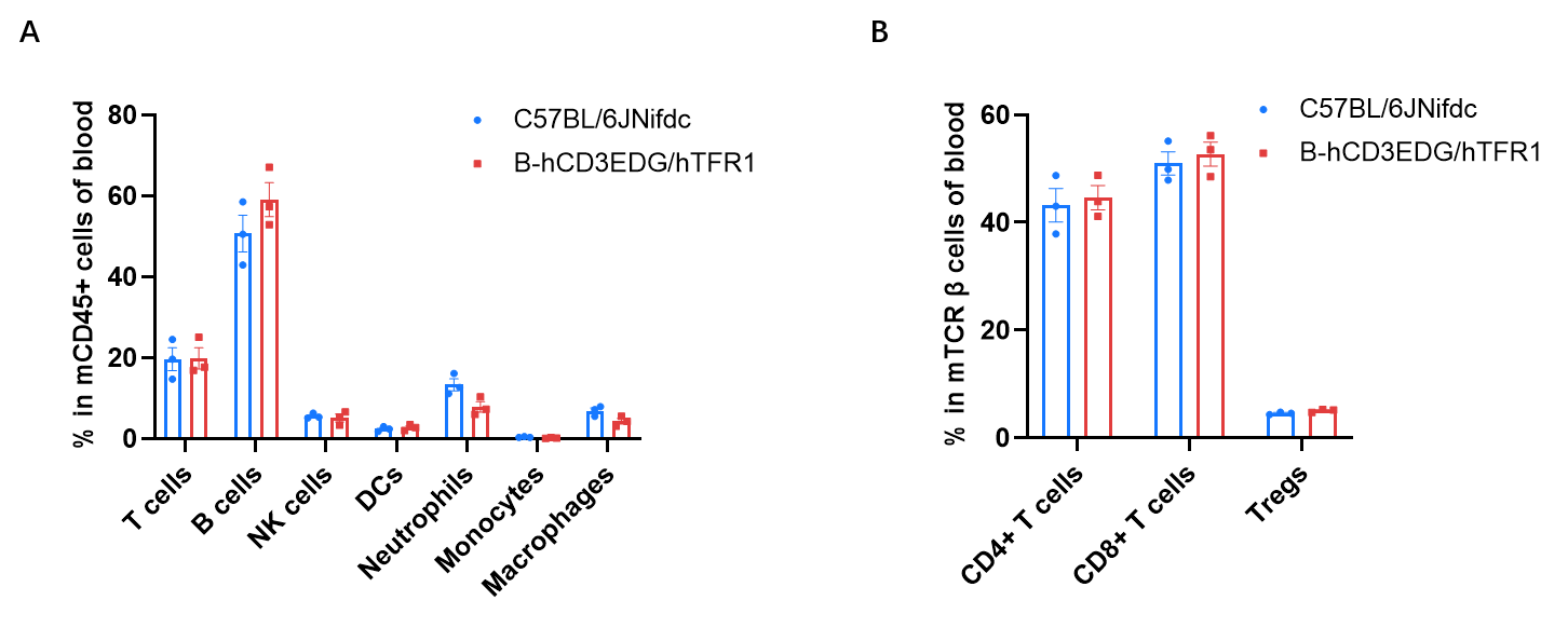
Frequency of leukocyte subpopulations in blood
Frequency of leukocyte subpopulations in blood by flow cytometry. Blood cells were isolated from wild-type C57BL/6JNifdc mice (male, n=3, 9-week-old) and homozygous B-hCD3EDG/hTFR1 mice (male, n=3, 9-week-old). A. Flow cytometry analysis of the blood cells was performed to assess the frequency of leukocyte subpopulations. B. Frequency of T cell subpopulations. Percentages of T cells, B cells, NK cells, dendritic cells, Neutrophils, monocytes, macrophages, CD4+ T cells, CD8+ T cells and Tregs in B-hCD3EDG/hTFR1 mice were similar to those in C57BL/6JNifdc mice. Values are expressed as mean ± SEM. Significance was determined by two-way ANOVA test. *P < 0.05, **P < 0.01, ***p < 0.001.
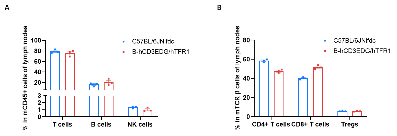
Frequency of leukocyte subpopulations in lymph nodes
Frequency of leukocyte subpopulations in Lymph nodes by flow cytometry. Lymph nodes cells were isolated from wild-type C57BL/6JNifdc mice (male, n=3, 9-week-old) and homozygous B-hCD3EDG/hTFR1 mice (male, n=3, 9-week-old). A. Flow cytometry analysis of the lymph nodes cells was performed to assess the frequency of leukocyte subpopulations. B. Frequency of T cell subpopulations. Percentages of T cells, B cells, NK cells, CD4+T cells, CD8+T cells and Tregs in B-hCD3EDG/hTFR1 mice were similar to those in C57BL/6JNifdc mice. Significance was determined by two-way ANOVA test. *P < 0.05, **P < 0.01, ***p < 0.001.
* When publishing results obtained using this animal model, please acknowledge the source as follows: The animal model [B-hCD3EDG/hTFR1 mice] (Cat# 113616) was purchased from Biocytogen.