Targeting strategy
Gene targeting strategy for B-hEGFR/hMET/hHGF mice.
The CDS that encodes the full-length human HGF protein, followed by mouse 3’UTR-STOP and polyA signal sequence was inserted into exon 3 of mouse Hgf to replace part of exon 3 to exon 6 of mouse Hgf gene. The promoter, 5’UTR and signal peptide region of the mouse Hgf gene were also retained. The human HGF protein expression will be driven by endogenous mouse Hgf promoter, while mouse Hgf gene transcription and translation will be disrupted.
The exons 3-14 of mouse Met gene that encode extracellular domain were replaced by human counterparts in B-hEGFR/hMET/hHGF mice. The genomic region of mouse Met gene that encodes transmembrane domain and cytoplasmic portion was retained. The promoter, 5’UTR and 3’UTR region, and signal peptide of the mouse gene were also retained. The chimeric MET expression was driven by endogenous mouse Met promoter, while mouse Met gene transcription and translation will be disrupted.
The exons 2-17 of mouse Egfr gene that encode the extracellular domain were replaced by human EGFR exons 2-17 in B-hEGFR/hMET/hHGF mice. The genomic region of mouse Egfr gene that encodes transmembrane domain and cytoplasmic portion was retained. The promoter, 5’UTR and 3’UTR region, and signal peptide of the mouse gene were also retained. The chimeric EGFR expression was driven by endogenous mouse Egfr promoter, while mouse Egfr gene transcription and translation will be disrupted.
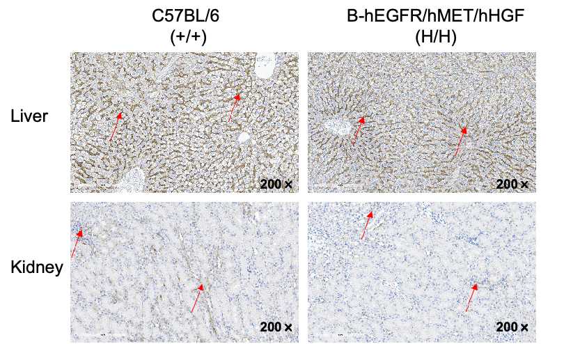
IHC analysis of EGFR expression
Immunohistochemical (IHC) analysis of EGFR expression in wild-type C57BL/6 mice and homozygous B-hEGFR/hMET/hHGF mice. The liver and kidney were collected from wild-type mice (+/+) and B-hEGFR/hMET/hHGF mice (H/H) and analyzed by IHC with anti-EGFR antibody(abcam, ab32198). EGFR was detectable in wild-type mice and homozygous B-hEGFR/hMET/hHGF mice due to the cross-reactivity of antibodies.
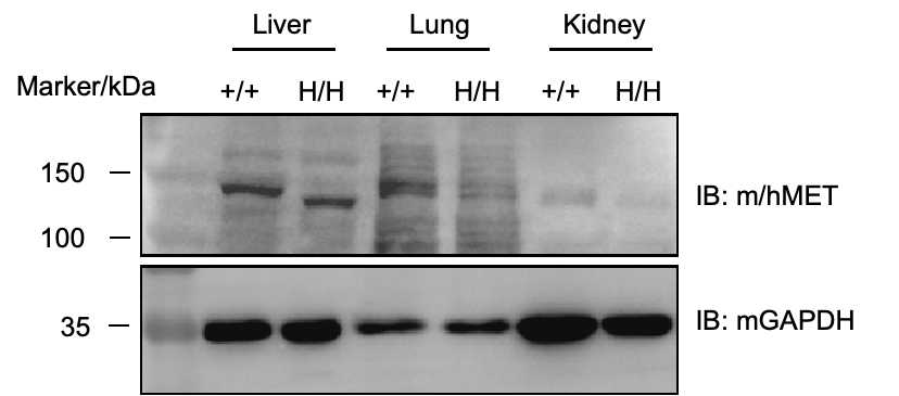
Protein expression analysis-MET
Western blot analysis of MET protein expression in homozygous B-hEGFR/hMET/hHGF mice. Liver, lung and kidney tissue lysates were collected from wild-type C57BL/6 mice (+/+) and homozygous B-hEGFR/hMET/hHGF mice (H/H), and then analyzed by western blot with anti-MET antibody(CST, 3127S). 60 μg total proteins were loaded for western blotting analysis. MET was detectable in wild-type mice and homozygous B-hEGFR/hMET/hHGF mice due to the cross-reactivity of antibodies.
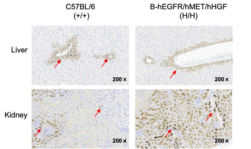
IHC analysis of MET expression
Immunohistochemical (IHC) analysis of MET expression in wild-type C57BL/6 mice and homozygous B-hEGFR/hMET/hHGF mice. The liver and kidney were collected from wild-type mice (+/+) and B-hEGFR/hMET/hHGF mice (H/H) and analyzed by IHC with anti-MET antibody(abcam, ab227637). MET was detectable in wild-type mice and homozygous B-hEGFR/hMET/hHGF mice due to the cross-reactivity of antibodies.
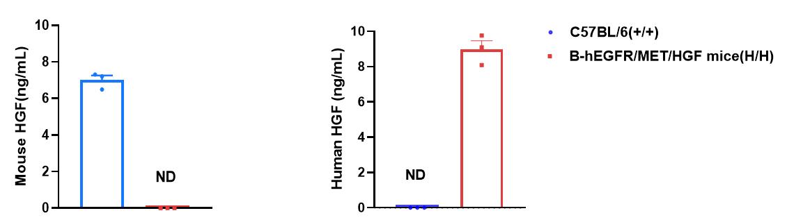
Protein expression analysis in serum-HGF
Strain specific HGF expression analysis in wild-type C57BL/6 mice and homozygous B-hEGFR/hMET/hHGF mice by ELISA. Serum was collected from wild-type C57BL/6 mice (+/+) and homozygous B-hEGFR/hMET/hHGF mice(H/H) (female, n=3, 9-week-old). Expression level of mouse and human HGF was analyzed by ELISA (anti-mouse HGF ELISA kit: R&D, MHG00; anti-human HGF ELISA kit: Abcam, ab275901). Mouse HGF was only detectable in wild-type C57BL/6 mice. Human HGF was exclusively detectable in homozygous B-hEGFR/hMET/hHGF mice. Values are expressed as mean ± SEM. ND: not detectable.
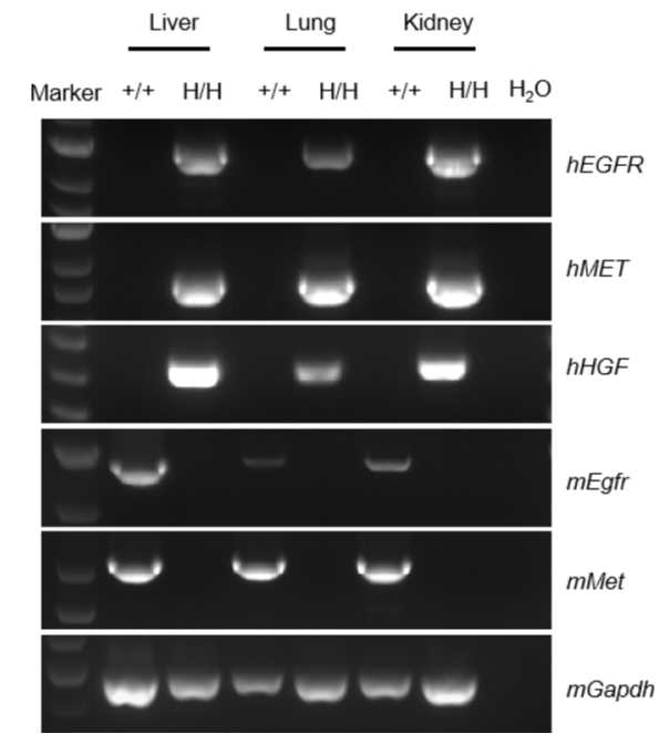
mRNA expression analysis
Strain specific analysis of EGFR, MET and HGF mRNA expression in wild-type C57BL/6 mice and homozygous B-hEGFR/hMET/hHGF mice by RT-PCR. Liver, lung and kidney RNA were isolated from wild-type C57BL/6 mice (+/+) and homozygous B-hEGFR/hMET/hHGF mice (H/H), then cDNA libraries were synthesized by reverse transcription, followed by PCR with mouse or human EGFR, MET and HGF primers. Mouse Egfr and Met mRNA were detectable only in wild-type C57BL/6 mice. Human EGFR, MET and HGF mRNA was exclusively detectable in homozygous B-hEGFR/hMET/hHGF mice but not in wild-type mice.
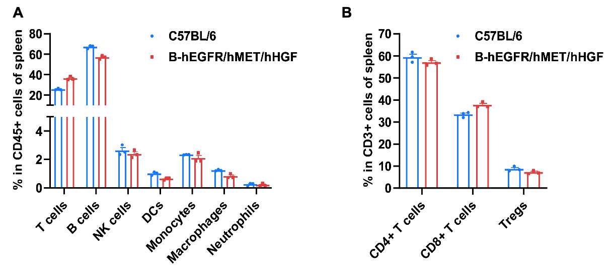
Frequency of leukocyte subpopulations in spleen
Frequency of leukocyte subpopulations in spleen by flow cytometry. Splenocytes were isolated from wild-type C57BL/6 mice and homozygous B-hEGFR/hMET/hHGF mice (Female,6-week-old, n=3). A. Flow cytometry analysis of the splenocytes was performed to assess the frequency of leukocyte subpopulations. B. Frequency of T cell subpopulations. Frequencies of T cells, B cells, NK cells, DCs, monocytes, macrophages, neutrophils, CD4+ T cells, CD8+ T cells and Tregs in B-hEGFR/hMET/hHGF mice were similar to those in C57BL/6 mice, demonstrating that humanization of EGFR, MET and HGF do not change the frequency or distribution of these cell types in spleen. Values are expressed as mean ± SEM.
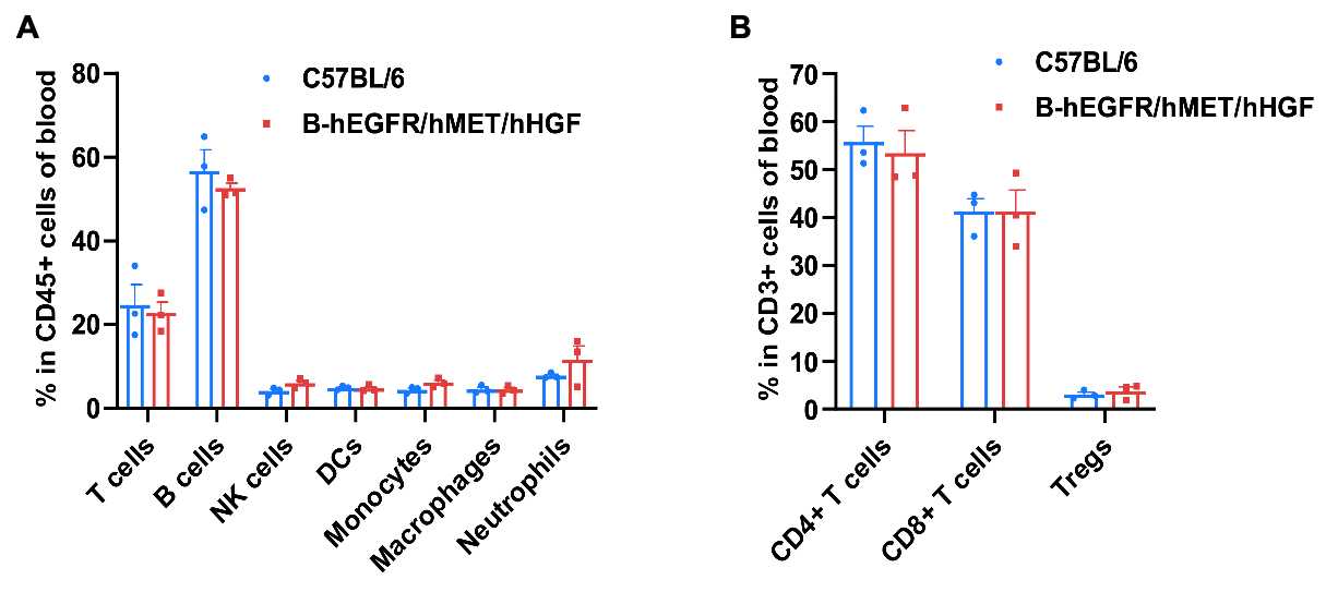
Frequency of leukocyte subpopulations in blood
Frequency of leukocyte subpopulations in blood by flow cytometry. Blood were isolated from wild-type C57BL/6 mice and homozygous B-hEGFR/hMET/hHGF mice (Female,6-week-old, n=3). A. Flow cytometry analysis of the blood was performed to assess the frequency of leukocyte subpopulations. B. Frequency of T cell subpopulations. Frequencies of T cells, B cells, NK cells, DCs, monocytes, macrophages, neutrophils, CD4+ T cells, CD8+ T cells and Tregs in B-hEGFR/hMET/hHGF mice were similar to those in C57BL/6 mice, demonstrating that humanization of EGFR, MET and HGF do not change the frequency or distribution of these cell types in blood. Values are expressed as mean ± SEM.
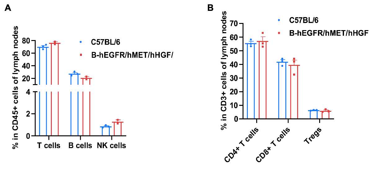
Frequency of leukocyte subpopulations in lymph node
Frequency of leukocyte subpopulations in lymph nodes by flow cytometry. Lymph nodes were isolated from wild-type C57BL/6 mice and homozygous B-hEGFR/hMET/hHGF mice (Female,6-week-old, n=3). A. Flow cytometry analysis of the lymph nodes was performed to assess the frequency of leukocyte subpopulations. B. Frequency of T cell subpopulations. Frequencies of T cells, B cells, NK cells, DCs, monocytes, macrophages, neutrophils, CD4+ T cells, CD8+ T cells and Tregs in B-hEGFR/hMET/hHGF mice were similar to those in C57BL/6 mice, demonstrating that humanization of EGFR, MET and HGF do not change the frequency or distribution of these cell types in lymph nodes. Values are expressed as mean ± SEM.
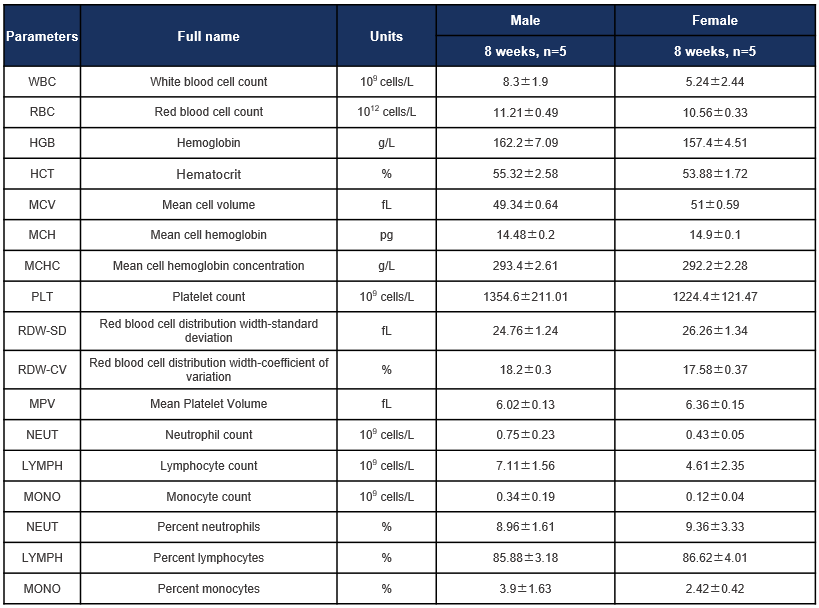
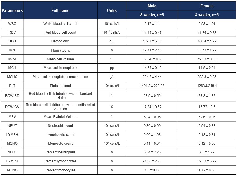
Hematology analysis
Complete blood count (CBC) of C57BL/6JNifdc mice. Values are expressed as mean ± SD.
Complete blood count (CBC) of B-hEGFR/hMET/hHGF mice. Values are expressed as mean ± SD.
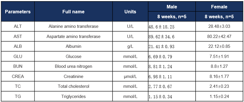
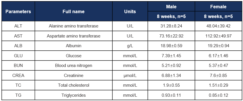
Biochemistry analysis
Biochemical test of C57BL/6JNifdc mice. Values are expressed as mean ± SD.
Biochemical test of B-hEGFR/hMET/hHGF mice. Values are expressed as mean ± SD.
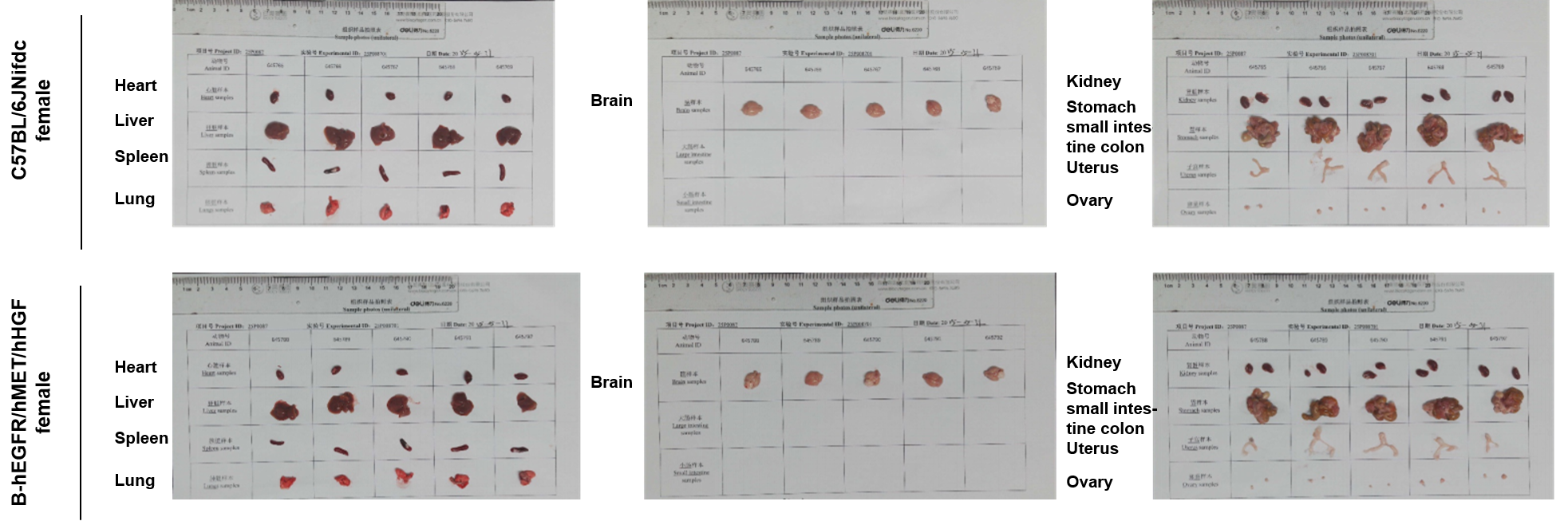
Gross anatomy of female mice
The organs of female C57BL/6JNifdc mice and B-hEGFR/hMET/hHGF mice (7-week-old, n=5).
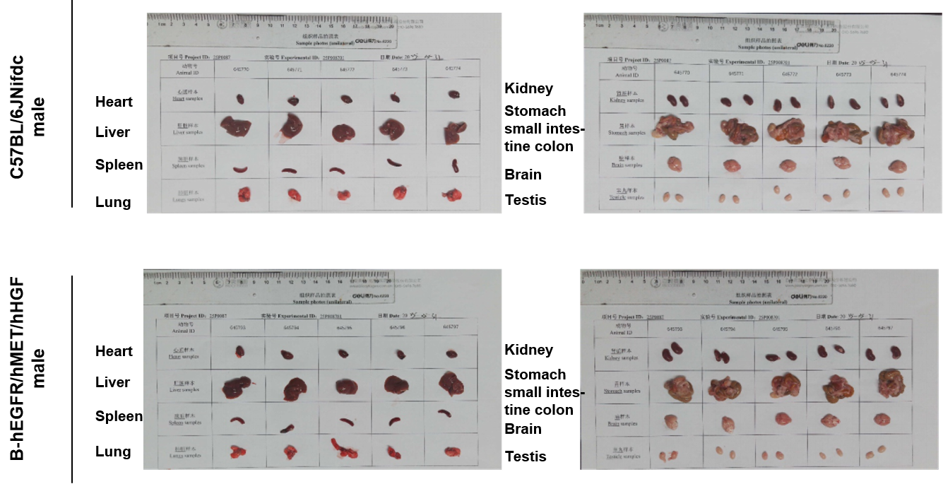
Gross anatomy of male mice
The organs of male C57BL/6JNifdc mice and B-hEGFR/hMET/hHGF mice (7-week-old, n=5).
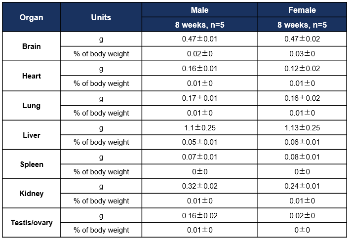
Organ Weight and Coefficient
Average weight of the main organs of C57BL/6JNifdc mice. Values are expressed as mean ± SD.
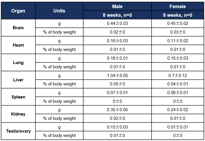
Organ weight and coefficient
Average weight of the main organs of B-hEGFR/hMET/hHGF mice. Values are expressed as mean ± SD.
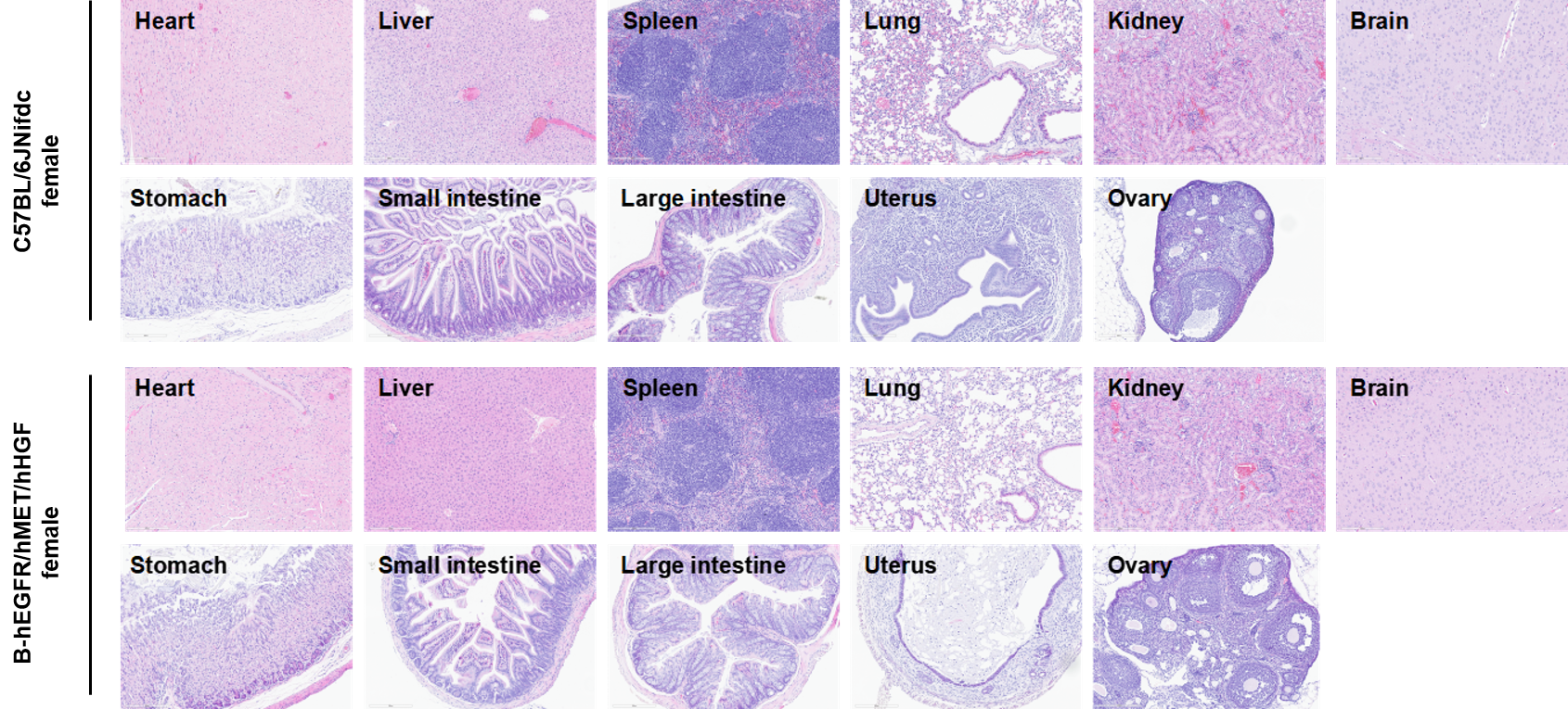
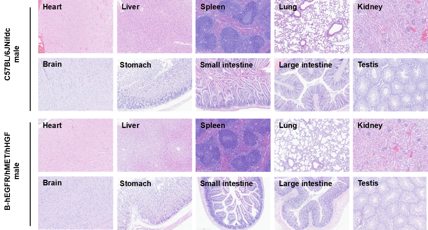
Histopathological analysis
Histopathological analysis of organs in B-hEGFR/hMET/hHGF mice. The main organs of B-hEGFR/hMET/hHGF mice were isolated at 8 weeks of age and analyzed with H&E staining (female, n=5). Results showed that no obvious abnormalities were found in all of the organs (heart, liver, spleen, lung, kidney, brain, stomach, small intestine, large intestine, kidney, uterus, ovary and testis). Scale bar: 200 μm.
Histopathological analysis of organs in B-hEGFR/hMET/hHGF mice. The main organs of B-hEGFR/hMET/hHGF mice were isolated at 8 weeks of age and analyzed with H&E staining (male, n=5). Results showed that no obvious abnormalities were found in all of the organs (heart, liver, spleen, lung, kidney, brain, stomach, small intestine, large intestine and testis). Scale bar: 200 μm.
* When publishing results obtained using this animal model, please acknowledge the source as follows: The animal model [B-hEGFR/hMET/hHGF mice] (Cat# 113292) was purchased from Biocytogen.