Q1. What are B-hPD-1/hPD-L1/hVEGFA mice mainly used for?
B-hPD-1/hPD-L1/hVEGFA mice are triple humanized PD-1/PD-L1/VEGFA mice designed for in vivo efficacy studies of human-specific PD-1/PD-L1 checkpoint inhibitors, anti-VEGFA antibodies and anti-PD-1/VEGF bispecific antibodies, including ivonescimab and bevacizumab/Keytruda combinations in B-hVEGFA MC38 syngeneic tumor models.
Q2. How do these humanized PD-1/PD-L1/VEGFA mice differ from single humanized PD-1 mice?
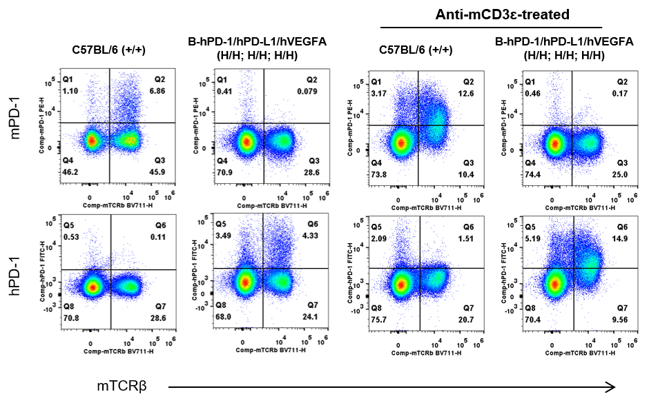
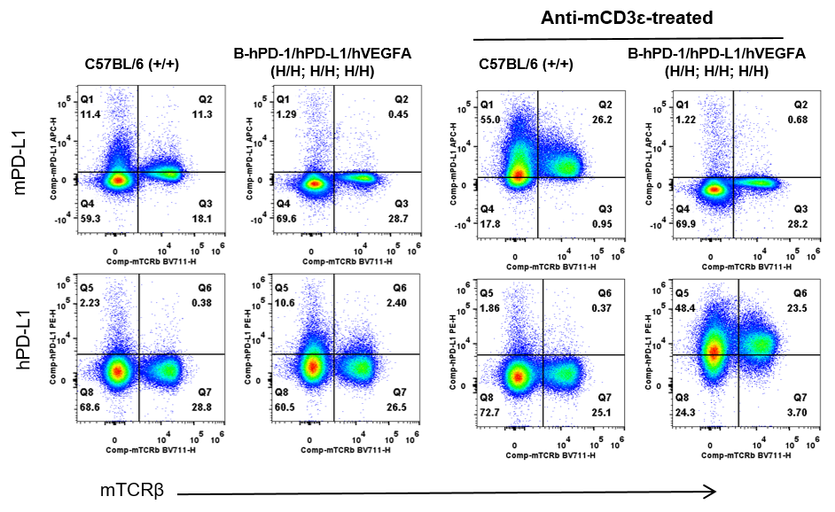
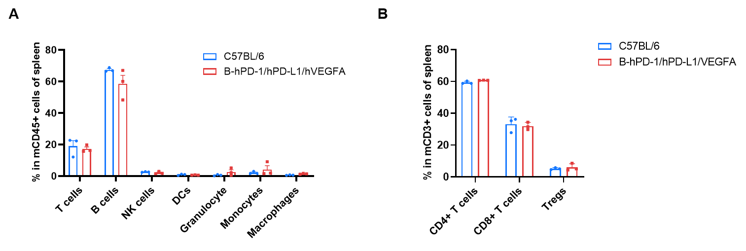
Compared with single-humanized PD-1 mice, this triple-humanized PD-1/PD-L1/VEGFA model simultaneously expresses human PD-1, PD-L1, and VEGFA. It better recapitulates the clinical setting in which anti–PD-1/PD-L1 and anti-VEGF/VEGFA agents act on multiple human targets, making it more predictive for combination therapies and bispecific antibody development.
Q3. Can I use B-hPD-1/hPD-L1/hVEGFA mice for ivonescimab (AK112) preclinical studies?
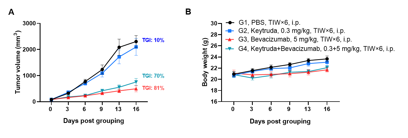
Yes. Ivonescimab (AK112) has been tested in B-hPD-1/hPD-L1/hVEGFA mice bearing B-hVEGFA/hPD-L1 MC38 tumors, where it showed significant antitumor activity, demonstrating that this model is suitable for ivonescimab and other anti-PD-1/VEGF bispecific antibody evaluation.
Q4. What tumor models are recommended with B-hPD-1/hPD-L1/hVEGFA mice?
B-hVEGFA MC38 and B-hVEGFA/hPD-L1 MC38 tumor models are recommended, as they express human VEGFA and human PD-L1, ensuring fully human ligand–receptor engagement with human PD-1/PD-L1/VEGFA in the host mice.