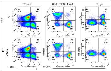
Analysis of spleen leukocyte subpopulations by FACS.
Splenocytes were isolated from male B-Foxp3-EGFP-DTR-Luc mice (n=3, 7-month-old) injected with PBS or DT (30ng/ per body weight) for two consecutive days. Flow cytometry analysis of the splenocytes was performed to assess leukocyte subpopulations.
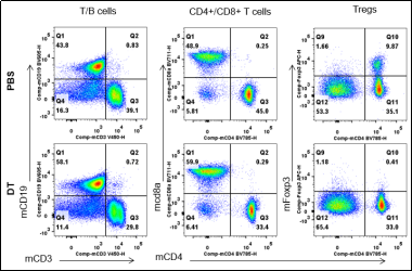
Analysis of lymph node leukocyte subpopulations by FACS.
Lymph node were isolated from male B-Foxp3-EGFP-DTR-Luc mice (n=3, 7-month-old) injected with PBS or DT (30ng/ per body weight) for two consecutive days. Flow cytometry analysis was performed to assess leukocyte subpopulations.
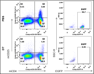
Analysis of EGFP expression in spleen of B-Foxp3-EGFP-DTR-Luc mice by FACS.
Splenocytes were isolated from B-Foxp3-EGFP-DTR-Luc mice mice(n=3, 7-month-old) injected with PBS or DT (30ng/per body weight) for two consecutive days. Flow cytometry analysis was performed to assess EGFP expression.
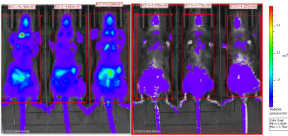
BLI analysis of homozygous B-Foxp3-EGFP-DTR-Luc mice after DT injection.
Homozygous B-Foxp3-EGFP-DTR-Luc mice i.p. injected with PBS (n=3) or DT (n=3) for two consecutive days were anaesthetized for the bioluminescence imaging. Mice were imaged 10min after i.p. injection of 150mg/kg D-Lucifenrin potassium salt using IVIS Lumina LT Inst Series IIIimaging system.The ratio of Treg cells in homozygous B-Foxp3-EGFP-DTR-Luc mice is comparable with that in wild type mice; EGFP was exclusively detectable in CD4+; CD25+ populations from Foxp3-EGFP-DTR-Luc mice and could be as a marker for Treg cells in vivo; besides Bioluminescence imaging could also be used for tracing Tregs cells; After DT injection, Treg cells showed dramatically decreased at EGFP, Foxp3 and bioluminescence imaging levels.
* When publishing results obtained using this animal model, please acknowledge the source as follows: The animal model [B-Foxp3-EGFP-DTR-Luc mice] (Cat# 112682) was purchased from Biocytogen.
