C57BL/6JNifdc-Apptm1(Abeta*K670N*M671L*G676R*F681Y*R684H*I716F)BcgenPsen1tm1Bcgen /Bcgen • 113247
| Product name | B-App NL-F/Psen1*E120K*M146L mice |
|---|---|
| Catalog number | 113247 |
| Strain name | C57BL/6JNifdc-Apptm1(Abeta*K670N*M671L*G676R*F681Y*R684H*I716F)BcgenPsen1tm1Bcgen /Bcgen |
| Strain background | C57BL/6JNifdc |
| NCBI gene ID | 11820,19164 (Mouse) |
| Aliases | Ag; Abpp; Adap; Cvap; Abeta; betaApp; E030013M08Rik; PS1; Ad3h; PS-1; S182 |
Gene targeting strategy for B-App NL-F/Psen1*E120K*M146L mice . B-App NL-F/Psen1*E120K*M146L mice carried some mutations on the mouse App gene, including R684H, F681Y, and G676R mutations on the Aβ region, and the KM670/671NL (Swedish) mutation in exon 16 as well as the I716F (Beyreuther/Iberian) mutation in exon 17. This mice expressed humanized Aβ with two familial AD mutations. B-App NL-F/Psen1*E120K*M146L mice also carried two mutations on the mouse Psen1 gene, including E120K and M146L mutations.
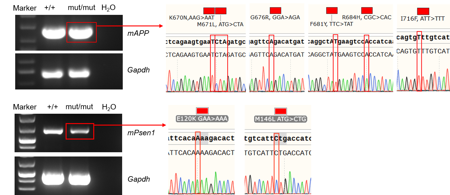
App and Psen1 mRNA expression in wild-type C57BL/6 mice and homozygous B-App NL-F/Psen1*E120K*M146L mice by RT-PCR. Brain RNA were isolated from wild-type C57BL/6 mice (+/+) and homozygous B-App NL-F/Psen1*E120K*M146L mice (mut/mut), then cDNA libraries were synthesized by reverse transcription, followed by PCR with mouse App and Psen1 primers. Mouse App and Psen1 mRNA were detectable in wild-type C57BL/6 and homozygous mice. And the point mutations were confirmed via Sanger Sequencing.
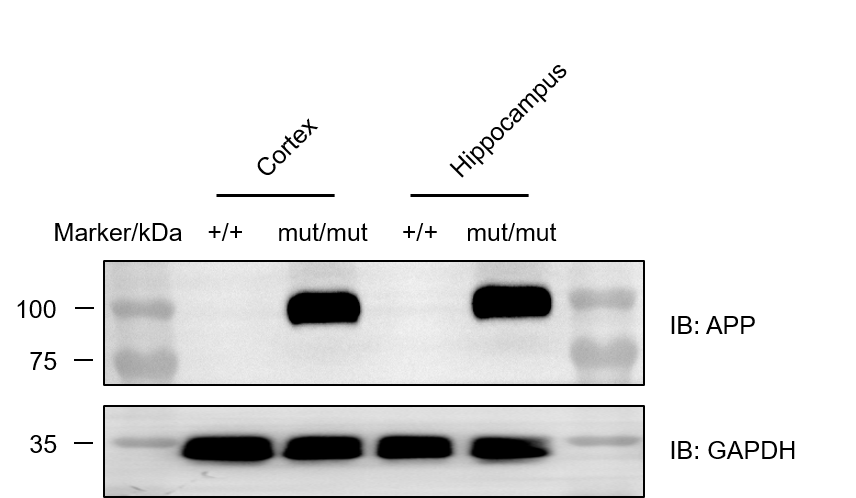
Western blot analysis of APP protein expression in homozygous B-App NL-F/Psen1*E120K*M146L mice . Various tissue lysates were collected from wild-type C57BL/6 mice (+/+) and homozygous B-App NL-F/Psen1*E120K*M146L mice (mut/mut), and then analyzed by western blot with species-specific anti-amyloid precursor antibody (Abcam, ab133588). 50 μg total proteins were loaded for western blotting analysis. Humanized Aβ was detected in the cortex and hippocampus of homozygous B-App NL-F/Psen1*E120K*M146L mice , but not in wild-type mice.
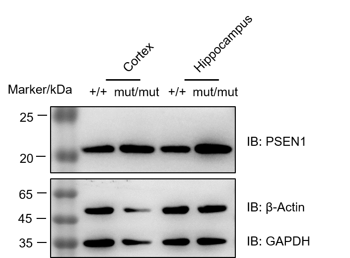
Western blot analysis of PSEN1 protein expression in homozygous B-App NL-F/Psen1*E120K*M146L mice. Cortex and hippocampus lysates were collected from wild-type C57BL/6 mice (+/+) and homozygous B-App NL-F/Psen1*E120K*M146L mice (mut/mut), and then analyzed by western blot with anti-PSEN1 antibody (CST, #5643). 50 μg total proteins were loaded for western blotting analysis. PSEN1 was detected in cortex and hippocampus both in wild-type and homozygous B-App NL-F/Psen1*E120K*M146L mice.
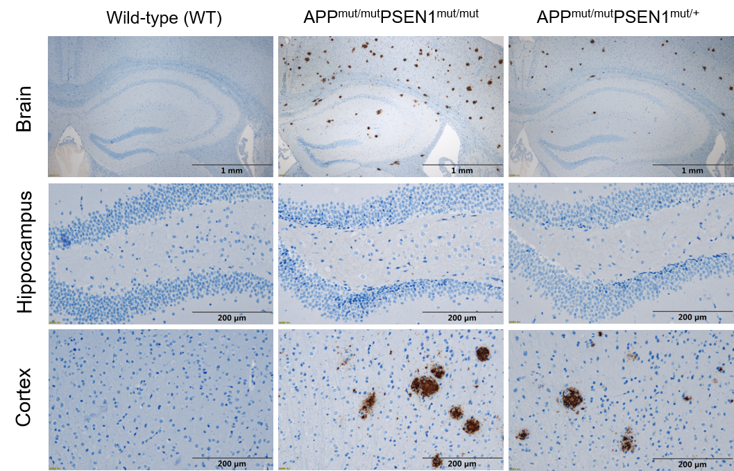
Histopathological analysis of human Aβ in B-App NL-F/Psen1*E120K*M146L mice. Brain was collected from wild-type mice (6-month-old) and B-App NL-F/Psen1*E120K*M146L mice (6-month-old) and processed into paraffin sections. The Aβ plaque in the cortex and hippocampus of C57BL/6 mice and B-App NL-F/Psen1*E120K*M146L mice was detected by IHC with anti-human β-Amyloid antibody (CST, #8243S). The Aβ plaque was exclusively detectable in B-App NL-F/Psen1*E120K*M146L mice, but not in wild-type mice. At the same age, Psen1 gene homozygous mice exhibit more pronounced Aβ deposition compared to heterozygous mice.
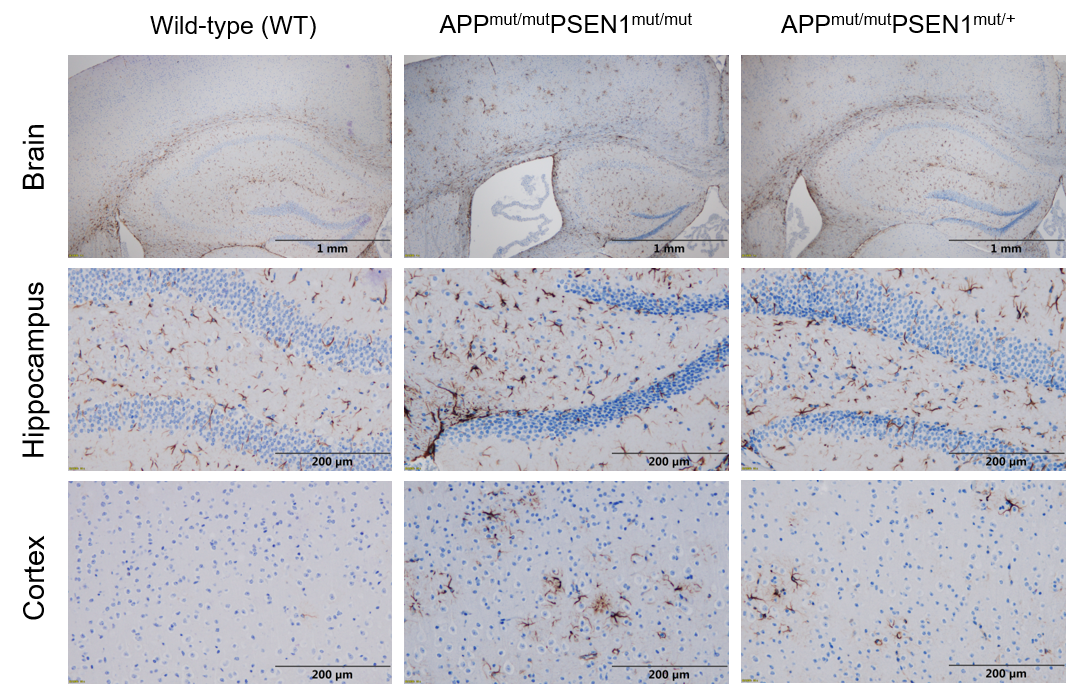
Histopathological analysis of astrocytes in B-App NL-F/Psen1*E120K*M146L mice. Brain was collected from wild-type mice (6-month-old) and B-App NL-F/Psen1*E120K*M146L mice (6-month-old) and processed into paraffin sections. The expression of GFAP in the cortex and hippocampus of C57BL/6 mice and B-App NL-F/Psen1*E120K*M146L mice was detected by IHC with anti-GFAP antibody (abcam, ab68428). Compared to wild-type mice, the number of activated astrocytes in the cortex and hippocampus of B-App NL-F/Psen1*E120K*M146L mice significantly increases, indicating the presence of inflammation in the brain.
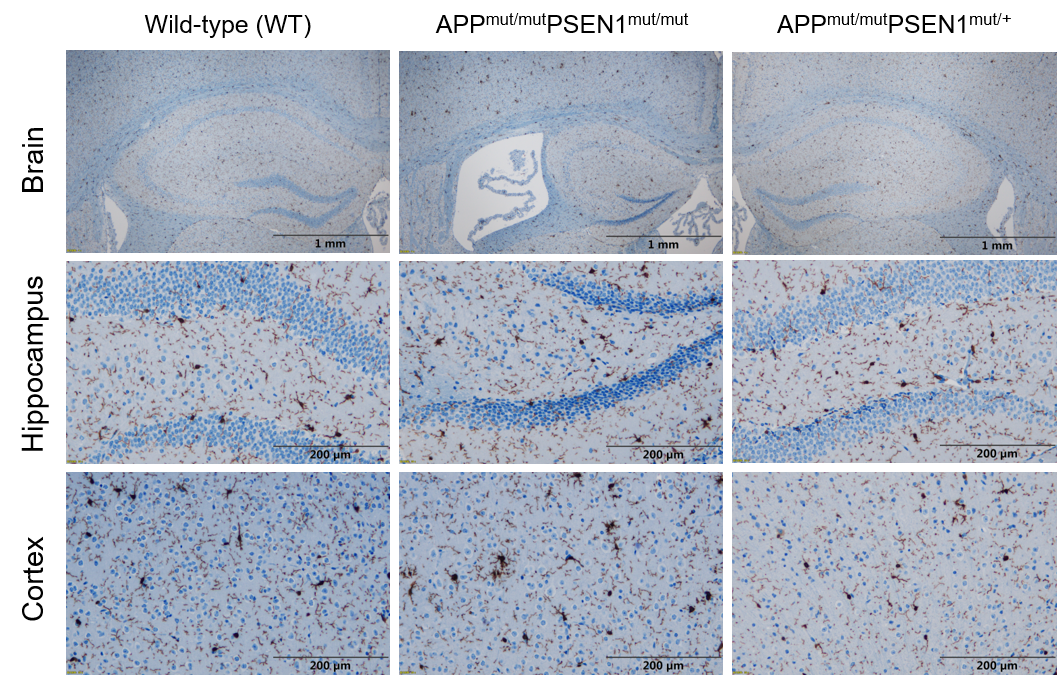
Histopathological analysis of microglia cells in B-App NL-F/Psen1*E120K*M146L mice. Brain was collected from wild-type mice (6-month-old) and B-App NL-F/Psen1*E120K*M146L mice (6-month-old) and processed into paraffin sections. The expression of Iba1 in the cortex and hippocampus of C57BL/6 mice and B-App NL-F/Psen1*E120K*M146L mice was detected by IHC with anti-Iba1 antibody (abcam, ab178846). Compared to wild-type mice, the number of activated microglia cells in the cortex and hippocampus of B-App NL-F/Psen1*E120K*M146L mice significantly increases, indicating the presence of inflammation in the brain.