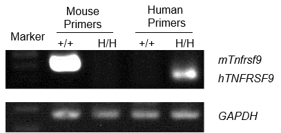
Expression by RT-PCR
- Mouse 4-1BB mRNA was detectable in wild-type C57BL/6 mice.
- Human 4-1BB mRNA was detectable only in homozygous B-h4-1BB mice but not in wild-type mice.
Strain specific analysis of 4-1BB mRNA expression in wild-type C57BL/6 mice and homozygous B-h4-1BB mice by RT-PCR. Spleen RNA was isolated from wild-type C57BL/6 mice (+/+) and homozygous B-h4-1BB mice (H/H), then cDNA libraries were synthesized by reverse transcription, followed by PCR with mouse or human 4-1BB primers.
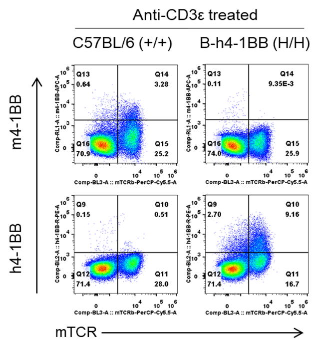
4-1BB Protein Expression
- Mouse 4-1BB was only detected on T cells in wild-type C57BL/6 mice.
- Human 4-1BB was detected on T cells in B-h4-1BB mice, but not in wild-type C57BL/6 mice.
Strain specific 4-1BB protein expression analysis in C57BL/6 mice and homozygous B-h4-1BB mice by flow cytometry. Splenocytes were collected from wild type C57BL/6 mice (+/+) and homozygous B-h4-1BB mice (H/H) that stimulated with anti-CD3ε in vivo, and analyzed by flow cytometry with species-specific anti-4-1BB antibodies.
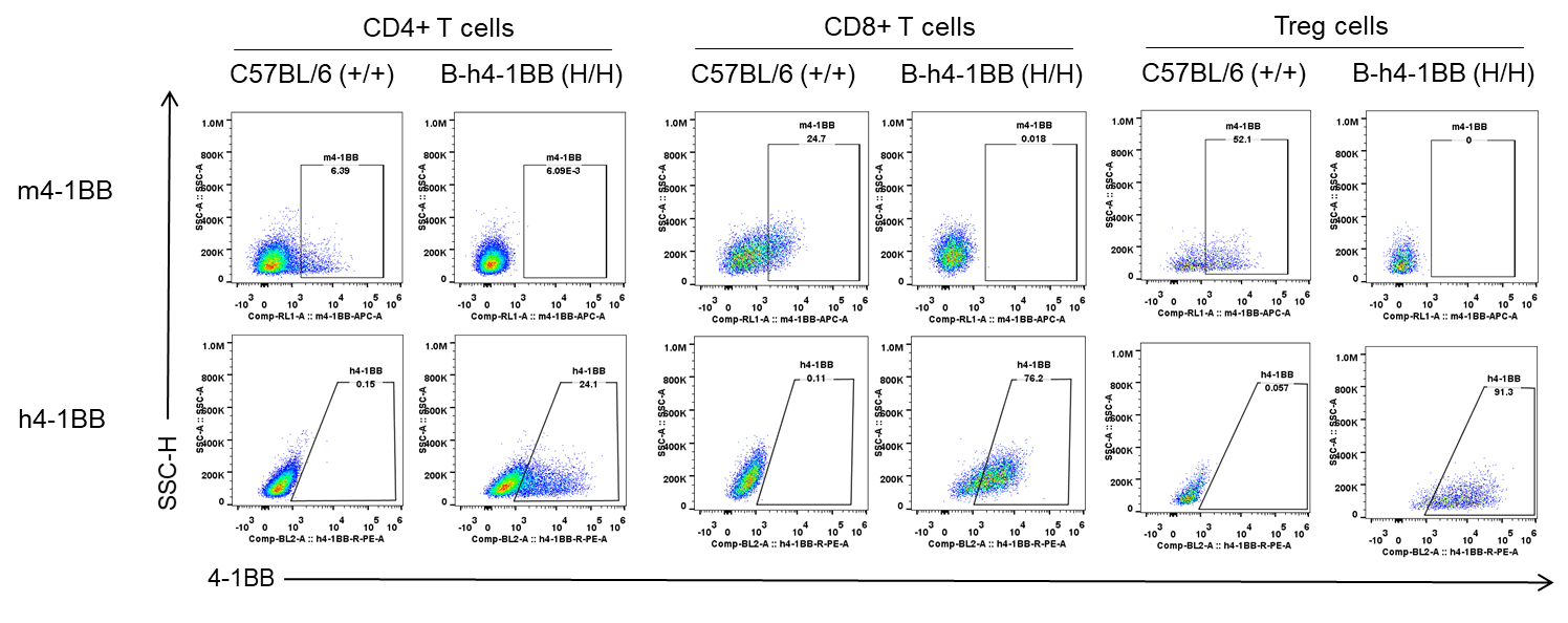
4-1BB Protein Expression in spleen
- Mouse 4-1BB was only detected on CD4+ T cells, CD8+ T cells and Treg cells in wild-type C57BL/6 mice.
- Human 4-1BB was detected on CD4+ T cells, CD8+ T cells and Treg cells in B-h4-1BB mice, but not in wild-type C57BL/6 mice.
Mouse and human 4-1BB expression analysis in splenocytes. Splenocytes were collected from wild-type C57BL/6 mice and homozygous B-h4-1BB mice that stimulated with anti-CD3ε in vivo. 4-1BB expression on T cells, CD4+ T cells, CD8+ T cells and Tregs was analyzed by flow cytometry using species-specific anti-4-1BB antibodies.
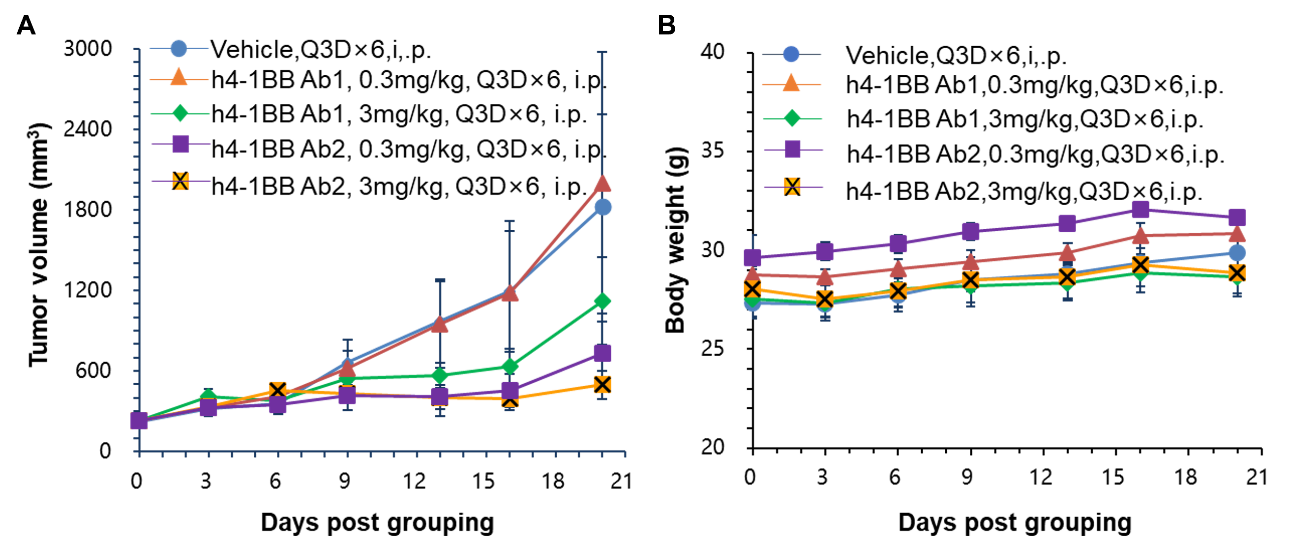
Efficacy Evaluation of anti-4-1BB antibody in the Treatment of the Subcutaneous MC38 Model in B-h4-1BB mice in vivo
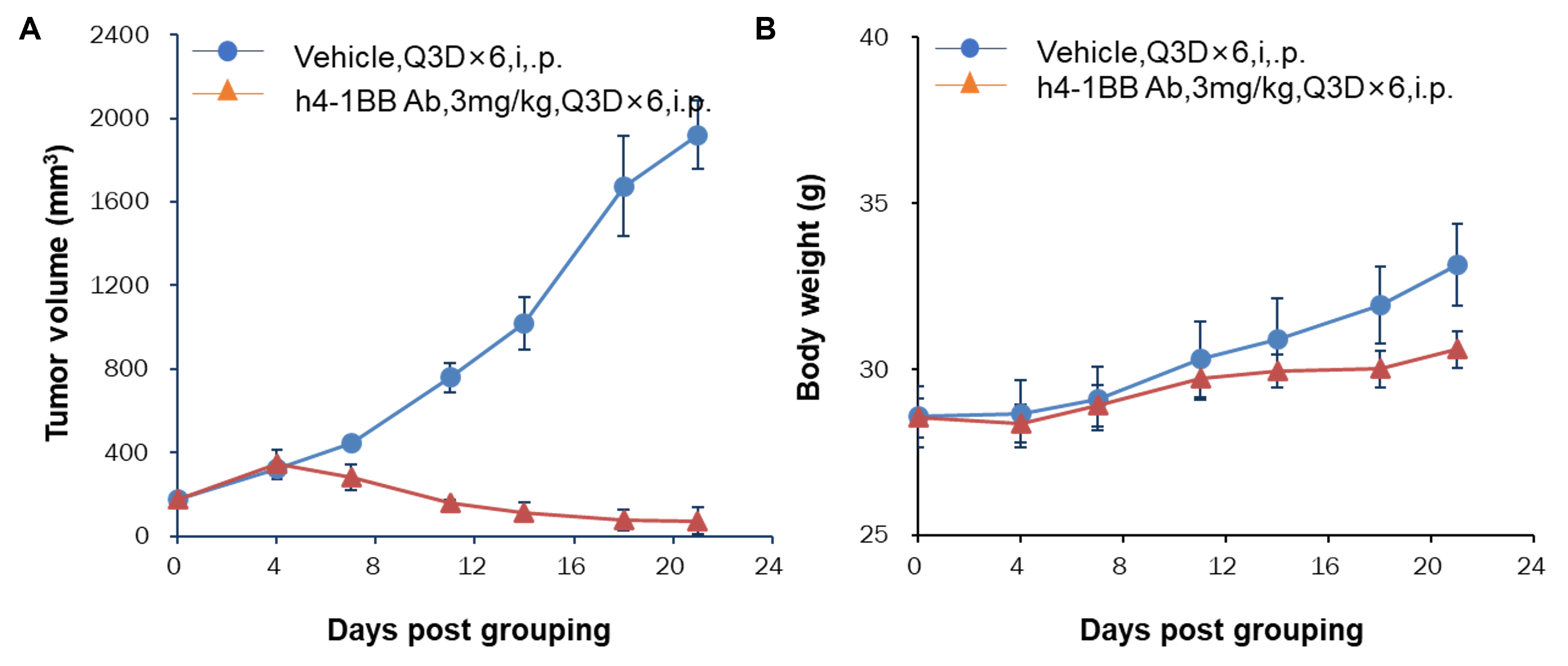
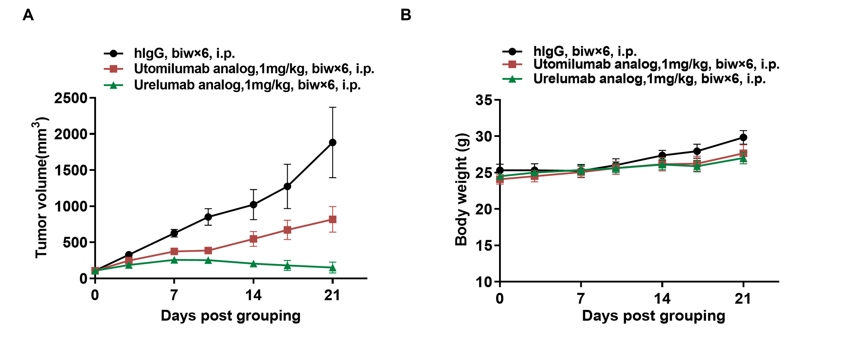
- Anti-human 4-1BB antibodies were efficacious in controlling tumor growth in B-h4-1BB mice.
- B-h4-1BB mice provide a powerful preclinical model for in vivo evaluation of anti-human 4-1BB antibodies.
Efficacy of anti-human 4-1BB antibodies in B-h4-1BB mice. (A) Anti-human 4-1BB antibodies inhibited MC38 tumor growth in B-h4-1BB mice. Murine colon cancer MC38 cells were subcutaneously implanted into homozygous B-h4-1BB mice (female, 6-7 week-old, n=5). Mice were grouped when tumor volume reached approximately 100 mm3, at which time they were treated with two anti-human 4-1BB antibodies with doses and schedules indicated in panel. (B) Body weight changes during treatment. Values are expressed as mean ± SEM.
- Anti-human 4-1BB antibodies were efficacious in controlling tumor growth in B-h4-1BB mice.
- B-h4-1BB mice provide a powerful preclinical model for in vivo evaluation of anti-human 4-1BB antibodies.
Efficacy of anti-human 4-1BB antibodies in B-h4-1BB mice. (A) Anti-human 4-1BB antibodies inhibited MC38 tumor growth in B-h4-1BB mice. Murine colon cancer MC38 cells were subcutaneously implanted into homozygous B-h4-1BB mice (male, 7-8 week-old, n=6). Mice were grouped when tumor volume reached approximately 100 mm3, at which time they were treated with an anti-human 4-1BB antibody with doses and schedules indicated in panel. (B) Body weight changes during treatment. Values are expressed as mean ± SEM.
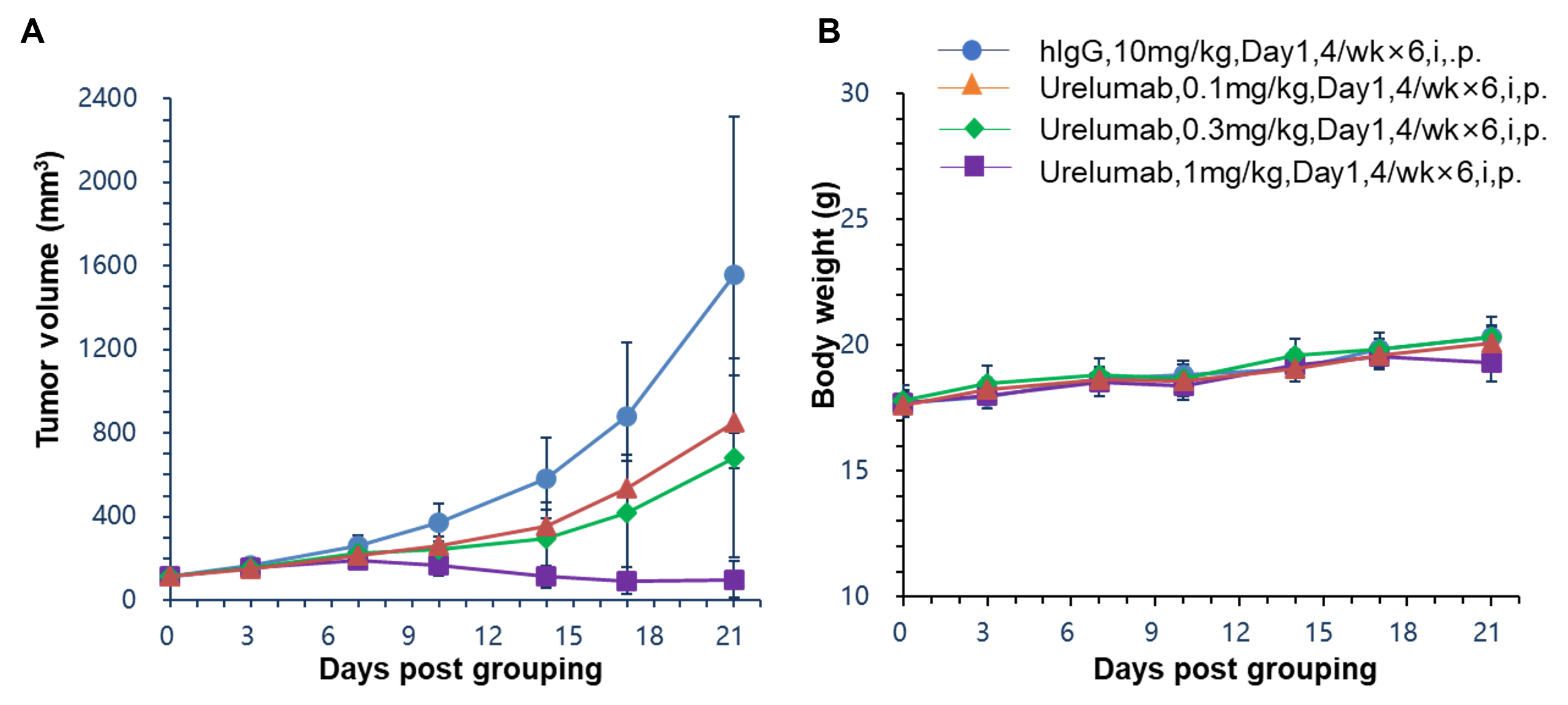
- Anti-human 4-1BB antibodies were efficacious in controlling tumor growth in B-h4-1BB mice.
- B-h4-1BB mice provide a powerful preclinical model for in vivo evaluation of anti-human 4-1BB antibodies.
Efficacy of anti-human 4-1BB antibodies in B-h4-1BB mice. (A) Anti-human 4-1BB antibodies inhibited MC38 tumor growth in B-h4-1BB mice. Murine colon cancer MC38 cells were subcutaneously implanted into homozygous B-h4-1BB mice (female, 6-7 week-old, n=6). Mice were grouped when tumor volume reached approximately 100 mm3, at which time they were treated with anti-human 4-1BB antibody Urelumab (in house) with doses and schedules indicated in panel. (B) Body weight changes during treatment. Values are expressed as mean ± SEM.
Efficacy Evaluation of anti-4-1BB antibody in the Treatment of the Subcutaneous MC38 Model in B-h4-1BB mice in vivo
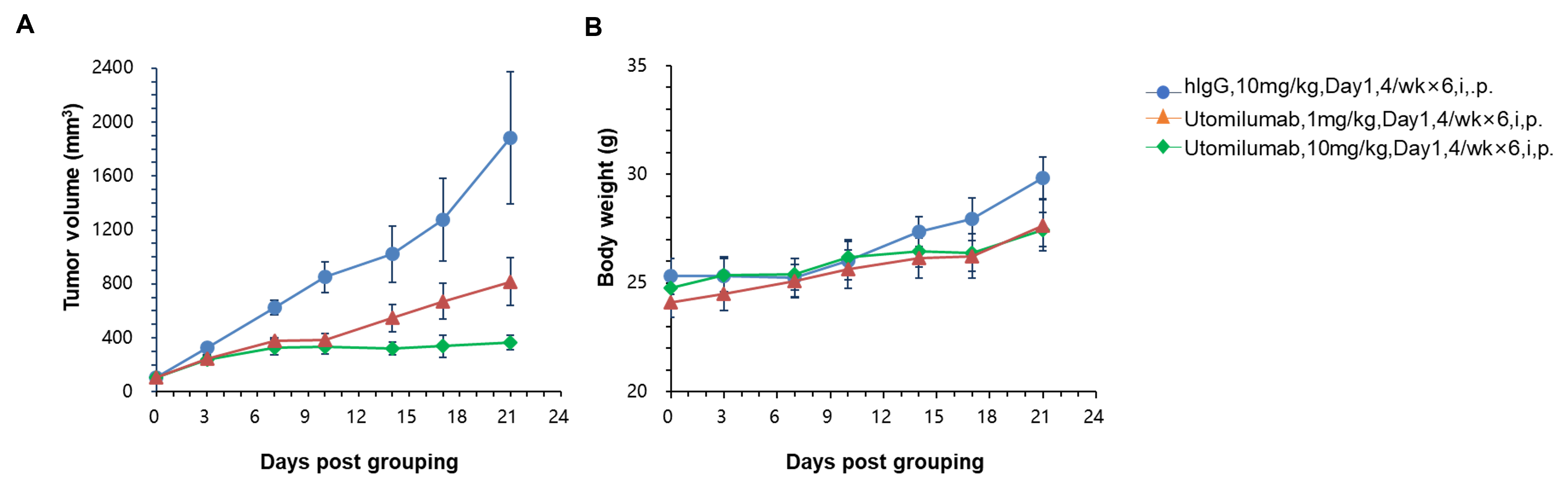
- Anti-human 4-1BB antibodies were efficacious in controlling tumor growth in B-h4-1BB mice.
- B-h4-1BB mice provide a powerful preclinical model for in vivo evaluation of anti-human 4-1BB antibodies.
Efficacy of anti-human 4-1BB antibodies in B-h4-1BB mice. (A) Anti-human 4-1BB antibodies inhibited MC38 tumor growth in B-h4-1BB mice. Murine colon cancer MC38 cells were subcutaneously implanted into homozygous B-h4-1BB mice (female, 6-7 week-old, n=5). Mice were grouped when tumor volume reached approximately 100 mm3, at which time they were treated with anti-human 4-1BB antibody utomilumab (in house) with doses and schedules indicated in panel. (B) Body weight changes during treatment. Values are expressed as mean ± SEM.
- Anti-human 4-1BB antibodies were efficacious in controlling tumor growth in B-h4-1BB mice.
- B-h4-1BB mice provide a powerful preclinical model for in vivo evaluation of anti-human 4-1BB antibodies.
Efficacy of anti-human 4-1BB antibodies in B-h4-1BB mice. (A) Anti-human 4-1BB antibodies inhibited MC38 tumor growth in B-h4-1BB mice. Murine colon cancer MC38 cells were subcutaneously implanted into homozygous B-h4-1BB mice (female, 6-7 week-old, n=8). Mice were grouped when tumor volume reached approximately 100 mm3, at which time they were treated with two anti-human 4-1BB antibodies with doses and schedules indicated in panel. (B) Body weight changes during treatment. Values are expressed as mean ± SEM.
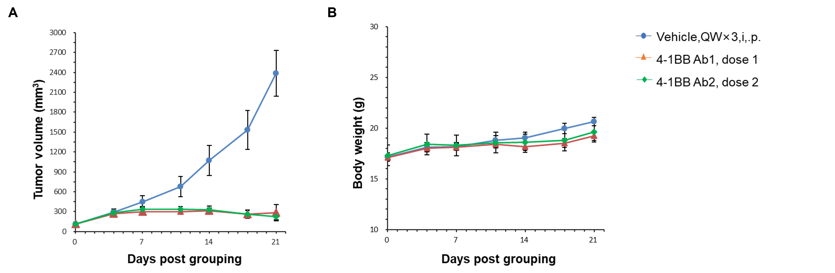
- Anti-human 4-1BB antibodies were efficacious in controlling tumor growth in B-h4-1BB mice.
- B-h4-1BB mice provide a powerful preclinical model for in vivo evaluation of anti-human 4-1BB antibodies.
Efficacy of anti-human 4-1BB antibodies in B-h4-1BB mice. (A) Anti-human 4-1BB antibodies inhibited MC38 tumor growth in B-h4-1BB mice. Murine colon cancer MC38 cells were subcutaneously implanted into homozygous B-h4-1BB mice (female, 6-7 week-old, n=5). Mice were grouped when tumor volume reached approximately 100 mm3, at which time they were treated with anti-human 4-1BB antibody utomilumab (in house) or Urelumab (in house) with doses and schedules indicated in panel. (B) Body weight changes during treatment. Values are expressed as mean ± SEM.
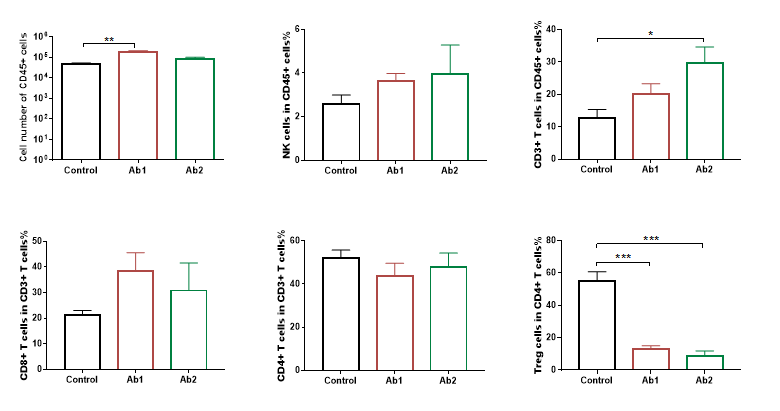
Analysis of tumor infiltrates lymphocytes(TILs) by FACS. Tumor cells were harvested at the endpoint of experiment (n=3). Flow cytometry analysis of the TILs were performed to assess cell number and proportion changes compared to the group with no anti-h4-1BB treated. CD45+ cells and CD3+ T cells were significantly increased when treated with anti-h4-1BB, while Treg cells were significantly reduced in the two groups treated with anti-h4-1BB compared to the control. Values are expressed as mean ± SEM.(*p<0.05, **p<0.01, ***p<0.001, ****p<0.0001)
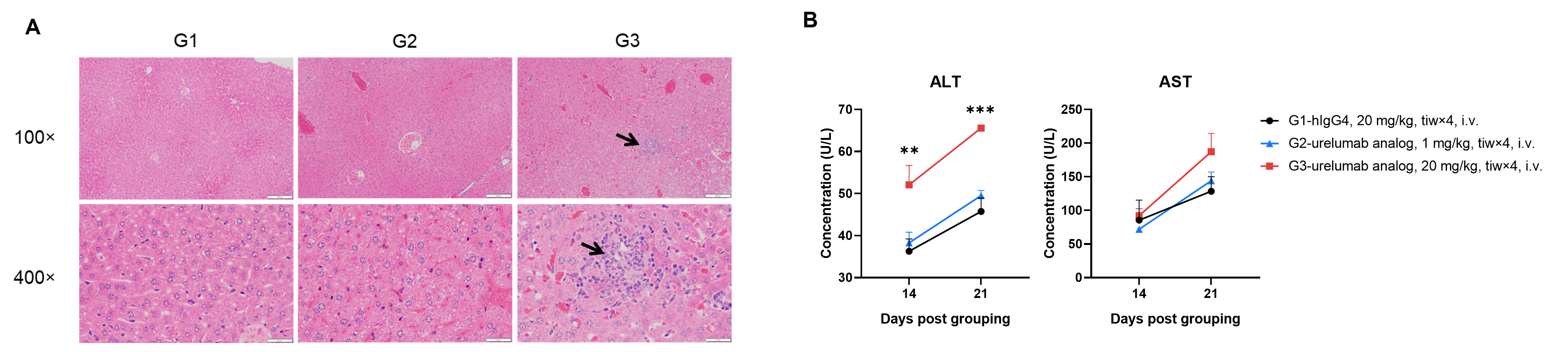
The Toxicity of anti-4-1BB Antibody
High-dose Urelumab analog (in-house) caused chronic inflammatory response in the liver of B-h4-1 BB mice. B-h4-1BB mice were divided into 3 groups and were treated with hIgG4 isotype antibody and Urelumab analog respectively. Blood were collected on days 14 and 21 after administration to measure the concentrations of ALT (B) and AST (B). On day 21, the mice were euthanized and liver tissues were taken for H&E staining (A). The results showed that in the high-dose group (G3, 20 mg/kg), 21 days after administration, there was a significant infiltration of inflammatory cells in the liver of B-h4-1 BB mice (A), and the ALT levels in the serum of B-h4-1 BB mice significantly increased on days 14 and 21 after administration, the AST level also showed an increasing trend 21 days after drug treatment (B). It is indicated that the high-dose Urelumab analog can cause significant liver toxicity in B-h4-1BB mice.
* When publishing results obtained using this animal model, please acknowledge the source as follows: The animal model [B-h4-1BB mice] (Cat# 110004) was purchased from Biocytogen.