Description
- MS4A6A is a protein-coding gene that encodes a member of the MS4A (transmembrane 4A) gene family. There is a potential relationship between alterations in MS4A6A and aging-related diseases, such as SNP rs610932, which is located in the 3‘ untranslated region of MS4A6A and is associated with cortical and hippocampal atrophy. Elevated expression of MS4A6A in late-onset Alzheimer's disease (AD) has been identified as an association with an elevated Braak Tangle score, a neuropathological indicator of AD onset and progression. In addition, MS4A6A can be used as a prognostic biomarker for macrophages in glioma patients, and it is upregulated in glioma tissues, leading to adverse clinical outcomes and adverse reactions to adjuvant chemotherapy. In castration-resistant advanced prostate cancer (CRPC) patients, MS4A6A was significantly associated with overall survival, and patients with lower gene expression survived longer.
- The exons 2-7 of mouse Ms4a6d gene that encode the whole molecule, including 3’UTR were replaced by human counterparts in B-hMS4A6A mice. The promoter and 5’UTR region of the mouse gene were retained. The human MS4A6A expression was driven by endogenous mouse Ms4a6d promoter, while mouse Ms4a6d gene transcription and translation will be disrupted.
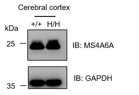
- MS4A6A was detectable in cerebral cortex of both wild-type C57BL/6N mice and homozygous B-hMS4A6A mice due to the cross-reactivity of antibody.
Targeting strategy
Gene targeting strategy for B-hMS4A6A mice. The exons 2-7 of mouse Ms4a6d gene that encode the whole molecule, including 3’UTR were replaced by human counterparts in B-hMS4A6A mice. The promoter and 5’UTR region of the mouse gene were retained. The human MS4A6A expression was driven by endogenous mouse Ms4a6d promoter, while mouse Ms4a6d gene transcription and translation will be disrupted.
Protein expression analysis
Western blot analysis of MS4A6A protein expression in homozygous B-hMS4A6A mice. Cerebral cortex lysates were collected from wild-type C57BL/6N mice (+/+) and homozygous B-hMS4A6A mice (H/H), and then analyzed by western blot with anti-MS4A6A antibody (abcam, ab189983). 40 μg total proteins were loaded for western blotting analysis. MS4A6A was detectable in cerebral cortex of both wild-type C57BL/6N mice and homozygous B-hMS4A6A mice due to the cross-reactivity of antibody.
* When publishing results obtained using this animal model, please acknowledge the source as follows: The animal model [B-hMS4A6A mice] (Cat# 113036) was purchased from Biocytogen.

