Description
Background:
- PRL (Prolactin) is a neuroendocrine hormone derived from the pituitary gland, which plays a crucial role in regulating the growth and differentiation of mammary tissues. It is also involved in other physiological processes such as water-electrolyte balance, endocrine and metabolic regulation, brain behavior, reproduction, immune modulation, and protection. PRL binds to its receptor PRLR to initiate signal transduction. PRLR is a transmembrane receptor belonging to the type I cytokine receptor family and is present in almost all organs and tissues.
- The active form of PRLR is a dimer. Upon binding to its ligand (PRL), it first activates JAK2, which further phosphorylates STAT5a, thereby stimulating the JAK/STAT signaling pathway to promote cell growth and differentiation. Depending on the cellular context, PRLR can also activate Src family kinases and focal adhesion kinase (FAK), subsequently triggering the MEK/ERK and PI3K/AKT signaling pathways to enhance cell proliferation and survival.
Targeting strategy:
- The exons 4-8 of mouse Prlr gene that encode signal peptide and extracellular domain are replaced by human counterparts in B-hPRLR mice. The genomic region of mouse Prlr gene that encodes transmembrane domain and cytoplasmic portion is retained. The promoter, 5’UTR and 3’UTR region of the mouse gene are also retained. The chimeric PRLR expression is driven by endogenous mouse Prlr promoter, while mouse Prlr gene transcription and translation will be disrupted.
Verification:
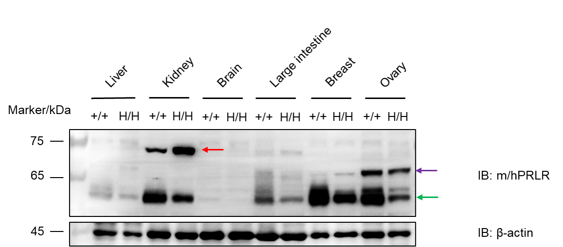
- Human PRLR was detectable in homozygous B-hPRLR mice and C57BL/6JNifdc mice.
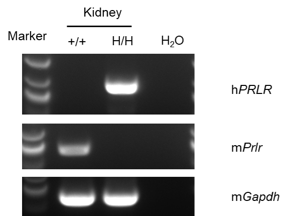
- Mouse Prlr mRNA was only detected in wild-type mice, human PRLR mRNA was only detected in homozygous B-hPRLR mice.
Application:
- This product can be used for efficacy testing and safety evaluation of target related drugs.
Targeting strategy
Gene targeting strategy for B-hPRLR mice. The exons 4-8 of mouse Prlr gene that encode signal peptide and extracellular domain are replaced by human counterparts in B-hPRLR mice. The genomic region of mouse Prlr gene that encodes transmembrane domain and cytoplasmic portion is retained. The promoter, 5’UTR and 3’UTR region of the mouse gene are also retained. The chimeric PRLR expression is driven by endogenous mouse Prlr promoter, while mouse Prlr gene transcription and translation will be disrupted.
Protein expression analysis
Western blot analysis of PRLR protein expression in homozygous B-hPRLR mice. Various tissue lysates were collected from wild-type C57BL/6JNifdc mice (+/+) and homozygous B-hPRLR mice (H/H), and then analyzed by western blot with anti-human PRLR antibody (R&D, MAB1167-SP). 40 μg total proteins were loaded for western blotting analysis. Human PRLR was detected in homozygous B-hPRLR mice and wild-type C57BL/6JNifdc mice. The red arrow points to the long isoform of PRLR, the purple arrow points to the intermediate isoform of PRLR, and the green arrow points to the short isoform of PRLR.
mRNA expression analysis
Strain specific analysis of PRLR mRNA expression in wild-type C57BL/6JNifdc mice and B-hPRLR mice by RT-PCR. Kidney RNA were isolated from wildtype C57BL/6JNifdc mice (+/+) and homozygous B-hPRLR mice (H/H), then cDNA libraries were synthesized by reverse transcription, followed by PCR with mouse or human PRLR primers. Mouse Prlr mRNA was only detectable in wild-type C57BL/6JNifdc mice. Human PRLR mRNA was only detectable in homozygous B-hPRLR mice, but not in wild-type mice.
* When publishing results obtained using this animal model, please acknowledge the source as follows: The animal model [B-hPRLR mice] (Cat# 111058) was purchased from Biocytogen.


