C57BL/6-Tfrctm1(TFRC)Bcgen Rag2tm1Bcgen/Bcgen • 112904

| Product name | B-hTFR1, Rag2 KO mice |
|---|---|
| Catalog number | 112904 |
| Strain name | C57BL/6-Tfrctm1(TFRC)Bcgen Rag2tm1Bcgen/Bcgen |
| Strain background | C57BL/6 |
| NCBI gene ID | 7037,5897 (Mouse) |
| Aliases | T9; TR; TFR; p90; CD71; TFR1; TRFR; IMD46; RAG-2 |
| Application | This product is used to evaluate the pharmacodynamics and safety of treatments for tumors and neurodegenerative diseases, as well as to assess the potential of drugs to penetrate the blood-brain barrier. |
Gene targeting strategy for B-hTFR1, Rag2 KO mice. The exons 4-19 of mouse Tfr1 gene that encode extracellular domain were replaced by human counterparts in B-hTFR1, Rag2 KO mice. The genomic region of mouse Tfr1 gene that encodes cytoplasmic and transmembrane portion was retained. The promoter, 5’UTR and 3’UTR region of the mouse gene are also retained. The chimeric TFR1 expression is driven by endogenous mouse Tfr1 promoter, while mouse Tfr1 gene transcription and translation will be disrupted. The exon 3 and 3’UTR region of mouse Rag2 were knocked out in B-hTFR1, Rag2 KO mice, resulting in a disruption of the Rag2 gene. This mouse model is obtained by crossing B-Rag2 KO mice (110809) with B-hTFR1 mice (110861).

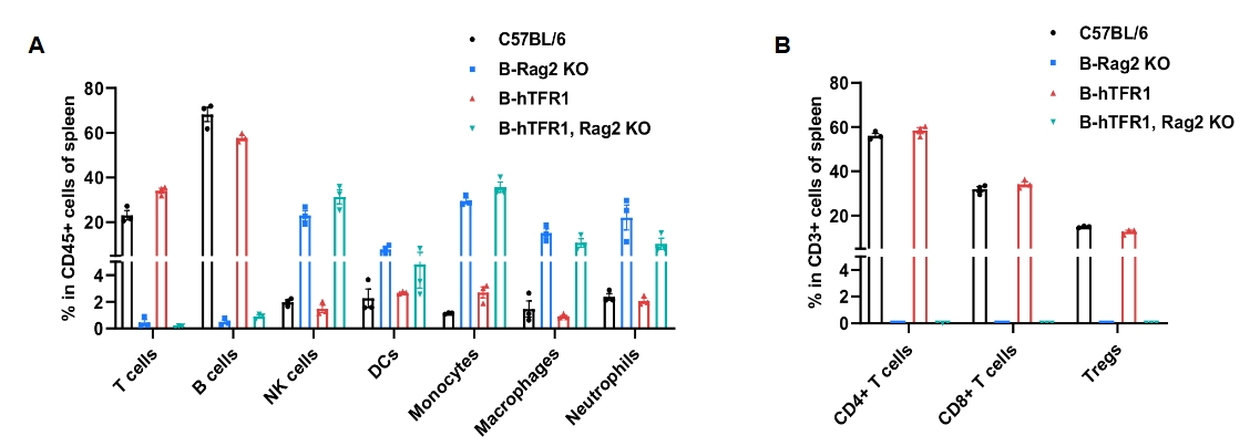
Frequency of leukocyte subpopulations in spleen by flow cytometry. Splenocytes were isolated from female wild-type C57BL/6 mice (n=3, 7 week-old), homozygous B-Rag2 KO mice (n=3, 6 week-old), homozygous B-hTFR1 mice (n=3, 9 week-old) and homozygous B-hTFR1/Rag2 KO mice (n=3, 10-week-old). A. Flow cytometry analysis of the splenocytes was performed to assess the frequency of leukocyte subpopulations. B. Frequency of T cell subpopulations. Percentages of T cells, B cells, NK cells, DCs, neutrophils, monocytes, macrophages, CD4+ T cells, CD8+ T cells and Tregs in B-hTFR1 mice were similar to those in C57BL/6 mice. Percentages of T cells, B cells, NK cells, DCs, neutrophils, monocytes, macrophages, CD4+ T cells, CD8+ T cells and Tregs in B-hTFR1/Rag2 KO mice were similar to those in B-Rag2 KO mice. Values are expressed as mean ± SEM.

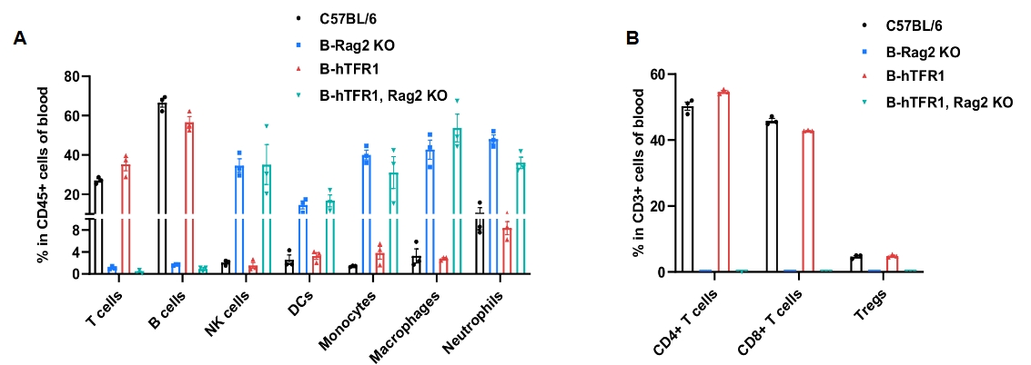
Frequency of leukocyte subpopulations in blood by flow cytometry. Blood cells were isolated from female wild-type C57BL/6 mice (n=3, 7 week-old), homozygous B-Rag2 KO mice (n=3, 6 week-old), homozygous B-hTFR1 mice (n=3, 9 week-old) and homozygous B-hTFR1/Rag2 KO mice (n=3, 10-week-old). A. Flow cytometry analysis of the splenocytes was performed to assess the frequency of leukocyte subpopulations. B. Frequency of T cell subpopulations. Percentages of T cells, B cells, NK cells, DCs, neutrophils, monocytes, macrophages, CD4+ T cells, CD8+ T cells and Tregs in B-hTFR1 mice were similar to those in C57BL/6 mice. Percentages of T cells, B cells, NK cells, DCs, neutrophils, monocytes, macrophages, CD4+ T cells, CD8+ T cells and Tregs in B-hTFR1/Rag2 KO mice were similar to those in B-Rag2 KO mice. Values are expressed as mean ± SEM.

In vivo pharmacokinetic (PK) evaluation of anti-human TFR1 nanobody. B-hTFR1, Rag2 KO mice were injected with Isotype hIgG (10 mpk) and anti-human TFR1 nanobody Ab.26 (5.5 mpk, produced in-house) via tail vein. Brain were taken for antibody concentration detection after 24 h. (A). Structure of isotype antibody and anti-TFR1 nanobody Ab.26 (This nanobody is independently developed by Biocytogen). (B) Antibody concentrations in brain parenchyma. The results confirmed that brain of B-hTFR1, Rag2 KO mice enables uptake of an intravenously administered anti-human TFR1 nanobody and B-hTFR1, Rag2 KO mice provide a powerful preclinical model for in vivo evaluation of effective delivery of protein therapeutics to the central nervous system (CNS). Graphs represent mean ± SEM.

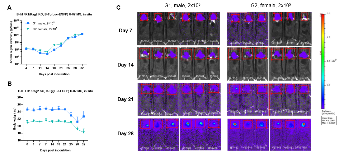
Growth kinetics of B-Tg(Luc-EGFP) U-87 MG tumors determined by bioluminescence imaging (BLI). B-Tg(Luc-EGFP) U-87 MG cells (2x10⁵) were injected into the brain of B-hTFR1, Rag2 KO mice (female and male, 8-week-old, n=6). Signal intensity and body weight were measured twice a week. (A) Signal intensity. (B) Body weight. (C) Raw bioluminescence images. These results indicate that B-Tg(Luc-EGFP) U-87 MG cells (Cat. No 322265) are able to establish brain orthotopic tumors in B-hTFR1, Rag2 KO mice and be used for efficacy studies. Values are expressed as mean ± SEM.