Description
- EGFR is expressed in various tissues. Upon binding of ligands like EGF to the EGFR receptor, EGFR dimers are formed. The activation of EGFR dimers stimulates intracellular tyrosine kinases, resulting in phosphorylation that triggers downstream signaling cascades, leading to cancer cell proliferation and the proliferation of new blood vessels within cancer cells. The mechanism of action of EGFR-targeting drugs primarily involves blocking or inhibiting the activity of EGFR, thereby preventing the growth and spread of cancer cells.
- This product is used for tumor pharmacology and safety evaluation of anti-human EGFR antibodies.
- Human EGFR mRNA was exclusively detectable in homozygous B-NDG hEGFR mice but not in wild-type mouse.
- EGFR protein was detectable in both B-NDG mice and homozygous B-NDG hEGFR mice because of antibody crossover.
Targeting strategy
Gene targeting strategy for B-NDG hEGFR mice. A chimeric CDS that encode mouse EGFR signal peptide, human EGFR extracellular domain, mouse transmembrane and cytoplasmic domain, followed by mouse 3’UTR-STOP was inserted right after mouse Egfr exon 2. The chimeric EGFR protein expression was driven by endogenous mouse Egfr promoter, while mouse Egfr gene transcription and translation will be disrupted.
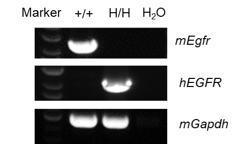
mRNA expression analysis
Strain specific analysis of EGFR mRNA expression in B-NDG mice and B-NDG hEGFR mice by RT-PCR. Liver RNA were isolated from B-NDG mice (+/+) (female, 8-week-old), and homozygous B-NDG hEGFR mice (H/H) (female, 9-week-old), then cDNA libraries were synthesized by reverse transcription, followed by PCR with mouse or human EGFR primers. mouse Egfr mRNA was only detectable in B-NDG mice. Human EGFR mRNA was exclusively detectable in homozygous B-NDG hEGFR mice but not in B-NDG mice.
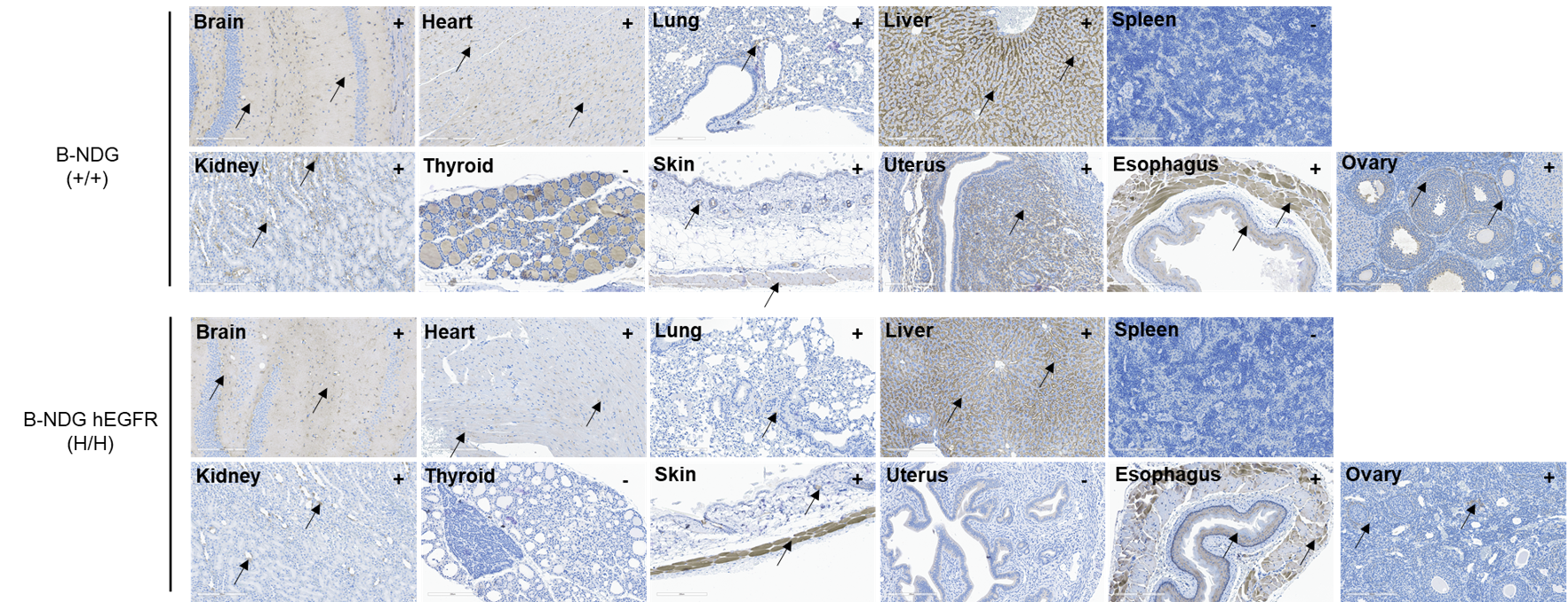
Protein expression analysis
Immunohistochemical (IHC) analysis of EGFR expression in homozygous B-NDG hEGFR mice. The brain, heart, lung, liver, spleen, kidney, thyroid, skin, uterus, esophagus and ovary were collected from B-NDG mice (+/+) (female, 8-week-old) and homozygous B-NDG hEGFR mice (H/H) (female, 9-week-old) , analyzed by IHC with anti-EGFR (abcam, ab32198). EGFR was detectable in both B-NDG mice and homozygous B-NDG hEGFR mice because of antibody crossover. The arrow indicates tissue cells with positive EGFR staining (brown). “+” indicates that the tissue is positive and “-” indicates that the tissue is negative.
* When publishing results obtained using this animal model, please acknowledge the source as follows: The animal model [B-NDG hEGFR mice] (Cat# 113309) was purchased from Biocytogen.