

C57BL/6-Tnfrsf1btm1(TNFRSF1B)Bcgen/Bcgen • 110032

Targeting the TNFR2: Biological Roles and Therapeutic Strategies

TNFR2

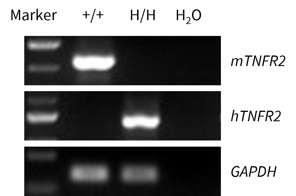
Strain specific analysis of TNFR2 gene expression in wild-type C57BL/6 mice and homozygous B-hTNFR2 mice by RT-PCR. The splenocytes RNA was isolated from wild-type C57BL/6 mice (+/+) and homozygous B-hTNFR2 mice (H/H), and then cDNA libraries were synthesized by reverse transcription, followed by PCR with mouse or human TNFR2 primers.
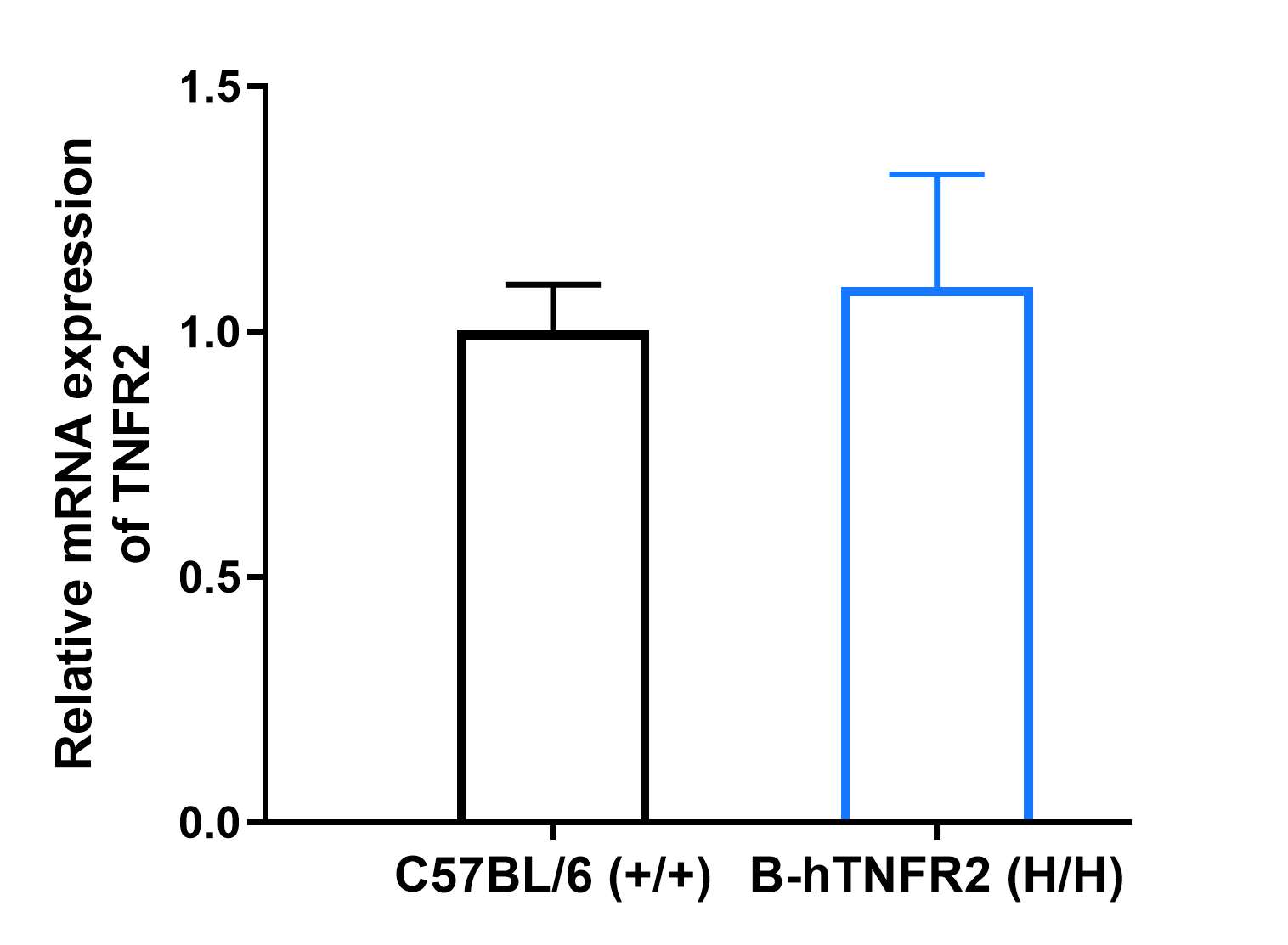
Strain specific analysis of TNFR2 gene expression in wild-type C57BL/6 mice and homozygous B-hTNFR2 mice by RT-PCR. The splenocytes RNA was isolated from wild-type C57BL/6 mice (+/+) and homozygous B-hTNFR2 mice (H/H), and then cDNA libraries were synthesized by reverse transcription, followed by PCR with mouse or human TNFR2 primers.
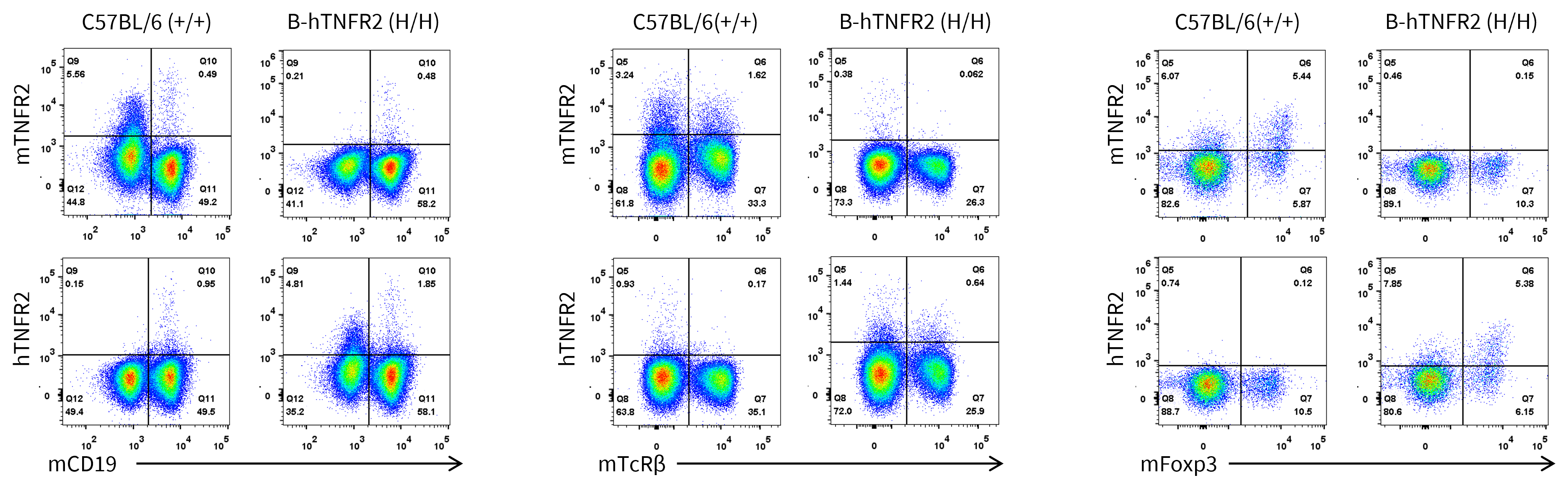
Strain specific TNFR2 expression analysis in wild-type C57BL/6 mice and homozygous B-hTNFR2 mice by flow cytometry. Splenocytes were collected from wild-type C57BL/6 mice (+/+) and homozygous B-hTNFR2 mice (H/H), and analyzed by flow cytometry with species-specific anti-TNFR2 antibody (anti-mouse CD120b (TNF R Type II/p75) Antibody, Biolegend, 113405; anti-human CD120b, Biolegend, 358403).
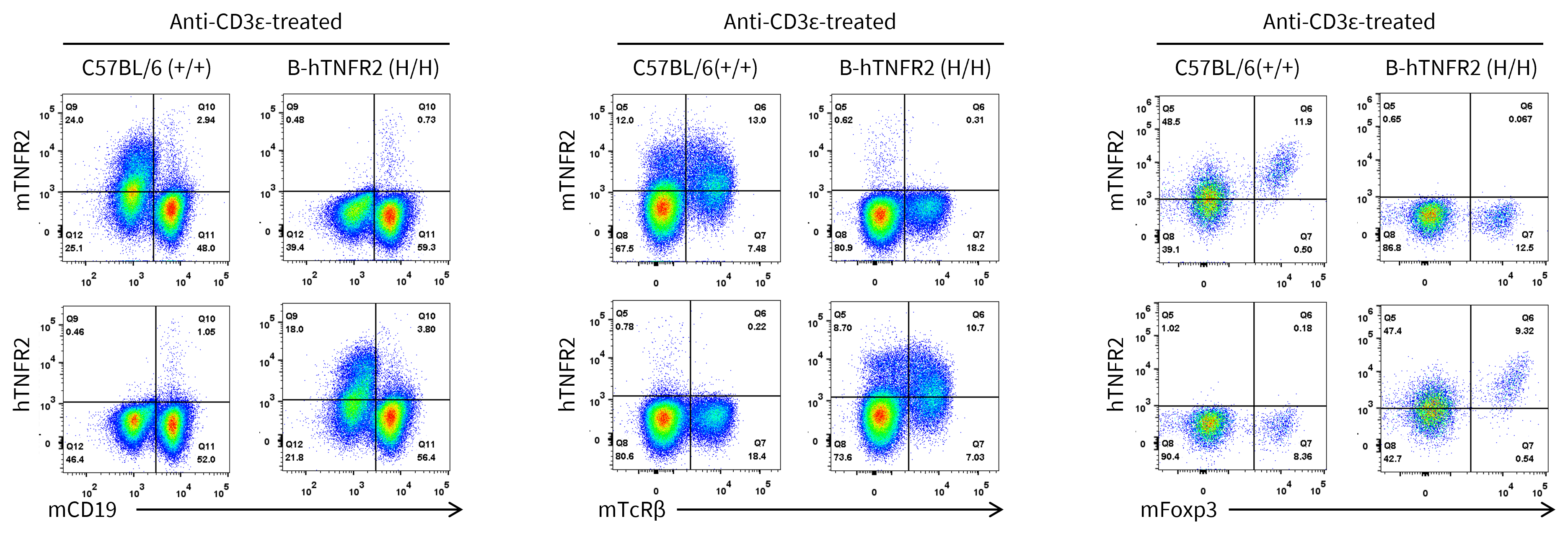
Strain specific TNFR2 expression analysis in wild-type C57BL/6 mice and homozygous B-hTNFR2 mice by flow cytometry. Splenocytes were collected from wild-type C57BL/6 mice (+/+) and homozygous B-hTNFR2 mice (H/H) stimulated with anti-CD3ε in vivo, and analyzed by flow cytometry with species-specific anti-TNFR2 antibody (anti-mouse CD120b (TNF R Type II/p75) Antibody, Biolegend, 113405; anti-human CD120b, Biolegend, 358403).
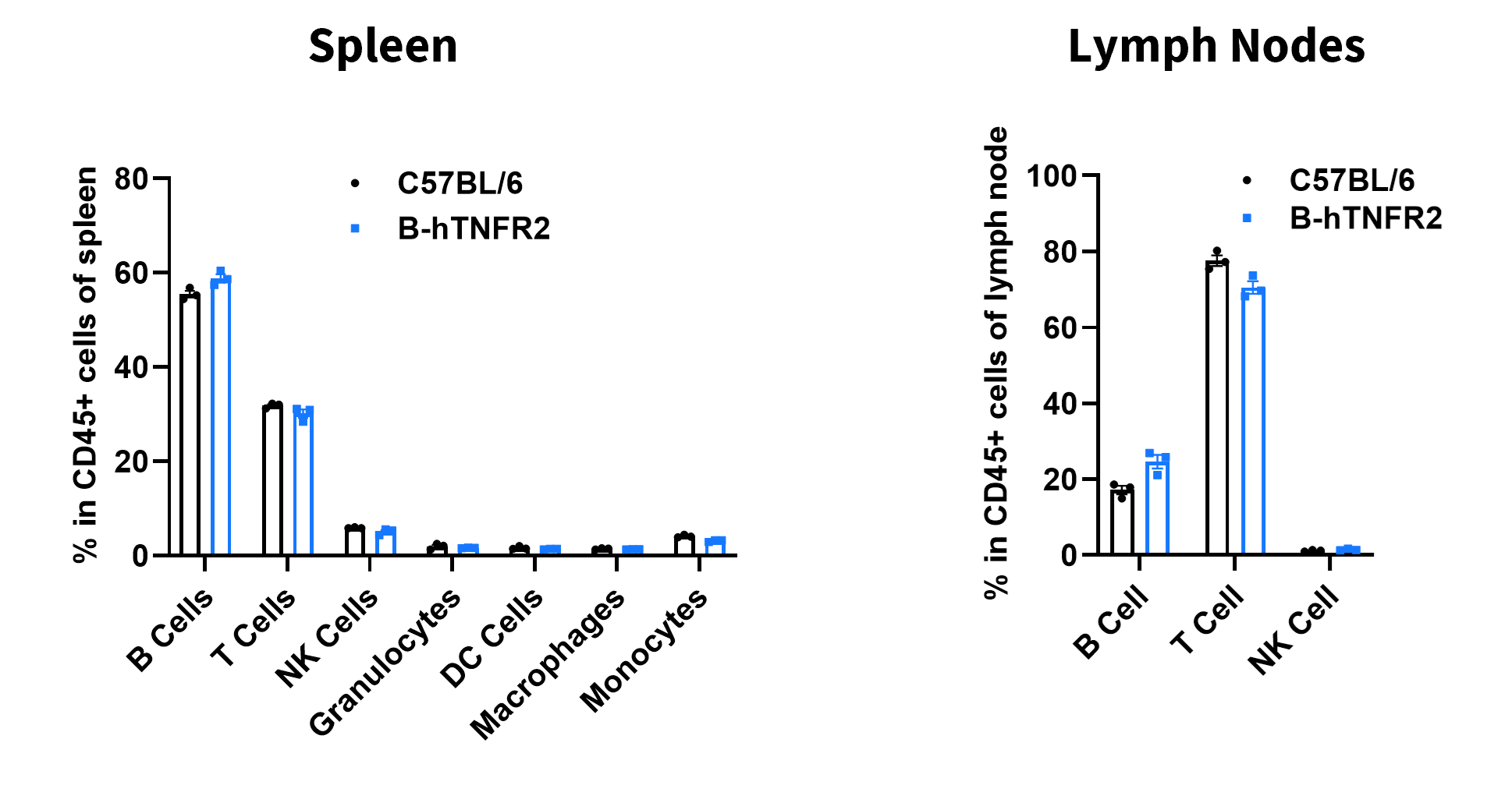
Analysis of leukocyte subpopulations by flow cytometry in immune organs. Splenocytes, and lymph nodes were isolated from female C57BL/6 and B-hTNFR2 mice (female, 6-week-old, n = 3). Single live cells were gated on the CD45⁺ population and analyzed by flow cytometry as indicated. Values are expressed as mean ± SEM.
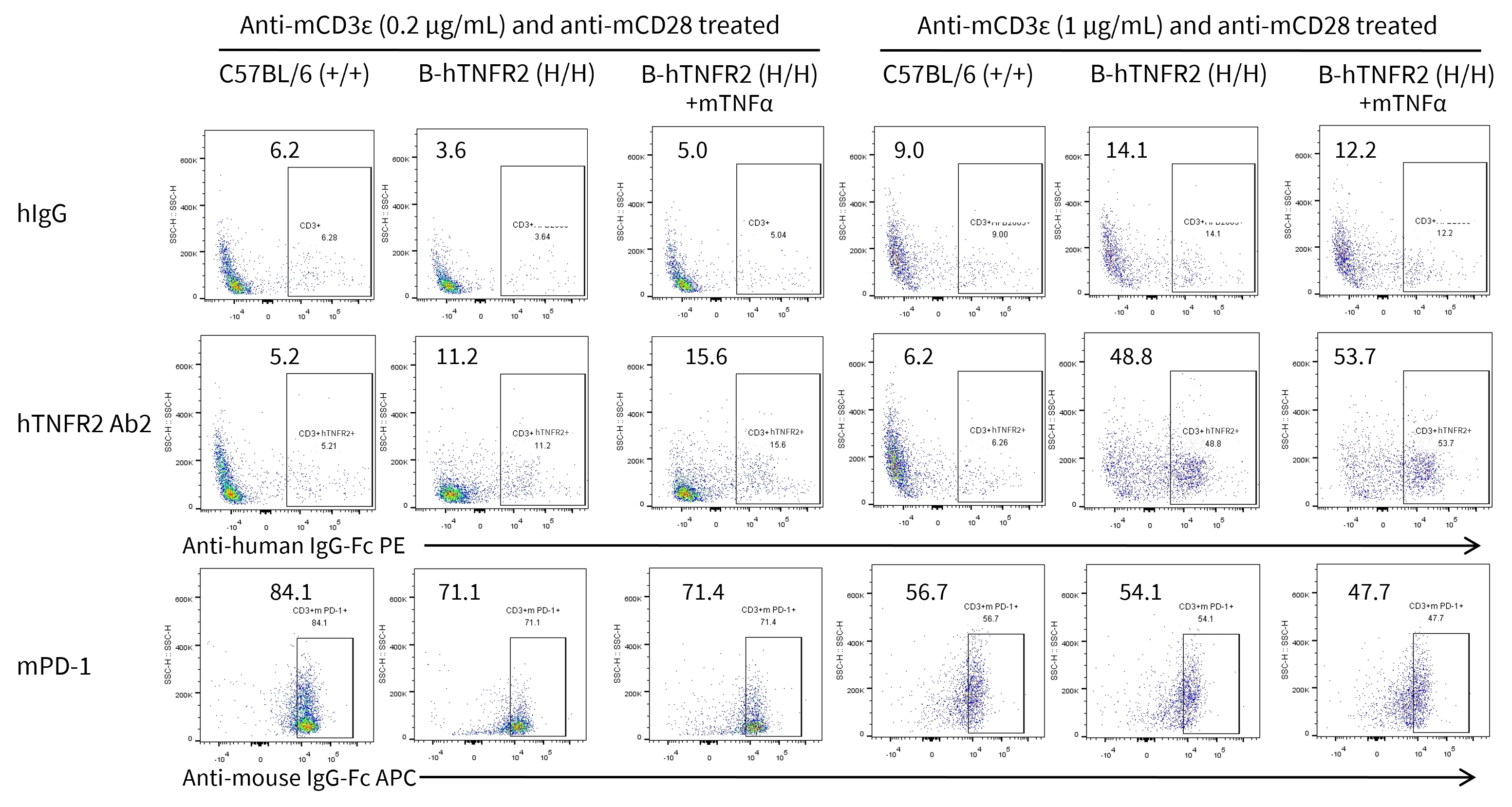
Analysis of splenocytes of B-hTNFR2 mice by FACS. Splenocytes were isolated from B-hTNFR2 mice (female, 6-week-old, n=3) treated with anti-mCD3ε (0.2 or 1 μg/mL) and anti-mCD28 (1 μg/mL) in vitro. Single live cells were gated for CD45 population and used for further analysis as indicated here. Human TNFR2 expression was detectable on CD3 T cells in B-hTNFR2 mice as evidenced by hTNFR2 Ab2 binding vs isotype control (hIgG). mTNFα enhanced hTNFR2 Ab2 binding under mild anti-mCD3ε (0.2 μg/mL) stimulation, suggesting that mTNFα/hTNFR2 signaling pathway also works well in the B-hTNFR2 mice.

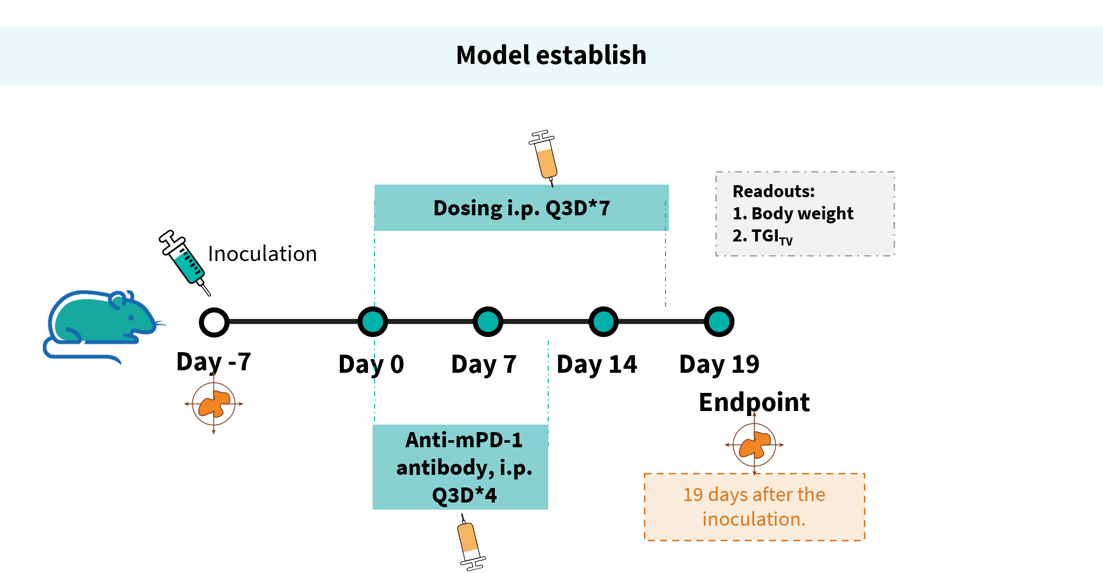
Establishment of a MC38 model and in vivo efficacy study of an anti-human TNFR2 antibodies. MC38 cells were implanted subcutaneously into homozygous B-hTNFR2 mice (male, 9-10-week-old, n=8).
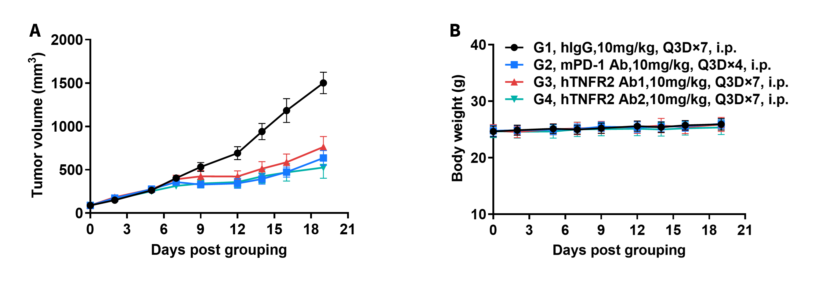
Antitumor activity of anti-human TNFR2 antibodies in B-hTNFR2 mice. (A) Tumor growth curves. (B) Body weight changes during treatment. These results demonstrate that B-hTNFR2 mice provide a powerful preclinical model for in vivo evaluation of anti-human TNFR2 antibodies.
The overage of this tumor model is 40%.

Antitumor activity of anti-human TNFR2 antibodies in B-hTNFR2 mice. Tumor cells growth of individual mouse. These results demonstrate that B-hTNFR2 mice provide a powerful preclinical model for in vivo evaluation of anti-human TNFR2 antibodies.
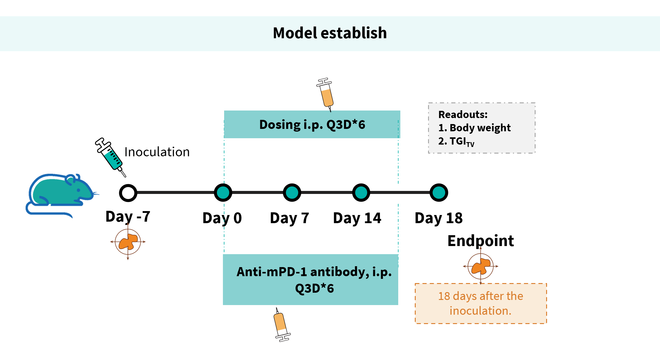
Establishment of a MC38 model and in vivo efficacy study of an anti-human TNFR2 antibodies. MC38 cells were implanted subcutaneously into homozygous B-hTNFR2 mice (male, 8-9-week-old, n=8).

Antitumor activity of anti-human TNFR2 antibodies in B-hTNFR2 mice. (A) Tumor growth curves. (B) Body weight changes during treatment. (C) Tumor cells growth of individual mouse. These results demonstrate that B-hTNFR2 mice provide a powerful preclinical model for in vivo evaluation of anti-human TNFR2 antibodies. The overage of this tumor model is 40%.
