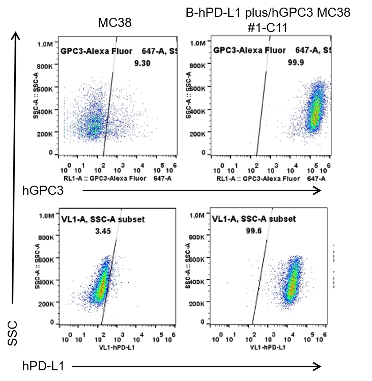
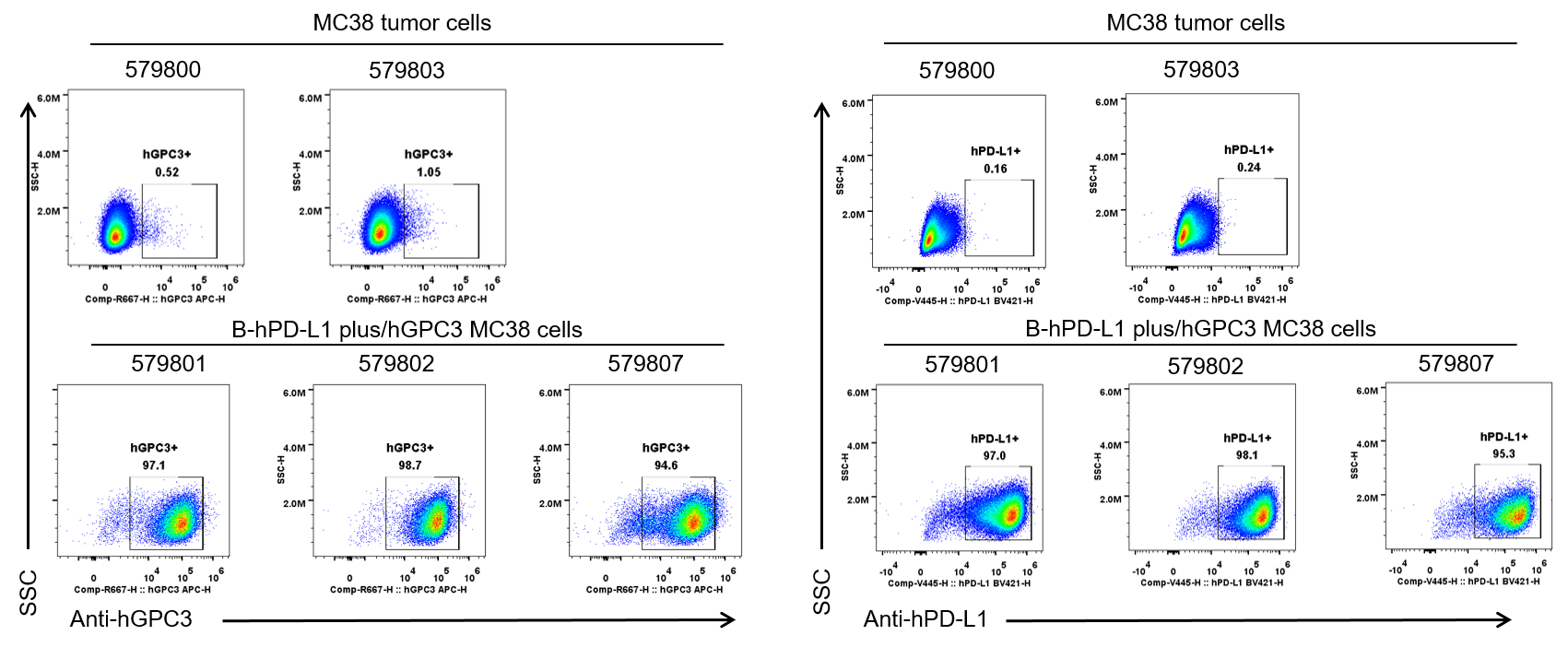
B-hPD-L1 plus/hGPC3 MC38
Catalog Number: 322418
Strain Name: NA
Strain Background: C57BL/6
NCBI gene ID: 60533,14734 (Human)
Aliases: B7h1; Pdl1; Pdcd1l1; Pdcd1lg1; A530045L16Rik; OCI-5
---
라이선스 옵션 제공 가능