

C57BL/6JNifdc-Abca4tm1Bcgen Rdh8tm1Bcgen/Bcgen • 114629


Gene targeting strategy for B-Abca4/Rdh8 DKO mice.
The exons 3-50 of mouse Abca4 gene were knocked out in B-Abca4/Rdh8 DKO mice.
The exons 2-6 of mouse Rdh8 gene were knocked out in B-Abca4/Rdh8 DKO mice.

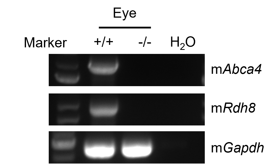
Strain specific analysis of Abca4 and Rdh8 mRNA expression in wild-type C57BL/6JNifdc and B-Abca4/Rdh8 DKO mice by RT-PCR. Eye RNA were isolated from wild-type C57BL/6JNifdc (+/+) and homozygous B-Abca4/Rdh8 DKO mice (-/-), then cDNA libraries were synthesized by reverse transcription, followed by PCR with mouse Abca4 and Rdh8 primers. Mouse Abca4 and Rdh8 mRNA was only detectable in wild-type mice but not in homozygous B-Abca4/Rdh8 DKO mice.
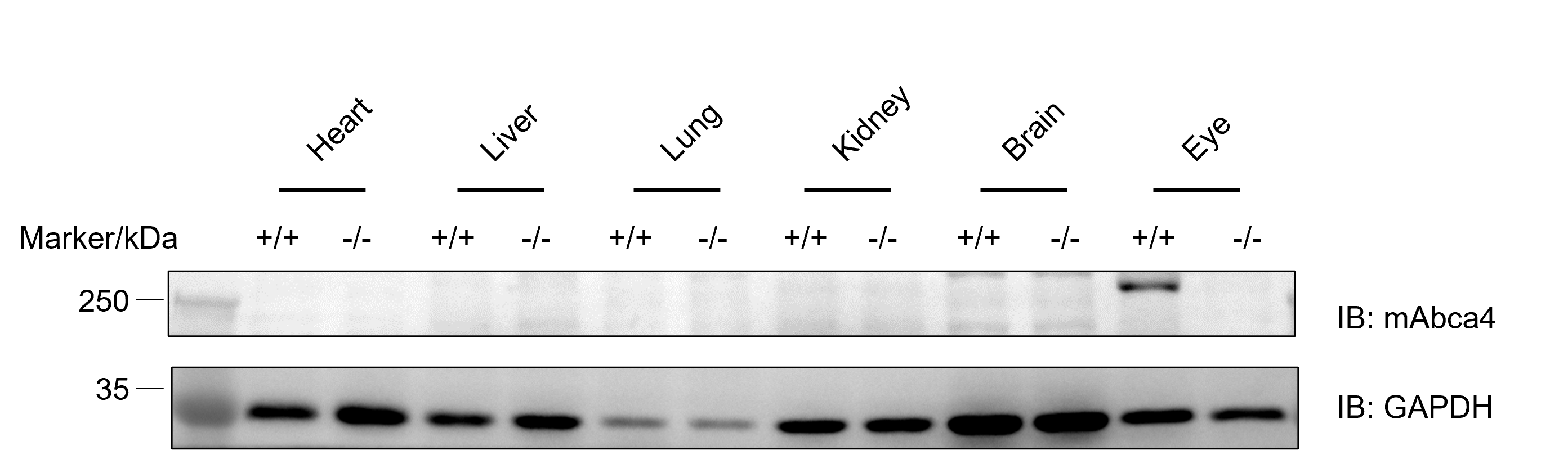
Western blot analysis of ABCA4 protein expression in homozygous B-Abca4/Rdh8 DKO mice. Various tissue lysates were collected from wild-type C57BL/6JNifdc mice (+/+) and homozygous B-Abca4/Rdh8 DKO mice (-/-), and then analyzed by western blot with anti-ABCA4 antibody (Abcam, ab72955). 40 μg total proteins were loaded for western blotting analysis. Mouse Abca4 was only detected in eye of wild-type mice but not in homozygous B-Abca4/Rdh8 DKO mice.
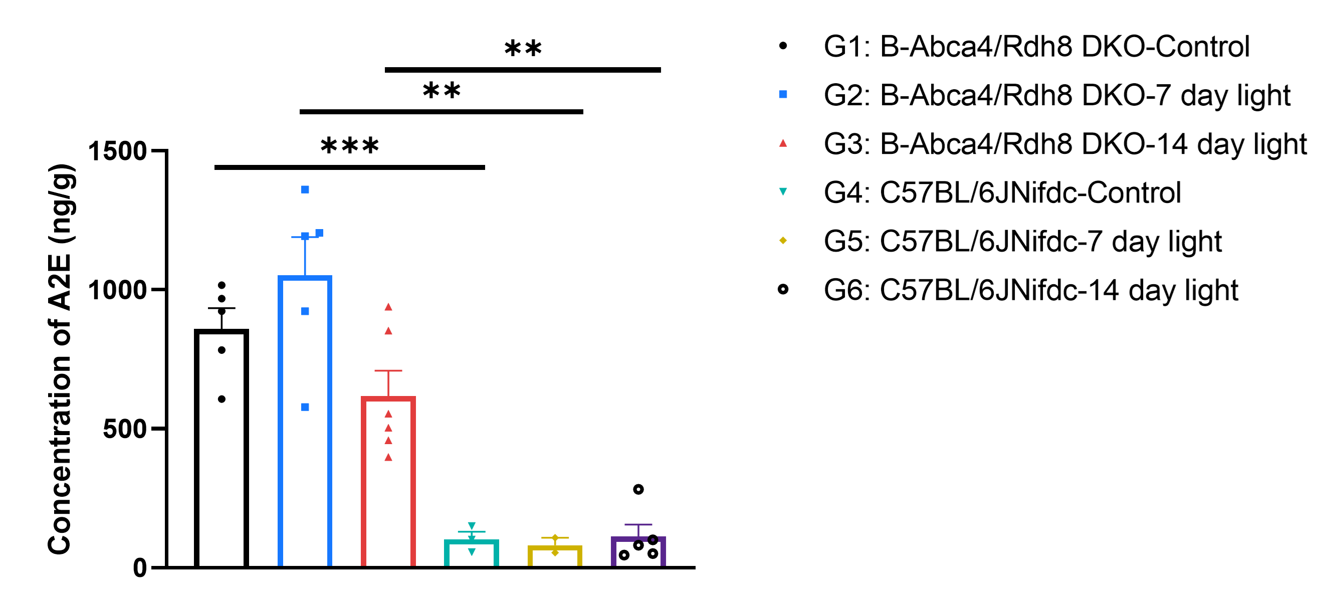
A2E analysis in wild-type C57BL/6JNifdc and B-Abca4/Rdh8 DKO mice. Eyes were isolated from wild-type C57BL/6JNifdc (+/+) and homozygous B-Abca4/Rdh8 DKO mice (-/-). LC-MS measurements of A2E showed that B-Abca4/Rdh8 DKO mice had substantially higher A2E levels than wild-type animals in both the absence and presence of light exposure, indicating that Abca4/Rdh8 deficiency alone drives robust bis-retinoid accumulation independent of illumination. Values are expressed as mean ± SEM. Significance was determined by t-test, *P<0.05, **P<0.01, ***P<0.001.
The co-validation data were provided by Pharmalegacy.
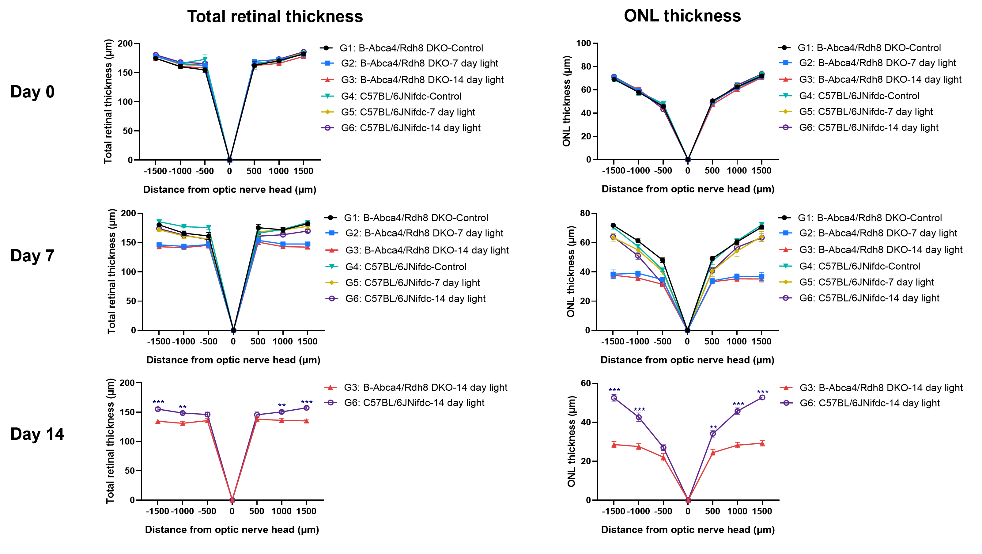
Total retinal thickness and ONL thickness analysis in wild-type C57BL/6JNifdc and homozygous B-Abca4/Rdh8 DKO mice. Left panels: Total retinal thickness; Right panels: ONL thickness. ONL thinning demonstrates that Abca4/Rdh8 double deletion exacerbates light-induced photoreceptor degeneration. Values are expressed as mean ± SEM. Significance was determined by t-test, *P<0.05, **P<0.01, ***P<0.001.
The co-validation data were provided by Pharmalegacy.
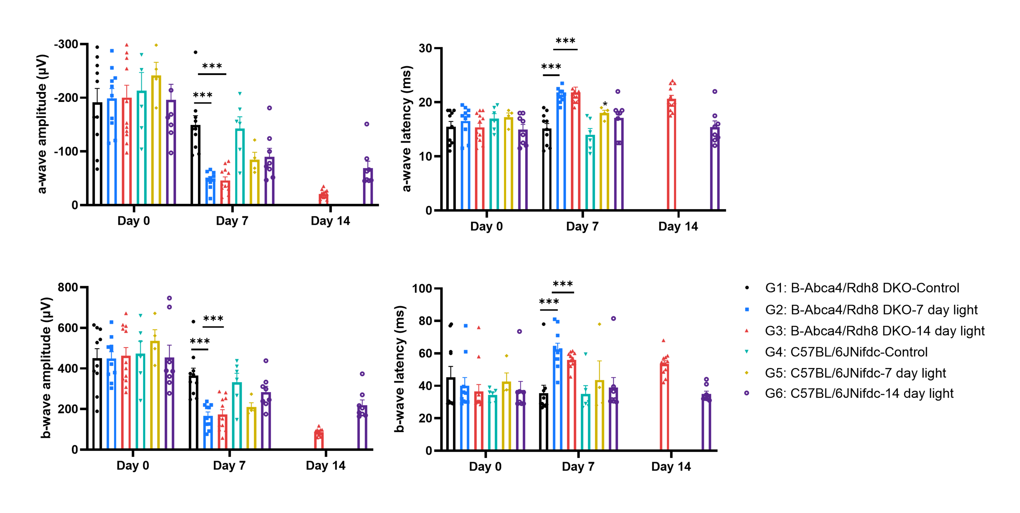
Dark-adapted ERG analysis of rod-mediated retinal function in B-Abca4/Rdh8 DKO and C57BL/6JNifdc mice after light exposure. Dark ERG recordings were performed at Day 0, Day 7 and Day 14 in control and light-exposed B-Abca4/Rdh8 DKO and C57BL/6JNifdc mice. a-wave amplitude, a-wave latency, b-wave amplitude and b-wave latency were quantified. Rod-mediated retinal function was markedly impaired in light-exposed B-Abca4/Rdh8 DKO mice compared with control groups. Values are expressed as mean ± SEM. Significance was determined by t-test, *P<0.05, **P<0.01, ***P<0.001.
The co-validation data were provided by Pharmalegacy.
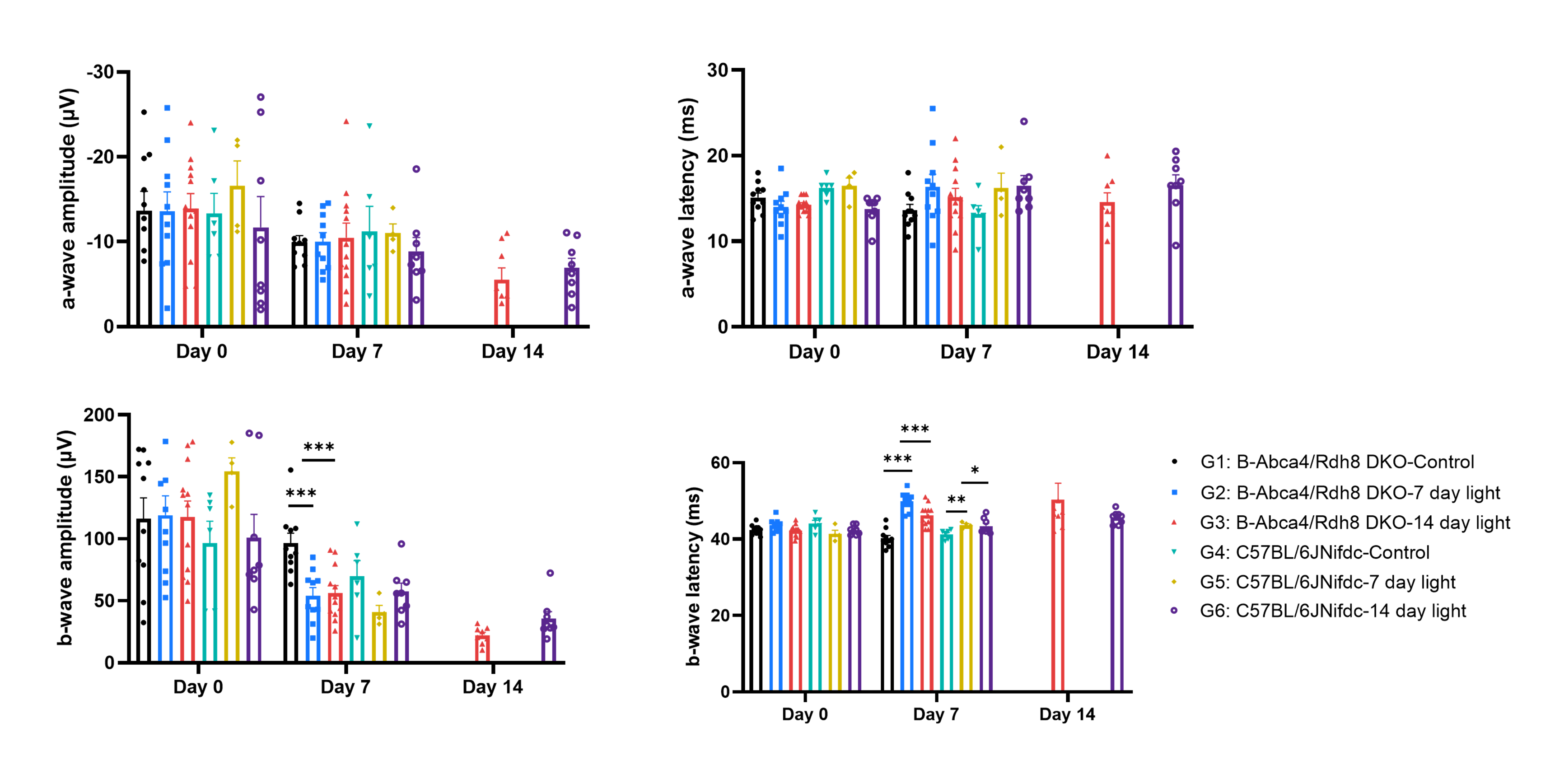
Light-adapted ERG analysis of cone-mediated retinal function in B-Abca4/Rdh8 DKO and C57BL/6JNifdc mice after light exposure. Light-adapted ERG recordings were performed at Day 0, Day 7 and Day 14 in control and light-exposed B-Abca4/Rdh8 DKO and C57BL/6JNifdc mice. a-wave amplitude, a-wave latency, b-wave amplitude and b-wave latency were quantified. Cone-mediated retinal function was markedly impaired in light-exposed B-Abca4/Rdh8 DKO mice compared with control groups. Values are expressed as mean ± SEM. Significance was determined by t-test, *P<0.05, **P<0.01, ***P<0.001.
The co-validation data were provided by Pharmalegacy.
