


Fibroblast activation protein (FAP) is a type II transmembrane serine protease predominantly expressed in activated fibroblasts, particularly cancer-associated fibroblasts (CAFs) within the tumor microenvironment. FAP plays a critical role in extracellular matrix remodeling, tumor progression, angiogenesis, and immunosuppression, making it an important therapeutic target in oncology.
In FAP humanized mice (B-hFAP mice), The exons 3-26 of mouse Fap gene that encode the extracellular domain were replaced by human FAP exons 3-26. This gene replacement strategy enables expression of human FAP in stromal compartments while disrupting endogenous mouse Fap expression, thereby providing a biologically relevant system for evaluating human-specific therapeutics in vivo.
Validation studies demonstrate species-specific expression in this model. Mouse Fap mRNA is detectable only in wild-type mice, whereas human FAP mRNA is exclusively detectable in FAP humanized mice (B-hFAP mice). At the protein level, human FAP is specifically detected in stromal cells (CD45⁻ populations) within tumor tissues and in MEF cells using flow cytometry, confirming successful humanization and functional expression.
Key Advantages
Validation
Applications
In vivo efficacy, pharmacodynamic, and translational evaluation of FAP-targeted therapies, including antibody-based therapeutics, stromal-targeting strategies, and tumor microenvironment modulation studies in oncology research.

FAP

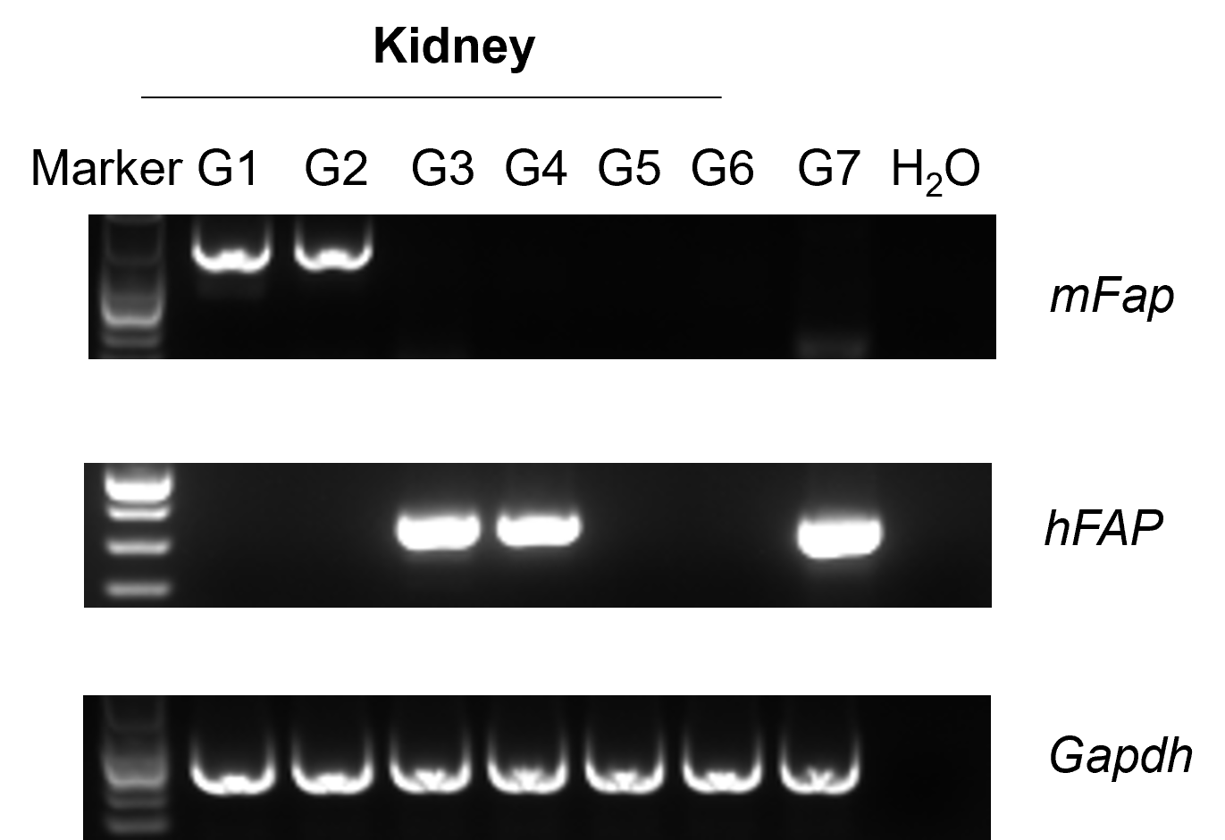
Strain-specific analysis of mouse and human FAP mRNA expression in wild-type C57BL/6 mice, FAP humanized mice, and Fap knockout mice by RT-PCR. Murine colon cancer MC38 cells (5 × 10⁵) and melanoma B16-F10 cells (2 × 10⁵) were separately implanted subcutaneously into wild-type C57BL/6 mice, homozygous FAP humanized mice, and homozygous Fap KO mice. The human FAP-overexpressing MC38 cell line (B-hFAP MC38) was used as a positive control (G7). Total RNA was isolated from kidney tissues of mice in groups G1–G6 for RT-PCR analysis of mouse Fap and human FAP mRNA expression.
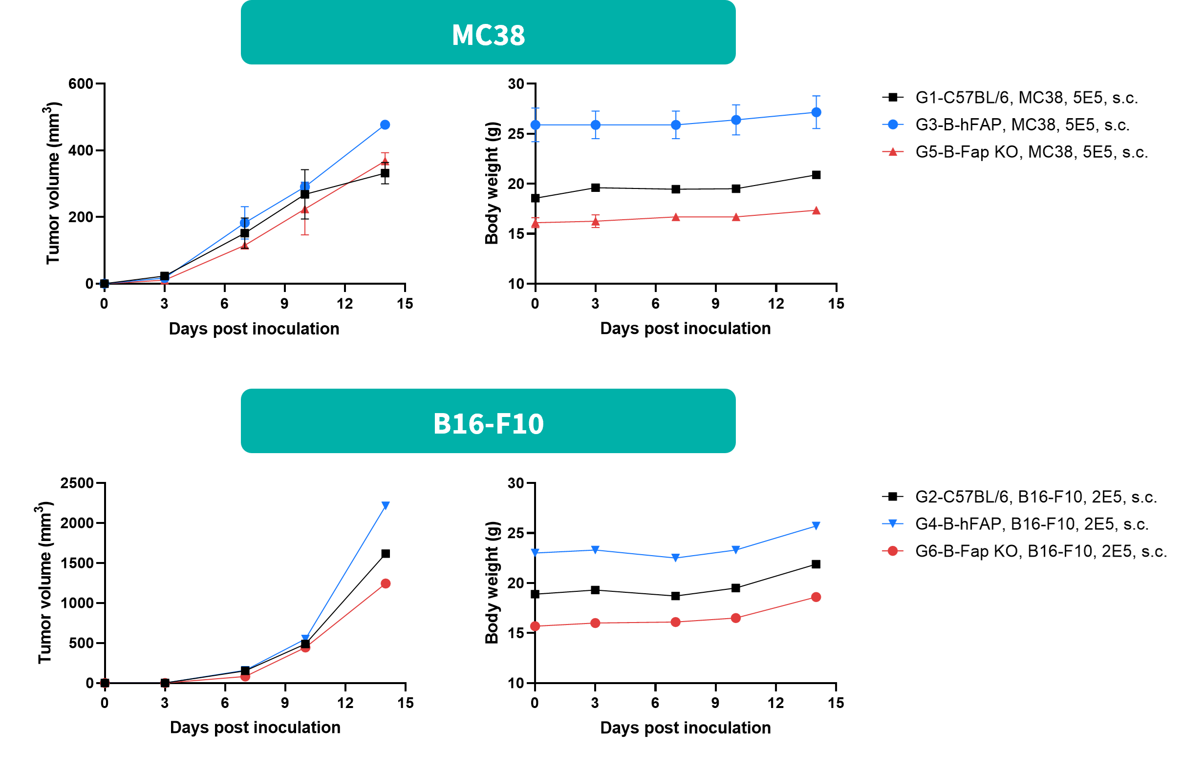
Subcutaneous tumor growth analysis of MC38 and B16-F10 cells in FAP humanized mice. Murine colon cancer MC38 cells (5 × 10⁵) were subcutaneously implanted into female FAP humanized mice (7 weeks old, n = 2). B16-F10 melanoma cells (2 × 10⁵) were subcutaneously implanted into female FAP humanized mice (7 weeks old, n = 1). Tumor volume and body weight were measured twice weekly. (A) Mean tumor volume ± SEM. (B) Body weight (mean ± SEM). Tumor volume was expressed in mm³ and calculated using the formula: V = 0.5 × long diameter × (short diameter)².
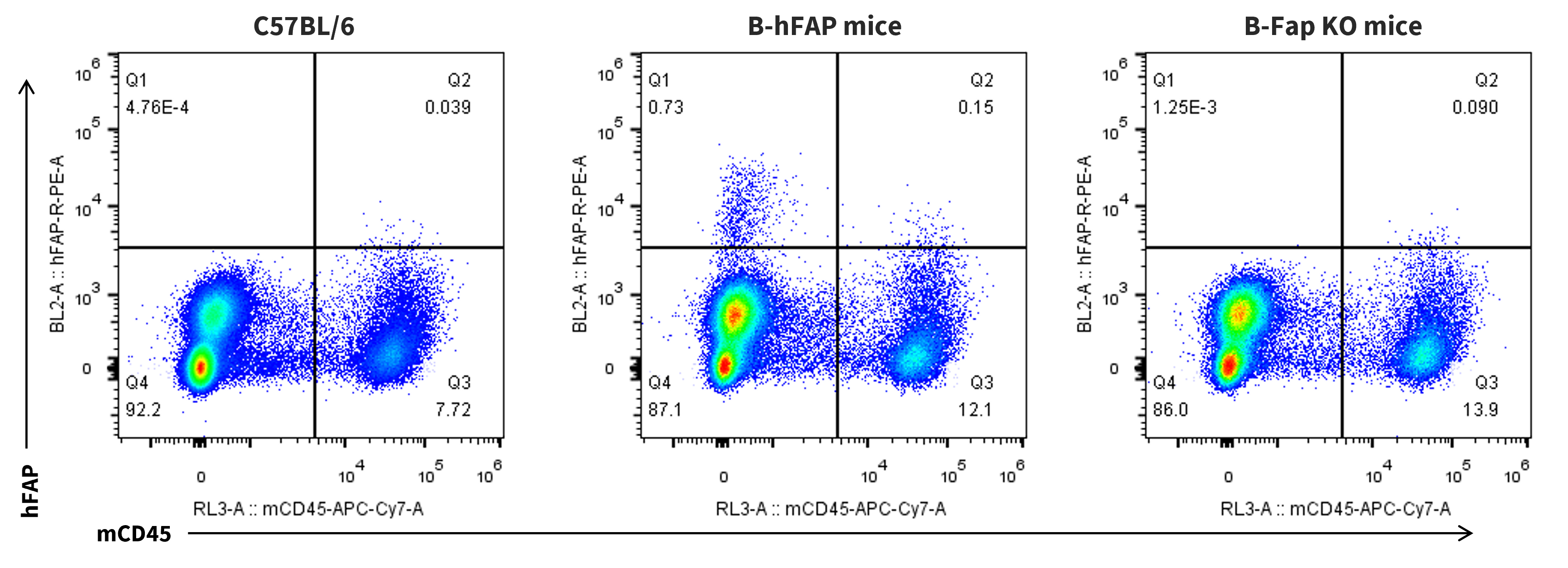
Strain-specific analysis of human FAP protein expression in B16-F10 tumors from wild-type C57BL/6 mice, FAP humanized mice, and Fap knockout mice by flow cytometry. Murine melanoma B16-F10 cells (2 × 10⁵) were subcutaneously implanted into wild-type C57BL/6 mice, homozygous FAP humanized mice, and homozygous B-Fap KO mice. Tumor tissues were collected from all three mouse strains, and human FAP protein expression was analyzed by flow cytometry using an anti-human FAP antibody (R&D Systems, FAB3715P-100).
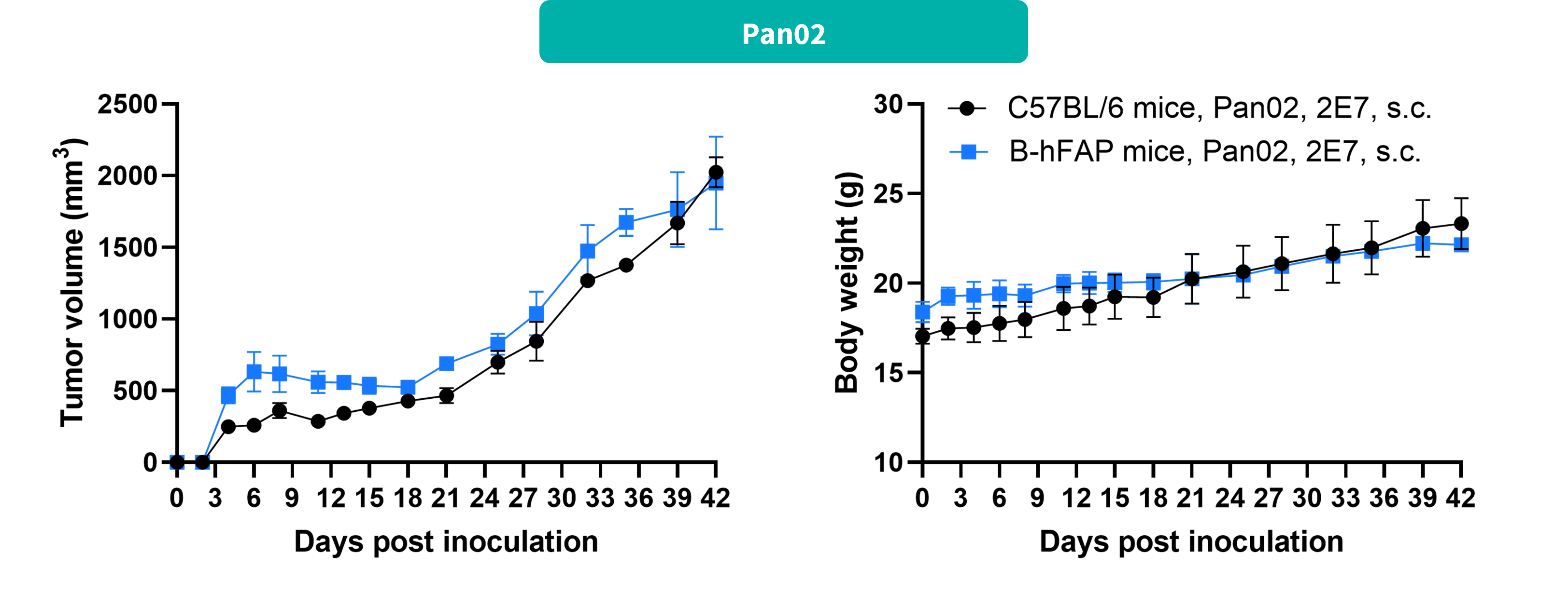
Subcutaneous tumor growth analysis of Pan02 pancreatic cancer cells in FAP humanized mice. Murine pancreatic cancer Pan02 cells (2 × 10⁷) were subcutaneously implanted into female FAP humanized mice (n = 3). Tumor volume and body weight were measured twice weekly. (A) Mean tumor volume ± SEM. (B) Body weight (mean ± SEM). Tumor volume was expressed in mm³ and calculated using the formula: V = 0.5 × long diameter × (short diameter)².
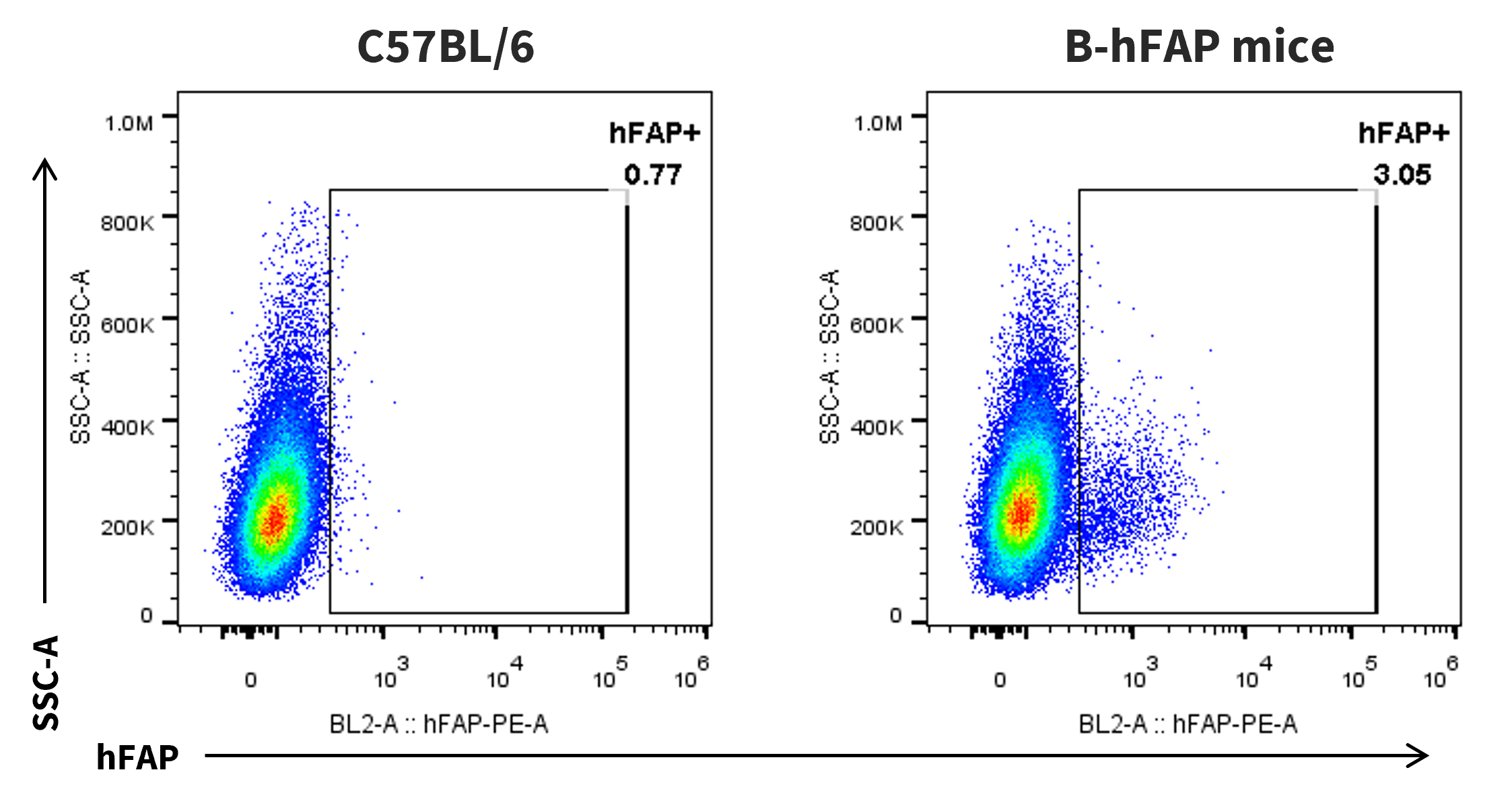
Strain-specific analysis of human FAP protein expression in Pan02 tumors from wild-type C57BL/6 mice and FAP humanized mice by flow cytometry. Murine pancreatic cancer Pan02 cells (2 × 10⁷) were subcutaneously implanted into wild-type C57BL/6 mice and homozygous FAP humanized mice. Tumor tissues were collected from both mouse strains, and human FAP protein expression was analyzed by flow cytometry using an anti-human FAP antibody (R&D Systems, FAB3715P-100).
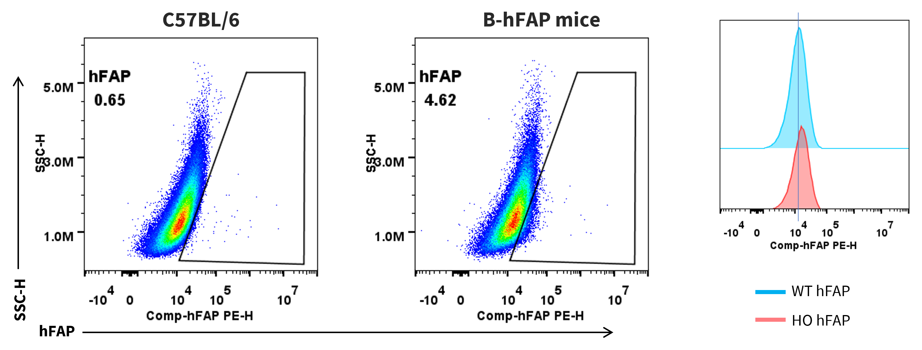
Strain-specific analysis of human FAP protein expression in mouse embryonic fibroblasts (MEFs) from wild-type C57BL/6 mice and FAP humanized mice by flow cytometry. Mouse embryonic fibroblasts (MEFs) were collected from wild-type C57BL/6 mice (+/+) and homozygous FAP humanized mice (H/H). Human FAP protein expression was analyzed by flow cytometry using an anti-human FAP antibody (R&D Systems, FAB3715P-100).

Strain-specific analysis of soluble mouse and human FAP (sFAP) protein expression in wild-type C57BL/6 mice and FAP humanized mice by ELISA. Plasma samples were collected from wild-type C57BL/6 mice and homozygous FAP humanized mice (female, 8 weeks old, n = 3). Human plasma was used as a positive control. Mouse and human soluble FAP (sFAP) protein levels were measured by ELISA using species-specific mouse FAP (Abcam, ab289903) and human FAP (Abcam, ab256404) ELISA kits, respectively. Values are expressed as mean ± SEM.
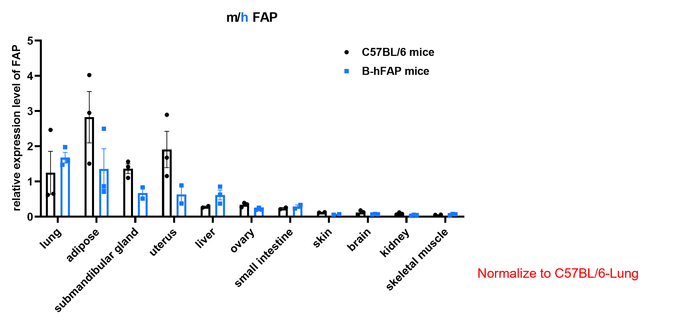
RT-qPCR analysis of relative FAP/Fap mRNA expression in different tissues of wild-type C57BL/6 mice and FAP humanized mice. Tissues were collected from wild-type C57BL/6 mice and homozygous FAP humanized mice (female, 8 weeks old, n = 3). Relative FAP/Fap mRNA expression levels were normalized to those in the lungs of wild-type C57BL/6 mice. The qPCR primers were designed to target an identical sequence shared by human FAP and mouse Fap, generating amplicons of the same length from both templates. Accordingly, the assay detects total FAP/Fap transcripts but does not distinguish between human FAP and mouse Fap mRNA. Values are expressed as mean ± SEM.
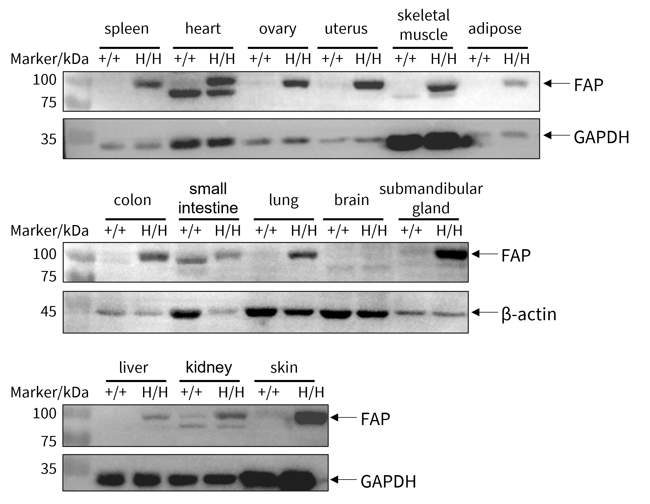
Western blot analysis of FAP protein expression in various tissues of wild-type C57BL/6 mice and FAP humanized mice. Tissues were collected from wild-type C57BL/6 mice (+/+) and homozygous FAP humanized mice (H/H). FAP protein expression was analyzed by Western blot using an anti-FAP antibody (R&D Systems, AF3715). In FAP humanized mice, FAP protein was detected in the spleen, heart, ovary, uterus, skeletal muscle, adipose tissue, colon, small intestine, lung, brain, submandibular gland, liver, kidney, and skin.


Experimental schedule for bleomycin-induced pulmonary fibrosis and in vivo efficacy evaluation of dexamethasone and talabostat mesylate in FAP humanized mice. Bleomycin (BLM) was administered intranasally to FAP humanized mice on days 0 and 1 to induce pulmonary fibrosis. Dexamethasone (DEX), a corticosteroid used as a positive control, was administered intraperitoneally according to the indicated dosing schedule, while talabostat mesylate (MCE, HY-13233A) was administered orally from day 7 to day 20. Mice were monitored throughout the study and euthanized at the experimental endpoint on day 21. BLM, bleomycin; DEX, dexamethasone; HYP, hydroxyproline.
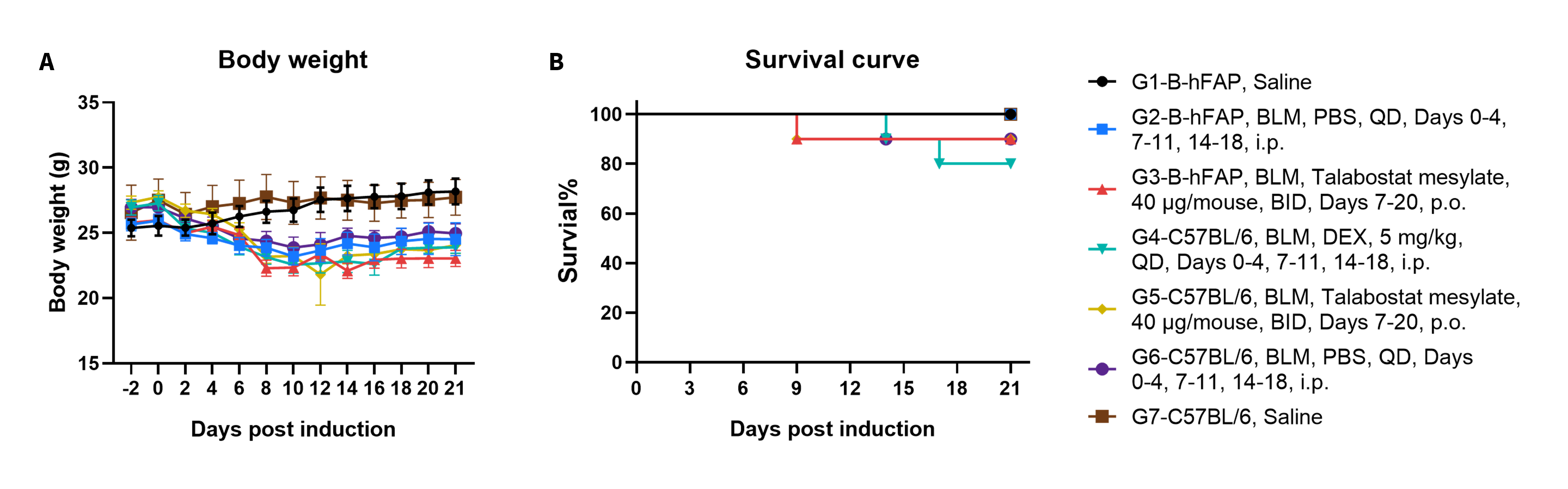
Body weight changes and survival analysis in a bleomycin-induced pulmonary fibrosis model using FAP humanized mice and wild-type C57BL/6 mice. FAP humanized mice and wild-type C57BL/6 mice were challenged intranasally with bleomycin (BLM) on days 0 and 1 to induce pulmonary fibrosis. FAP humanized mice were treated with PBS or talabostat mesylate, while wild-type C57BL/6 mice received PBS, dexamethasone, or talabostat mesylate according to the indicated dosing schedules. (A) Body weight changes during treatment. (B) Survival curves. Values are expressed as mean ± SEM.

Hydroxyproline analysis of collagen deposition in a bleomycin-induced pulmonary fibrosis model using FAP humanized mice and wild-type C57BL/6 mice. Lung tissues were collected following bleomycin (BLM) induction and treatment with PBS, dexamethasone, or talabostat mesylate. Hydroxyproline (HYP) content was measured to assess collagen deposition and the severity of pulmonary fibrosis. (A) Total HYP content per lung. (B) HYP content normalized to wet lung weight. Bleomycin treatment increased lung HYP levels compared with saline controls, indicating successful induction of pulmonary fibrosis. Values are expressed as mean ± SEM. Statistical significance was determined using one-way ANOVA. *p < 0.05, **p < 0.01.
