Q1: What are B-hIGF1R mice plus?
B-hIGF1R mice plus are target-humanized mice expressing a chimeric IGF1R protein with human IGF1R extracellular and transmembrane regions, enabling evaluation of human IGF1R-targeted therapeutics in vivo.
Q2: Why is IGF1R an important therapeutic target?
IGF1R is a receptor tyrosine kinase involved in IGF signaling, cell growth, survival, metabolism, and transformation, making IGF1R relevant to oncology, metabolic disease, obesity, diabetes, and CNS delivery research.
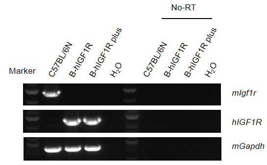
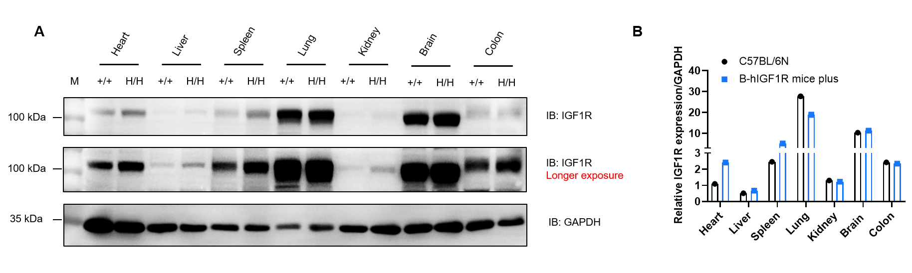
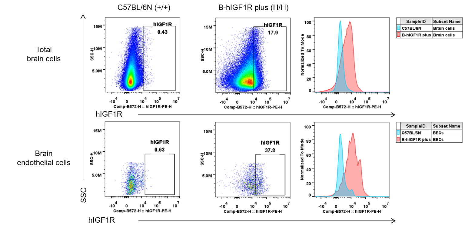
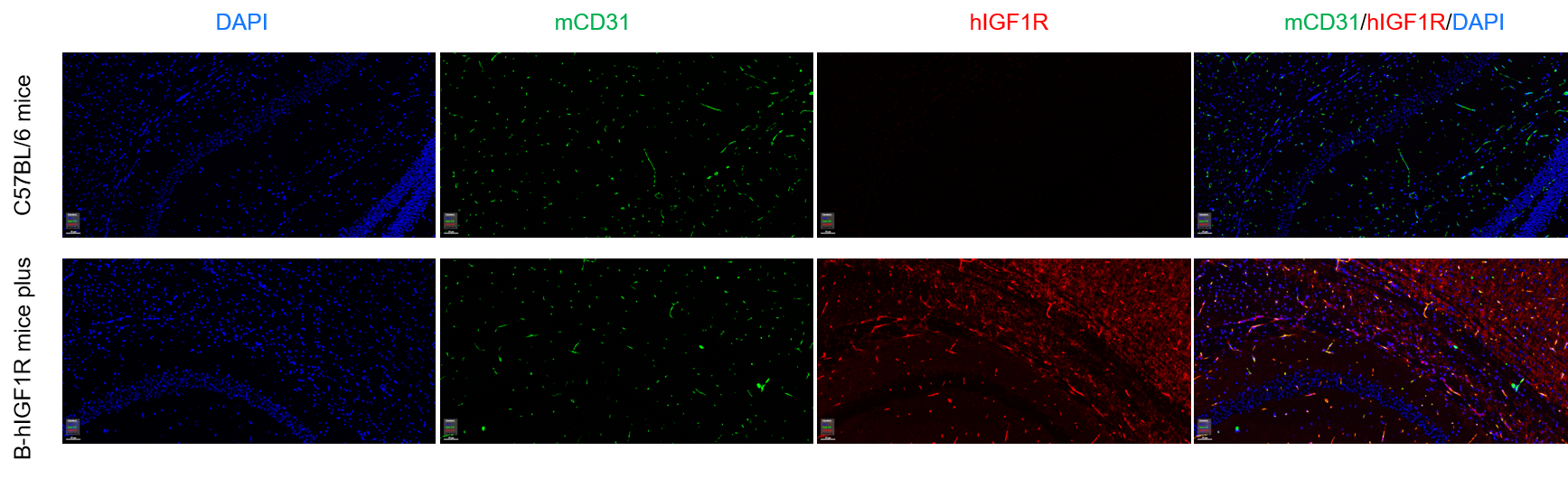
Q3: How was IGF1R expression validated in B-hIGF1R mice plus?
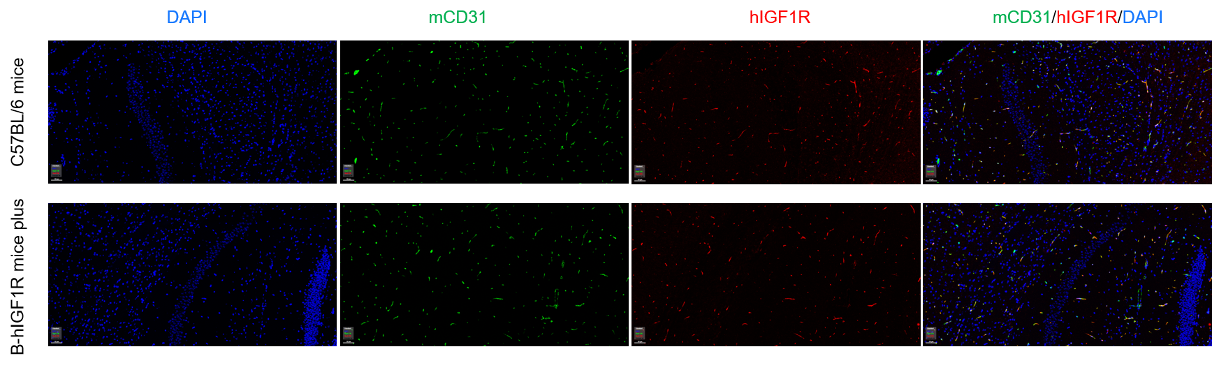
Human IGF1R mRNA was validated by RT-PCR, IGF1R protein was detected by western blot across multiple tissues, and human IGF1R expression was confirmed in brain endothelial cells and brain micro-vessels.
Q4: Can B-hIGF1R mice plus be used for functional antibody studies?
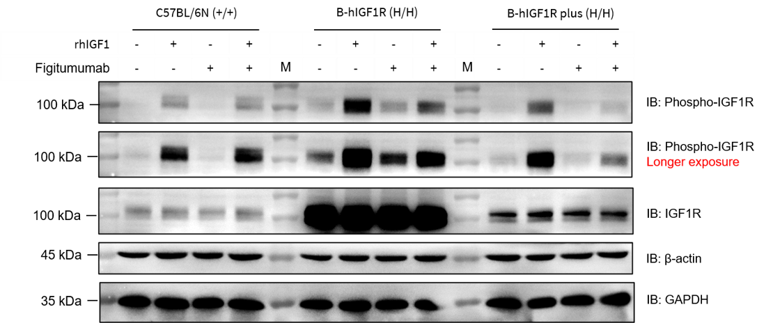
Yes. Figitumumab-analog suppressed IGF1-induced activation of humanized IGF1R in B-hIGF1R mice plus, supporting functional evaluation of human IGF1R-targeted antibodies.
Q5: What are the main applications of B-hIGF1R mice plus?
Applications include anti-IGF1R antibody efficacy studies, IGF1R pathway pharmacology, obesity and metabolic disease research, oncology studies, brain endothelial IGF1R research, and preclinical safety evaluation.