B-hIL23A/hIL12B mice

C57BL/6-Il23atm1(IL23A)Bcgen Il12btm1(IL12B)Bcgen/Bcgen • 120553

B-hIL23A/hIL12B mice

Catalog Number: 120553
Strain Name: C57BL/6-Il23atm1(IL23A)Bcgen Il12btm1(IL12B)Bcgen/Bcgen
Strain Background: C57BL/6
NCBI gene ID: 51561,3593 (Human)
Aliases: P19; SGRF; IL-23; IL-23A; IL23P19; CLMF; NKSF; CLMF2; IMD28; IMD29; NKSF2; IL-12B
---
라이선스 옵션 제공 가능
B-hIL23A/hIL12B mice

이 페이지에서

  • Description
  • Targeting strategy
  • Phenotypic analysis
  • Efficacy

포스터

모두 보기

    출판물

      Description

      IL-12 and IL-23: Shared Subunit Cytokines that Govern T-Cell Fate

      • Gene Information: The IL12B gene (chromosome 5q33) encodes the IL-12p40 subunit shared by both IL-12 and IL-23. IL12A (p35 subunit, chromosome 3p12) combines with p40 to form IL-12, while IL23A (p19 subunit, chromosome 12q13) pairs with p40 to create IL-23.
      • Protein Expression: Both are covalently-linked heterodimeric proteins (IL-12: p35/p40, IL-23: p19/p40). They are produced by activated antigen-presenting cells (e.g., dendritic cells, macrophages) and act as powerful instructors of CD4+ T-cell differentiation.
      • Signaling Pathway: IL-12 signals via a specific receptor (IL-12Rβ1/IL-12Rβ2), activating STAT4 to promote Th1 cell development. IL-23 binds a related receptor (IL-12Rβ1/IL-23R) and activates STAT3, essential for the expansion and stabilization of pathogenic Th17 cells.
      • Therapeutic Inhibition: The shared p40 subunit makes it an effective target for simultaneous pathway inhibition (e.g., Ustekinumab), a therapy widely used in psoriasis and inflammatory bowel disease. More recent, specific inhibitors target only the IL-23 p19 subunit, proving highly effective with a favorable safety profile (e.g., Guselkumab, Risankizumab).
      Targeting strategy

      IL23A

      • The exons 1-4 of mouse Il23a gene that encode the full length of encoding regions were replaced by human IL23A counterpart gene sequences in B-hIL23A/hIL12B mice.
      • The endogenous mouse promoter, 5′ UTR, and 3′ UTR regions are retained, allowing human IL23A expression to be driven by the native mouse Il23a promoter, while endogenous mouse Il23a transcription and translation are abolished.

      IL12B

      • The exons 2-8 of mouse Il12b gene that encode the full length of encoding regions and 3’UTR were replaced by human IL12B counterpart gene sequences in B-hIL23A/hIL12B mice.
      • The endogenous mouse promoter and 5′ UTR regions are retained, allowing human IL12B expression to be driven by the native mouse Il12b promoter, while endogenous mouse Il12b transcription and translation are abolished.
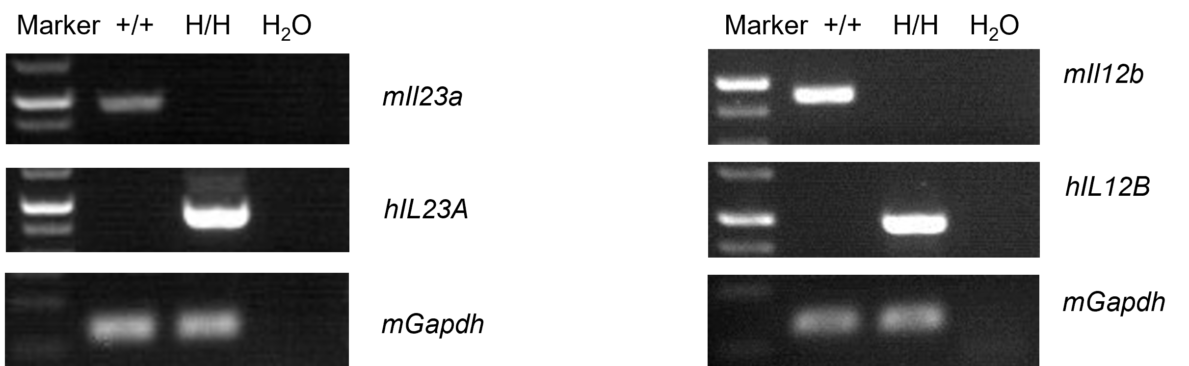
      mRNA Expression Analysis in Thymus
      • Mouse Il23a, Il12b mRNA were detectable in wild-type mice.
      • Human IL23A, IL12B mRNA were detectable in homozygous B-hIL23A/hIL12B mice but not in wild-type mice.

      Strain specific analysis of IL23A, IL12B gene expression in wild-type mice and B-hIL23A/hIL12B mice by RT-PCR. Thymus RNA was isolated from wild-type C57BL/6JNifdc mice (+/+) and homozygous B-hIL23A/hIL12B mice (H/H, H/H), then cDNA libraries were synthesized by reverse transcription, followed by PCR with mouse or human IL23A, IL12B primers. Mouse Il23a, Il12b mRNA were detectable in wild-type mice. Human IL23A, IL12B mRNA were detectable in homozygous B-hIL23A/hIL12B mice but not in wild-type mice.

      Protein Expression Analysis in Supernatants
      • Mouse IL23 was only detectable in wild-type C57BL/6 mice.
      • Human IL23 was exclusively detectable in homozygous B-hIL23A/hIL12B mice and B-hTL1A/hIL23A/hIL12B mice.

      Strain specific IL23 (IL23A/IL12B) expression analysis in wild-type C57BL/6 mice, homozygous B-hIL23A/hIL12B mice and B-hTL1A/hIL23A/hIL12B mice by ELISA. Bone marrow derived dendritic cells were produced by culturing the bone marrow from wild-type C57BL/6 mice, homozygous B-hIL23A/hIL12B mice and B-hTL1A/hIL23A/hIL12B mice (female, 6-week-old, n=3), which were stimulated with 10 μg/mL LPS in vivo for 24 h . After stimulation, the supernatants were collected and the levels of mouse IL23 and human IL23 were analyzed by ELISA. Mouse IL23 was only detectable in wild-type C57BL/6 mice. Human IL23 was exclusively detectable in homozygous B-hIL23A/hIL12B mice and B-hTL1A/hIL23A/hIL12B mice. Values are expressed as mean ± SEM. ND: not detectable.
      Analysis of Leukocyte Subpopulations
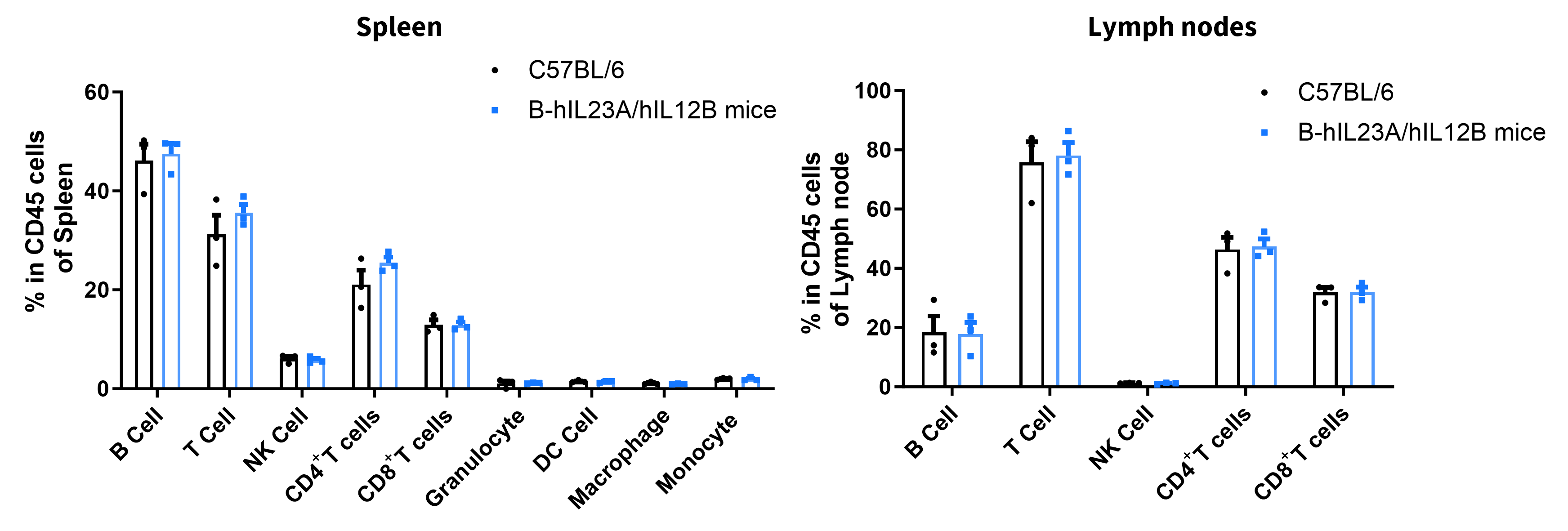
      • Percent of T cells, B cells, NK cells, dendritic cells, granulocytes, monocytes and macrophages in homozygous B-hIL23A/hIL12B mice was similar to those in the C57BL/6 mice.
      • Humanization of IL23A/IL12B does not affect normal immune cell development or splenic distribution.

      Analysis of subpopulation of leukocytes by FACS. Splenocytes and lymph nodes were isolated from female C57BL/6 and B-hIL23A/hIL12B mice (n=3, 7-week-old). Flow cytometry analysis of the splenocytes and lymph nodes were performed to assess leukocyte subpopulations. Percent of T cells, B cells, NK cells, dendritic cells, granulocytes, monocytes and macrophages in homozygous B-hIL23A/hIL12B mice were similar to those in the C57BL/6 mice, demonstrating that introduction of hIL23A and hIL12B in place of its mouse counterpart does not change the overall development, differentiation or distribution of these cell types. Values are expressed as mean ± SEM.
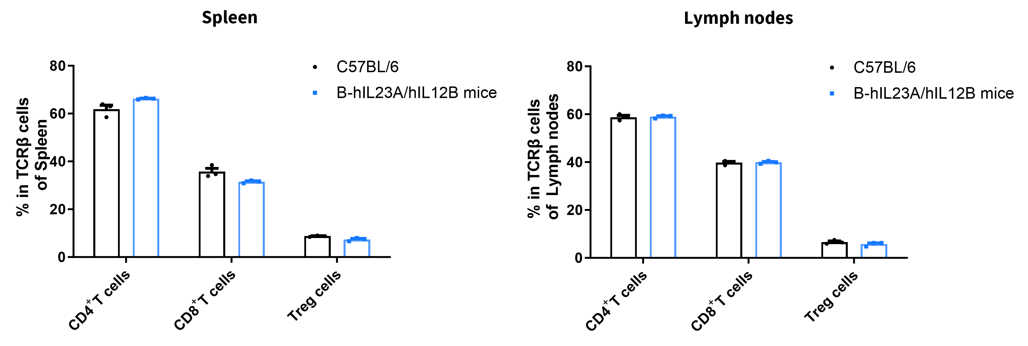
      • Percent of CD8+ T cells, CD4+ T cells and Treg cells in homozygous B-hIL23A/hIL12B mice were similar to those in the C57BL/6 mice.
      • Humanization of IL23A/IL12B does not change the overall development, differentiation or distribution of these T cell subtypes.

      Analysis of T cell subpopulations by FACS. Splenocytes and lymph nodes were isolated from female C57BL/6 and B-hIL23A/hIL12B mice (n=3, 7-week-old). Flow cytometry analysis of the splenocytes and lymph nodes were performed to assess leukocyte subpopulations. Percent of CD4+ T cells, CD8+ T cells and Treg cells in homozygous B-hIL23A/hIL12B mice were similar to those in the C57BL/6 mice, demonstrating that introduction of hIL23A and hIL12B in place of its mouse counterpart does not change the overall development, differentiation or distribution of these T cell subtypes. Values are expressed as mean ± SEM.
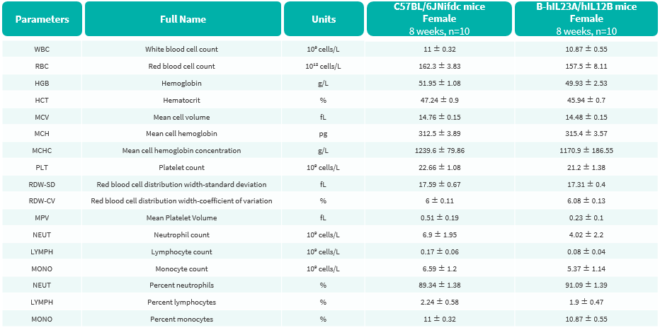
      Hematology Analysis
      • No significant differences were observed compared with wild-type mice.

      Complete blood count (CBC) of B-hIL23A/hIL12B mice. Values are expressed as mean ± SD.
      Blood Biochemical Analysis
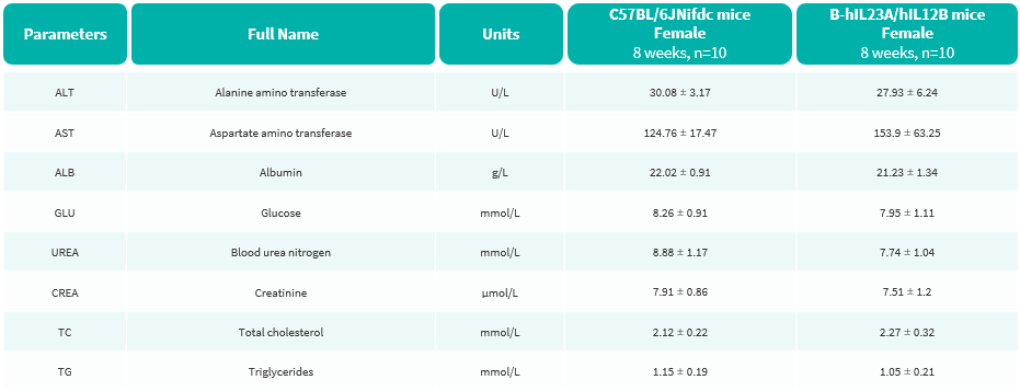
      • No significant differences were observed compared with wild-type mice.

      Blood biochemical parameters of B-hIL23A/hIL12B mice are shown. Values are expressed as mean ± SD.

      In vivo Efficacy of Anti-human IL17A Antibodies in Psoriasis Model Induced in B-hIL23A/hIL12B Mice

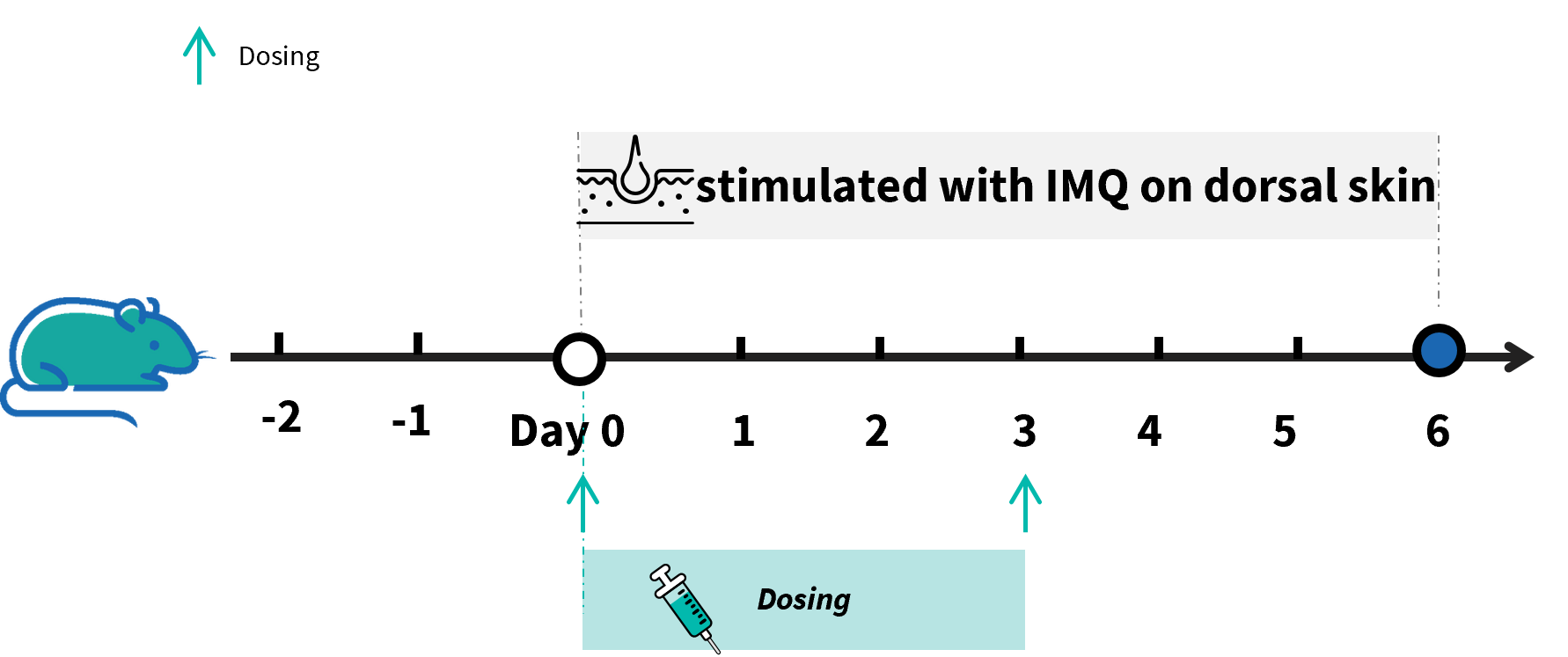
      Experimental schedule for induction of IMQ-induced psoriasis model in B-hIL23A/hIL12B mice. Mice at 7 week-old of age received a daily topical of commercially available IMQ cream on the shaved back for 6 consecutive days to induce psoriasis. Control mice were treated similarly with Vaseline cream. Severity of skin inflammation was daily scored and back skin was collected at the endpoint. IMQ: imiquimod.
      In Vivo Efficacy in Psoriasis Model Induced in B-hIL23A/hIL12B Mice
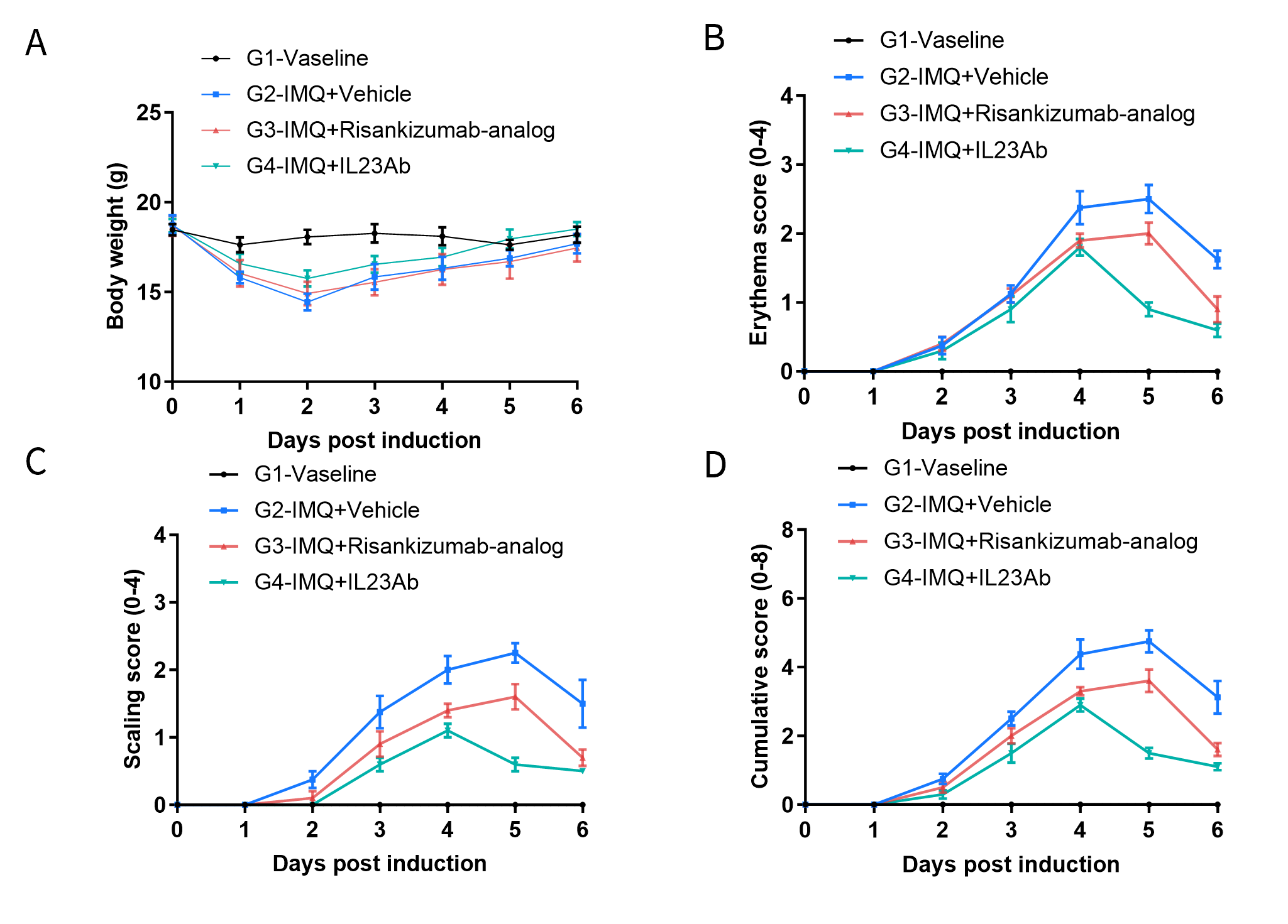
      • After treatment of anti-human IL23A antibody , the phenotypical presentation level and erythema and scaling score of the back were much lower than the control in homozygous B-hIL23A/hIL12B mice.

      Effects of anti-human IL23A antibody in IMQ-induced psoriasis model of B-hIL23A/hIL12B mice. (A) Body weight changes during treatment. (B-C) Erythema and the scaling score of the back were scored daily on a scale from 0 to 4. Additionally, the cumulative score (erythema plus scaling) is depicted (D). Results indicated that structural features of IMQ-induced skin inflammation is visible and increased in severity up to day 5. Anti-human IL23A antibody can efficiently decrease the development of the psoriasis-like disease in B-hIL23A/hIL12B mice.

      * When publishing results obtained using this animal model, please acknowledge the source as follows: The animal model [B-hIL23A/hIL12B mice] (Cat# 120553) was purchased from Biocytogen.