Description
IL-12 has a distinct, specialized role in driving the Th1 (Type 1 helper T cell) immune response.
- Gene Information: IL12A encodes the interleukin-12 subunit alpha and functions as a pro-inflammatory cytokine subunit that joins with IL12B to form IL-12. This cytokine is secreted by antigen-presenting cells and links innate and adaptive immunity by driving Th1 cell differentiation and interferon-gamma production.
- Protein Expression: IL12 is primarily produced by antigen-presenting cells (DCs, macrophages, neutrophils) of the innate immune system. Its expression is usually a "danger signal" triggered by the presence of bacteria, viruses, or intracellular parasites.
- Signaling Pathway: IL-12 stimulation of JAK2 and TYK2 activity leads to phosphorylation of STAT4 and other STAT molecules. IL-12 could induce the production of IFN-γ, which is required for the development of TH1 immune response.
- Therapeutic Inhibition: By inhibiting IL-12 and IL-23 signaling, these inhibitors suppress pro-inflammatory cytokine production and modulate the immune response to alleviate clinical symptoms.
Targeting strategy
IL12A
- The exons 1-7 of mouse Il12a gene that encode the full length of encoding regions were replaced by human IL12A counterpart gene sequences.
- The promoter, 5’UTR and 3’UTR region of the mouse gene were also retained. The human IL12A expression was driven by endogenous mouse Il12a promoter, while mouse Il12a gene transcription and translation will be disrupted.
IL12B
- The exons 2-8 of mouse Il12b gene that encode the full length of encoding regions and 3’UTR were replaced by human IL12B counterpart gene sequences.
- The promoter and 5’UTR region of the mouse gene were also retained. The human IL12B expression was driven by mouse Il12b promoter, while mouse IL12B gene transcription and translation will be disrupted.
IL12RB1
- The exons 1-14 of mouse Il12rb1 gene that encode signal peptide and extracellular domain were replaced by human counterparts. The genomic region of mouse Il12rb1 gene that encodes transmembrane domain and cytoplasmic portion was retained.
- The promoter, 5’UTR and 3’UTR region of the mouse gene were also retained. The human IL12RB1 expression was driven by endogenous mouse Il12rb1 promoter, while mouse Il12rb1 gene transcription and translation will be disrupted.
IL12RB2
- A chimeric CDS that encodes mouse IL12RB2 signal peptide, human IL12RB2 extracellular domain, mouse IL12RB2 transmembrane and cytoplasmic domain, followed by mouse 3’UTR-STOP was inserted after exon 2 of mouse Il12rb2.
- The chimeric IL12RB2 protein expression will be driven by endogenous mouse Il12rb2 promoter.
mRNA Expression Analysis
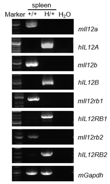
- Mouse Il12a, Il12b, Il12rb1, Il12rb2 mRNA were detectable in wild-type mice.
- Human IL12A, IL12B, IL12RB1, IL12RB2 mRNA were detectable in homozygous B-hIL12A/hIL12B/hIL12RB1 plus/hIL12RB2 ad mice but not in wild-type mice.
Strain specific analysis of IL12A, IL12B, IL12RB1, IL12RB2 mRNA expression in wild-type C57BL/6JNifdc mice and homozygous B-hIL12A/hIL12B/hIL12RB1 plus/hIL12RB2 ad mice by RT-PCR. Spleen RNA was isolated from wild-type C57BL/6JNifdc mice (+/+) and homozygous B-hIL12A/hIL12B/hIL12RB1 plus/hIL12RB2 ad mice (H/H, H/H, H/H, H/H), then cDNA libraries were synthesized by reverse transcription, followed by PCR with mouse or human IL12A, IL12B, IL12RB1, IL12RB2 primers. Mouse Il12a, Il12b, Il12rb1, Il12rb2 mRNA were detectable in wild-type mice. Human IL12A, IL12B, IL12RB1, IL12RB2 mRNA were detectable in homozygous B-hIL12A/hIL12B/hIL12RB1 plus/hIL12RB2 ad mice but not in wild-type mice.
IL12 Protein Expression Analysis
- Mouse IL12 was only detectable in wild-type C57BL/6JNifdc mice after LPS stimulation.
- Human IL12 was exclusively detectable in homozygous B-hIL12A/hIL12B/hIL12RB1 plus/hIL12RB2 ad mice after LPS stimulation.
Strain specific IL12 expression analysis in wild-type C57BL/6JNifdc mice and homozygous B-hIL12A/hIL12B/hIL12RB1 plus/hIL12RB2 ad mice by ELISA. Bone marrow-derived dendritic cells were isolated from wild-type C57BL/6JNifdc mice (+/+) and homozygous B-hIL12A/hIL12B/hIL12RB1 plus/hIL12RB2 ad mice (H/H, H/H, H/H, H/H) stimulated with LPS in vitro for 24 hrs, then cell supernatants were collected and analyzed by ELISA (Mouse IL-12(p70) ELISA MAXTM Deluxe Set , Biolegend, 433607; Human IL12 p70 SimpleStep ELISA kit, Abcam, ab223592). Mouse IL12 was only detectable in wild-type C57BL/6JNifdc mice and human IL12 was exclusively detectable in homozygous B-hIL12A/hIL12B/hIL12RB1 plus/hIL12RB2 ad mice after LPS stimulation. Values are expressed as mean ± SEM.
Functional validation
- Expression of mouse IFNγ was increased in wild-type C57BL/6JNifdc mice and homozygous B-hIL12A/hIL12B/hIL12RB1 plus/hIL12RB2 ad mice after mIL12, hIL12 stimulation.
- Humanized IL12A, IL12B, IL12RB1, and IL12RB2 are able to perform their functions normally.
Strain specific IFNγ expression analysis in wild-type C57BL/6JNifdc mice and homozygous B-hIL12A/hIL12B/hIL12RB1 plus/hIL12RB2 ad mice by ELISA. Spleen CD4+ T cells supernatant was collected from wild-type C57BL/6JNifdc mice (+/+) and homozygous B-hIL12A/hIL12B/hIL12RB1 plus/hIL12RB2 ad mice (H/H, H/H, H/H, H/H) stimulated with mIL12, hIL12 in vitro for 48 hrs. Expression level mouse mIFNγ were analyzed by ELISA (Mouse IFNγ ELISA MAXTM Deluxe Set, 430804). Expression of mouse IFNγ was increased in wild-type C57BL/6JNifdc mice and homozygous B-hIL12A/hIL12B/hIL12RB1 plus/hIL12RB2 ad mice after mIL12, hIL12 stimulation. Values are expressed as mean ± SEM.
* When publishing results obtained using this animal model, please acknowledge the source as follows: The animal model [B-hIL12A/hIL12B/hIL12RB1 plus/hIL12RB2 ad mice] (Cat# 113241) was purchased from Biocytogen.