TL1A binds to death receptor 3 (DR3) and regulates immune signaling pathways involved in effector cell proliferation, activation, apoptosis, and cytokine production. Soluble decoy receptor 3 (DcR3) can bind TL1A and modulate TL1A–DR3-mediated inflammatory responses.
IL-23 is a heterodimeric cytokine composed of IL23A (p19) and IL12B (p40) subunits that is primarily produced by macrophages and dendritic cells. IL-23 signaling regulates downstream inflammatory cytokines and plays a critical role in chronic intestinal inflammation and autoimmune disease progression.
Integrin α4β7 is a heterodimer composed of ITGA4 and ITGB7 subunits that mediates lymphocyte adhesion and gut homing through interactions with MAdCAM-1, VCAM-1, and fibronectin. The α4β7 pathway is an important therapeutic target in inflammatory bowel disease and intestinal immune regulation.
TL1A/IL23A/IL12B/α4β7 humanized mice were generated by replacing the extracellular or full-length coding regions of mouse Tl1a, Il23a, Il12b, Itga4, and Itgb7 genes with their human counterparts while retaining endogenous mouse regulatory elements. Human TL1A, IL23A, IL12B, ITGA4, and ITGB7 proteins were specifically expressed in homozygous TL1A/IL23A/IL12B/α4β7 humanized mice.
This model provides a translational preclinical platform for evaluating anti-human TL1A, IL23, and α4β7 therapeutics in inflammatory bowel disease, autoimmune disease, and mucosal immunology research.
Key Advantages
Validation
Applications

TL1A
IL23A
IL12B
ITGA4
ITGB7
Note: TL1A/IL23A/IL12B/α4β7 humanized mice were obtained by mating B-hTL1A/hIL23A/hIL12B mice (113038) and B-hTL1A/hα4β7 mice (113037).

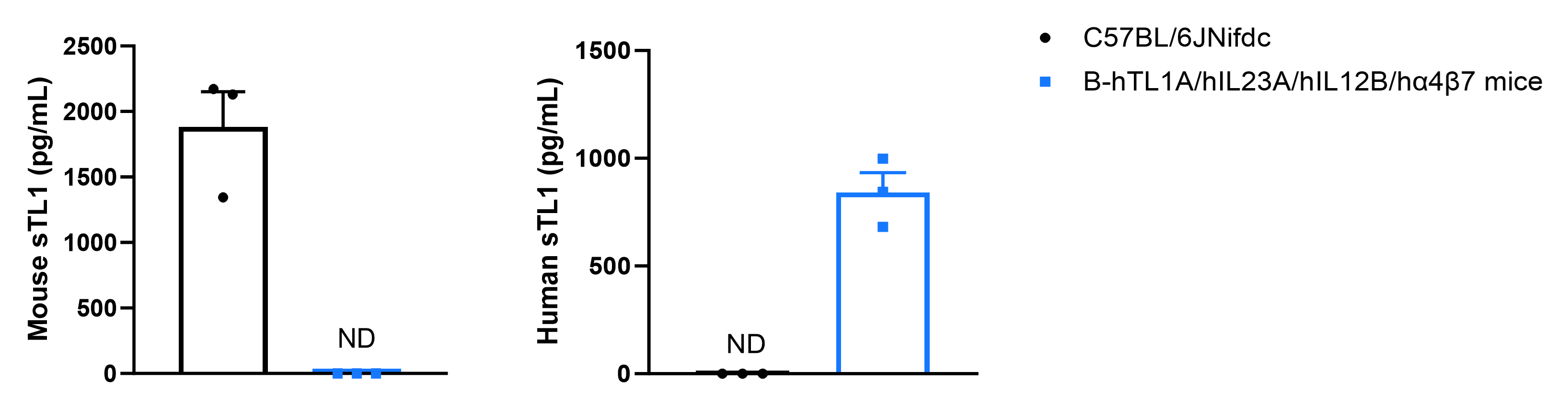
Soluble TL1A expression analysis in B-hTL1A/hIL23A/hIL12B/hα4β7 mice by ELISA. Bone marrow derived dendritic cells (BMDC) were produced by culturing the bone marrow from wild-type C57BL/6JNifdc mice (+/+) and homozygous B-hTL1A/hIL23A/hIL12B/hα4β7 mice (H/H;H/H;H/H;H/H;H/H), which were stimulated with LPS in vitro. After stimulation, the supernatants were collected and the level of soluble TL1A was analyzed by ELISA. Soluble human TL1A was exclusively detectable in homozygous B-hTL1A/hIL23A/hIL12B/hα4β7 mice, but not in wild-type C57BL/6JNifdc mice. Values are expressed as mean ± SEM. ND: not detectable.
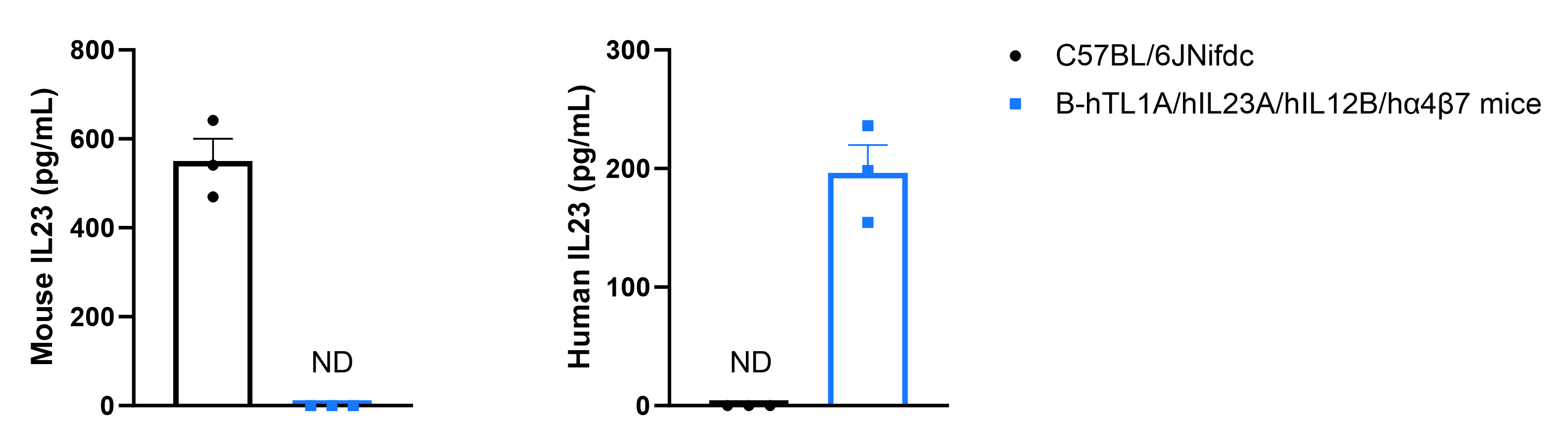
Mouse IL23 and human IL23 expression analysis in B-hTL1A/hIL23A/hIL12B/hα4β7 mice by ELISA. BMDCs were produced by culturing the bone marrow from wild-type C57BL/6JNifdc mice (+/+) and homozygous B-hTL1A/hIL23A/hIL12B/hα4β7 mice (H/H;H/H;H/H;H/H;H/H), which were stimulated with LPS in vitro. After stimulation, the supernatants were collected and the levels of mouse and human IL23 were analyzed by ELISA (R&D, M2300; R&D, D2300B). Mouse IL23 was only detectable in wild-type C57BL/6JNifdc mice. Human IL23 was exclusively detectable in homozygous B-hTL1A/hIL23A/hIL12B/hα4β7 mice. Values are expressed as mean ± SEM. ND: not detectable.
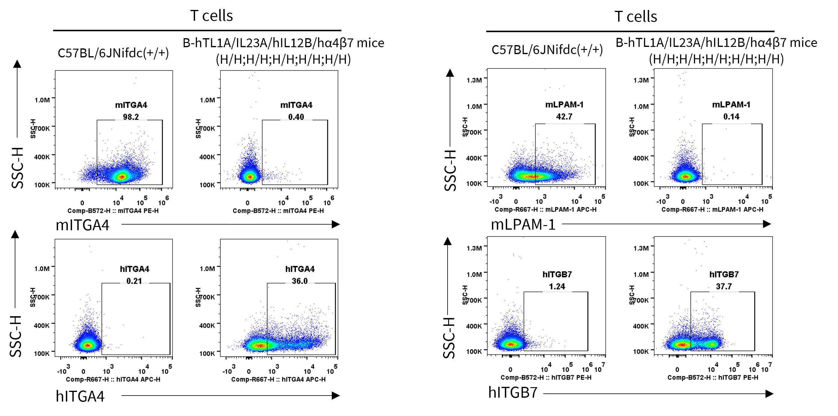
Strain specific ITGA4 and ITGB7 expression analysis in wild-type C57BL/6JNifdc mice and homozygous humanized B-hTL1A/hIL23A/hIL12B/hα4β7 mice by flow cytometry. Splenocytes were collected from wild-type C57BL/6JNifdc mice (+/+) and homozygous B-hTL1A/hIL23A/hIL12B/hα4β7 mice (H/H;H/H;H/H;H/H;H/H). Protein expression was analyzed with anti-mouse ITGA4 antibody (Biolegend, 103705), anti-mouse ITGB7 antibody (Biolegend, 120607), anti-human ITGA4 antibody (Biolegend, 304307) anti-human ITGB7 antibody (Invitrogen, MA5-23541) by flow cytometry. Mouse ITGA4 and ITGB7 were only detectable in T cells of wild-type C57BL/6JNifdc mice. Human ITGA4 and ITGB7 were exclusively detectable in T cells of homozygous B-hTL1A/hIL23A/hIL12B/hα4β7 mice.
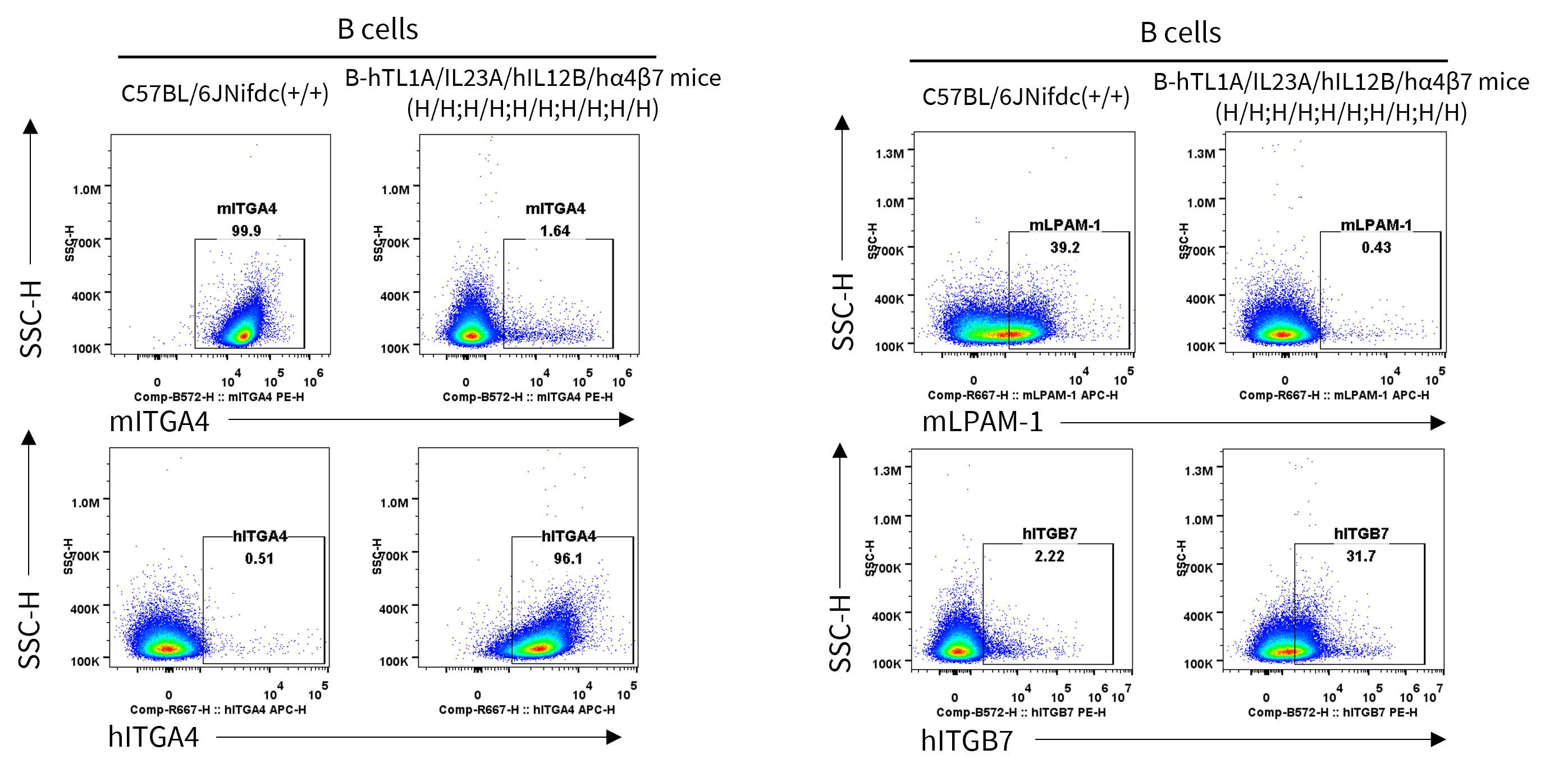
Strain specific ITGA4 and ITGB7 expression analysis in wild-type C57BL/6JNifdc mice and homozygous humanized B-hTL1A/hIL23A/hIL12B/hα4β7 mice by flow cytometry. Splenocytes were collected from wild-type C57BL/6JNifdc mice (+/+) and homozygous B-hTL1A/hIL23A/hIL12B/hα4β7 mice (H/H;H/H;H/H;H/H;H/H). Protein expression was analyzed with anti-mouse ITGA4 antibody (Biolegend, 103705), anti-mouse ITGB7 antibody (Biolegend, 120607), anti-human ITGA4 antibody (Biolegend, 304307) anti-human ITGB7 antibody (Invitrogen, MA5-23541) by flow cytometry. Mouse ITGA4 and ITGB7 were only detectable in B cells of wild-type C57BL/6JNifdc mice. Human ITGA4 and ITGB7 were exclusively detectable in B cells of homozygous B-hTL1A/hIL23A/hIL12B/hα4β7 mice.
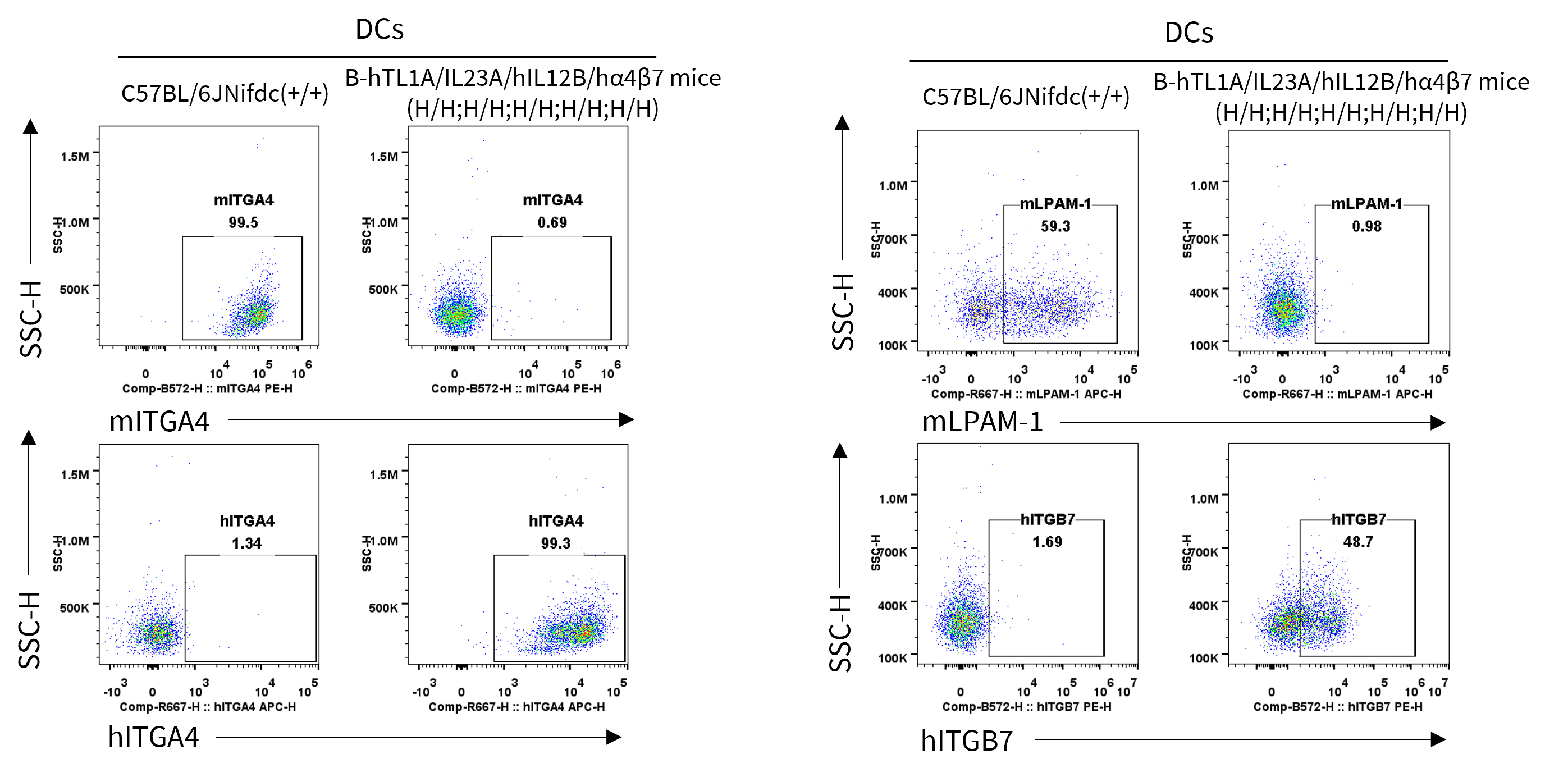
Strain specific ITGA4 and ITGB7 expression analysis in wild-type C57BL/6JNifdc mice and homozygous humanized B-hTL1A/hIL23A/hIL12B/hα4β7 mice by flow cytometry. Splenocytes were collected from wild-type C57BL/6JNifdc mice (+/+) and homozygous B-hTL1A/hIL23A/hIL12B/hα4β7 mice (H/H;H/H;H/H;H/H;H/H). Protein expression was analyzed with anti-mouse ITGA4 antibody (Biolegend, 103705), anti-mouse ITGB7 antibody (Biolegend, 120607), anti-human ITGA4 antibody (Biolegend, 304307) anti-human ITGB7 antibody (Invitrogen, MA5-23541) by flow cytometry. Mouse ITGA4 and ITGB7 were only detectable in DCs of wild-type C57BL/6JNifdc mice. Human ITGA4 and ITGB7 were exclusively detectable in DCs of homozygous B-hTL1A/hIL23A/hIL12B/hα4β7 mice.
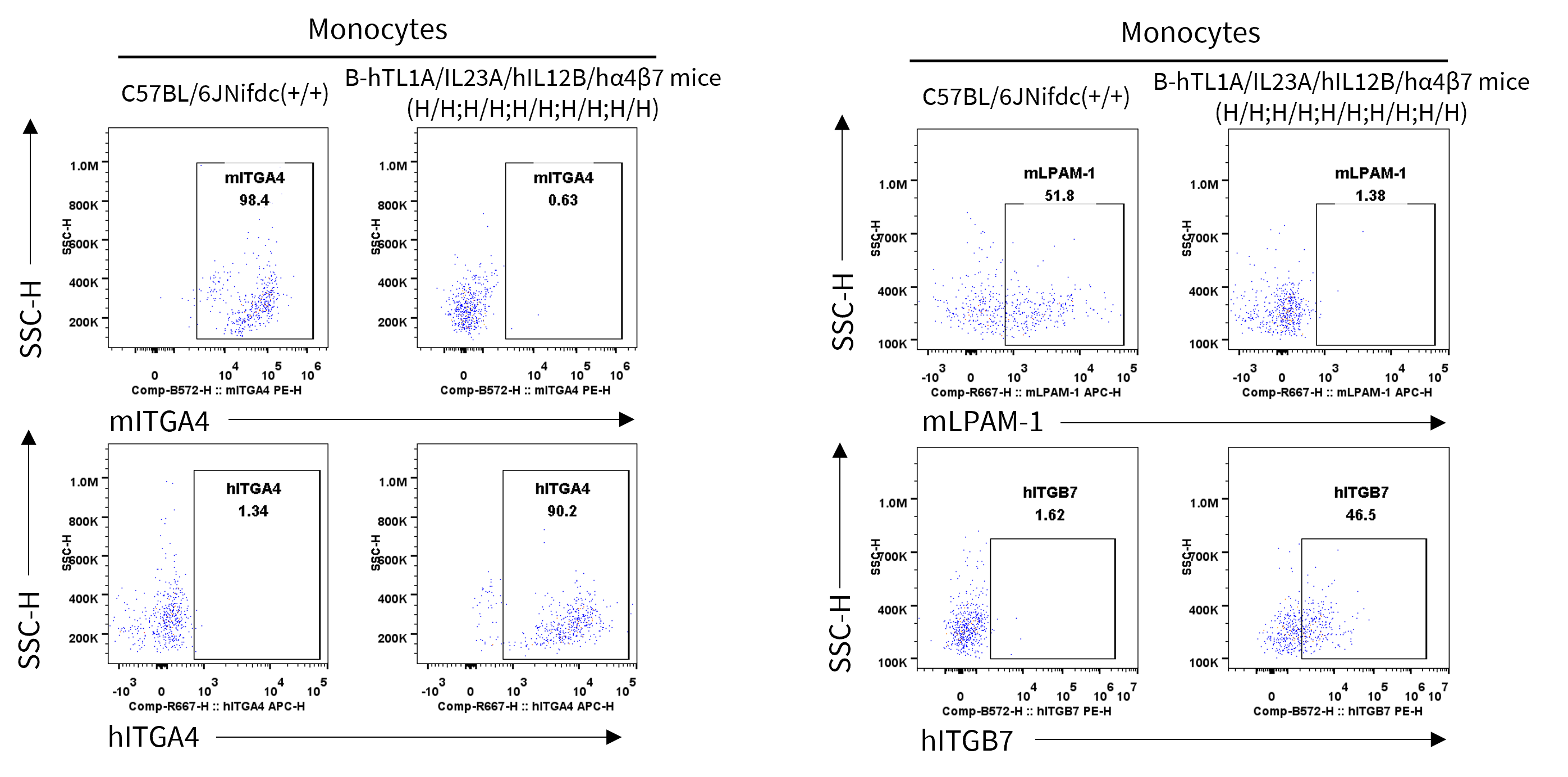
Strain specific ITGA4 and ITGB7 expression analysis in wild-type C57BL/6JNifdc mice and homozygous humanized B-hTL1A/hIL23A/hIL12B/hα4β7 mice by flow cytometry. Splenocytes were collected from wild-type C57BL/6JNifdc mice (+/+) and homozygous B-hTL1A/hIL23A/hIL12B/hα4β7 mice (H/H;H/H;H/H;H/H;H/H). Protein expression was analyzed with anti-mouse ITGA4 antibody (Biolegend, 103705), anti-mouse ITGB7 antibody (Biolegend, 120607), anti-human ITGA4 antibody (Biolegend, 304307) anti-human ITGB7 antibody (Invitrogen, MA5-23541) by flow cytometry. Mouse ITGA4 and ITGB7 were only detectable in monocytes of wild-type C57BL/6JNifdc mice. Human ITGA4 and ITGB7 were exclusively detectable in monocytes of homozygous B-hTL1A/hIL23A/hIL12B/hα4β7 mice.
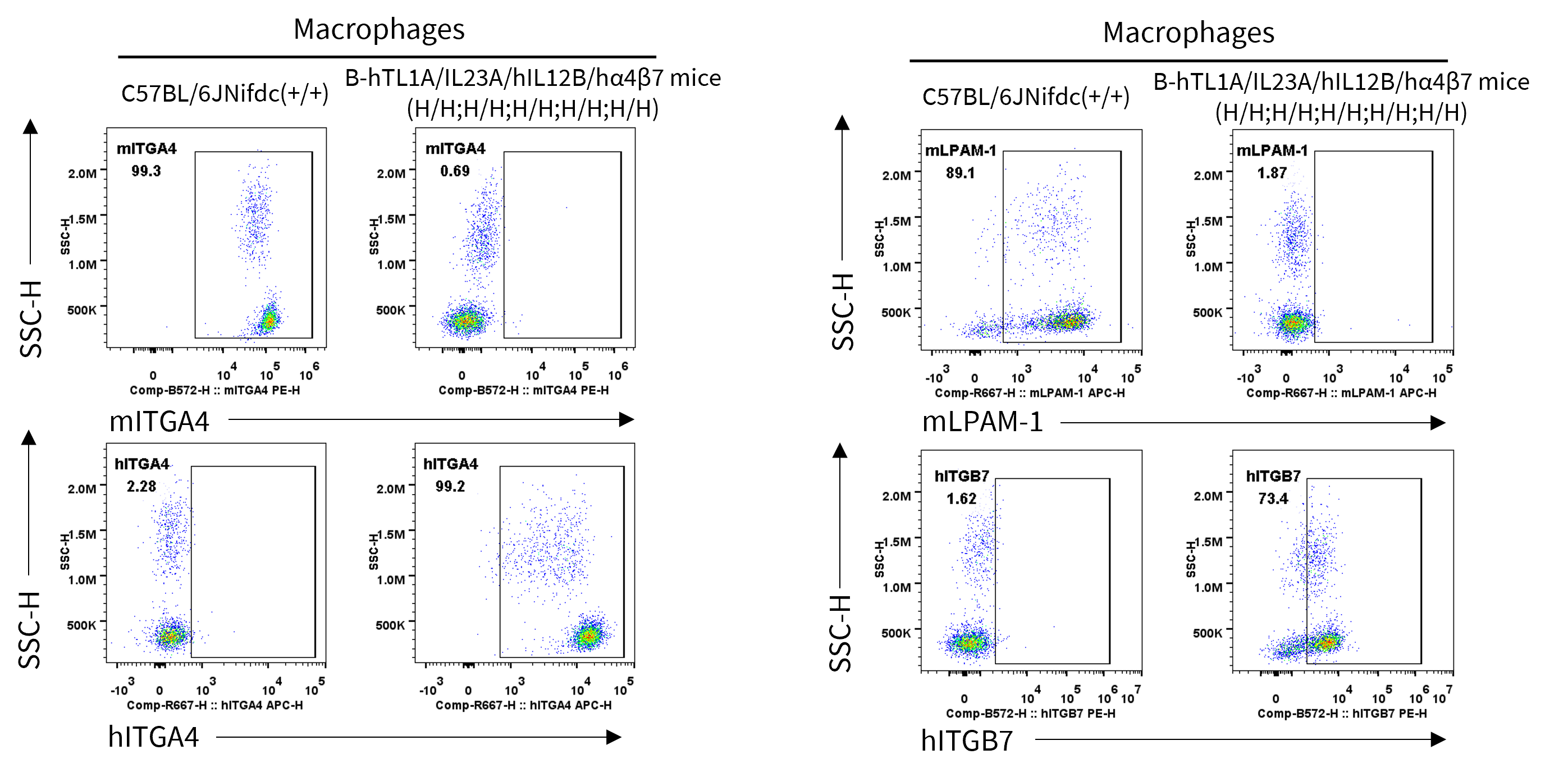
Strain specific ITGA4 and ITGB7 expression analysis in wild-type C57BL/6JNifdc mice and homozygous humanized B-hTL1A/hIL23A/hIL12B/hα4β7 mice by flow cytometry. Splenocytes were collected from wild-type C57BL/6JNifdc mice (+/+) and homozygous B-hTL1A/hIL23A/hIL12B/hα4β7 mice (H/H;H/H;H/H;H/H;H/H). Protein expression was analyzed with anti-mouse ITGA4 antibody (Biolegend, 103705), anti-mouse ITGB7 antibody (Biolegend, 120607), anti-human ITGA4 antibody (Biolegend, 304307) anti-human ITGB7 antibody (Invitrogen, MA5-23541) by flow cytometry. Mouse ITGA4 and ITGB7 were only detectable in macrophages of wild-type C57BL/6JNifdc mice. Human ITGA4 and ITGB7 were exclusively detectable in macrophages of homozygous B-hTL1A/hIL23A/hIL12B/hα4β7 mice.
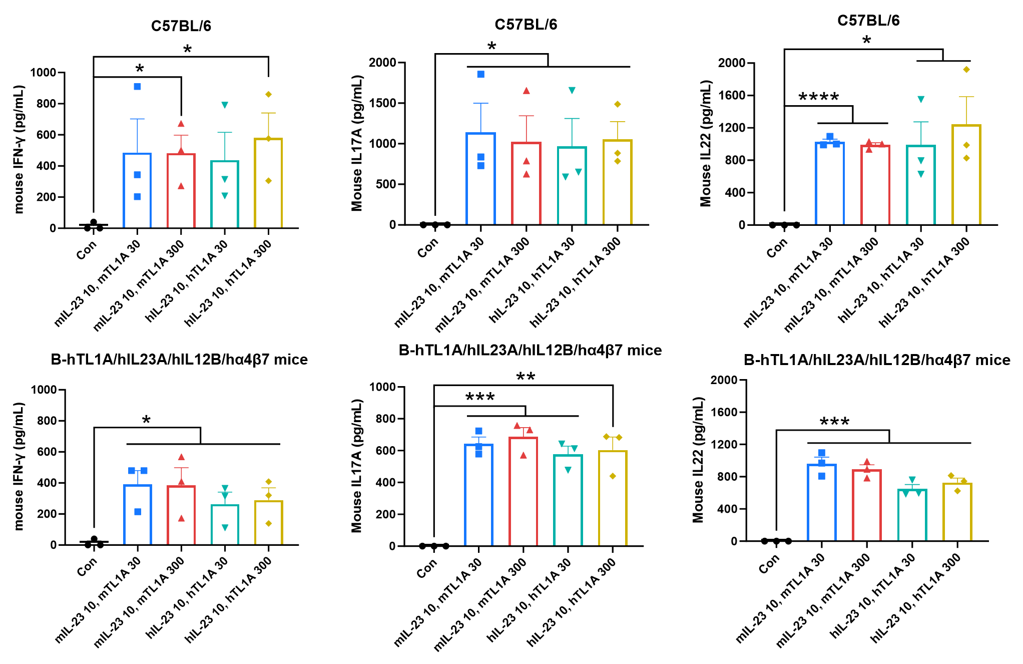
Ex vivo functional analysis in B-hTL1A/hIL23A/hIL12B/hα4β7 mice. Splenocytes were collected from wild-type C57BL/6 mice (+/+) and homozygous B-hTL1A/hIL23A/hIL12B/hα4β7 mice (H/H;H/H;H/H;H/H;H/H), then the production of mouse IFN-γ, mouse IL17A, and mouse IL22 in supernatants were assessed after 72 h of incubation with mIL23 (10 ng/mL), mTL1A (30, 300 ng/mL), hIL23 (10 ng/mL) and hTL1A (30, 300 ng/mL) in vitro.
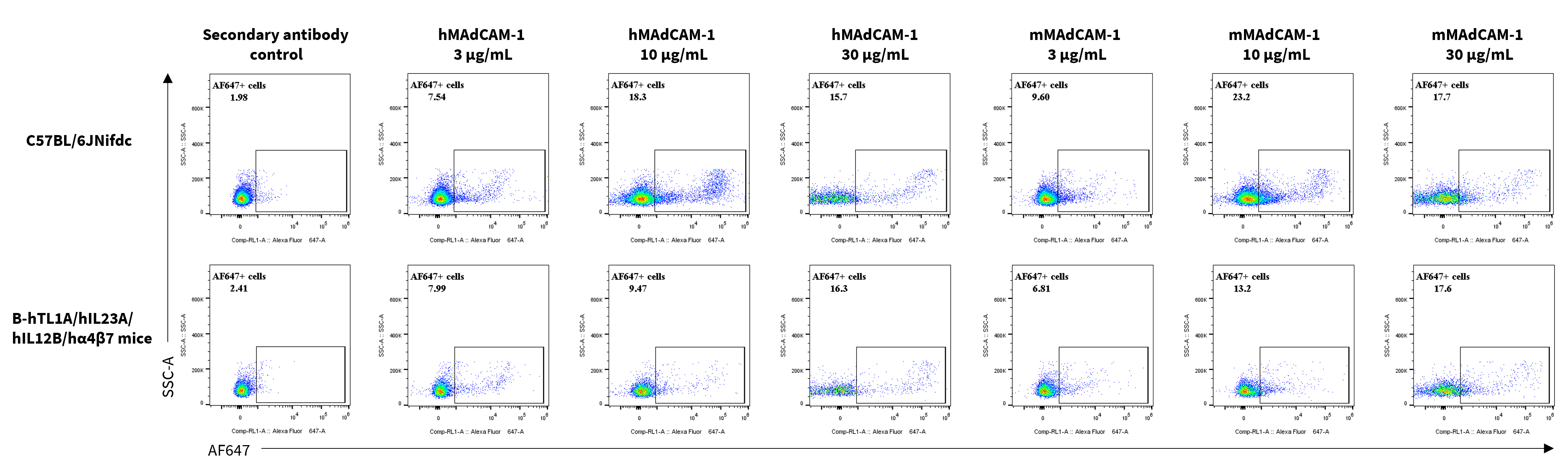
Assessment of MAdCAM-1 binding in C57BL/6 and B-hTL1A/hIL23A/hIL12B/hα4β7 mice. Splenocytes were collected from homozygous B-hTL1A/hIL23A/hIL12B/hα4β7 mice (H/H;H/H;H/H;H/H;H/H), and were incubated with human MAdCAM-1 protein (3, 10, 30 μg/mL) or mouse MAdCAM-1 protein (3, 10, 30 μg/mL) in vitro for 1 h. The binding of MAdCAM-1 to α4β7 were detected by flow cytometry.
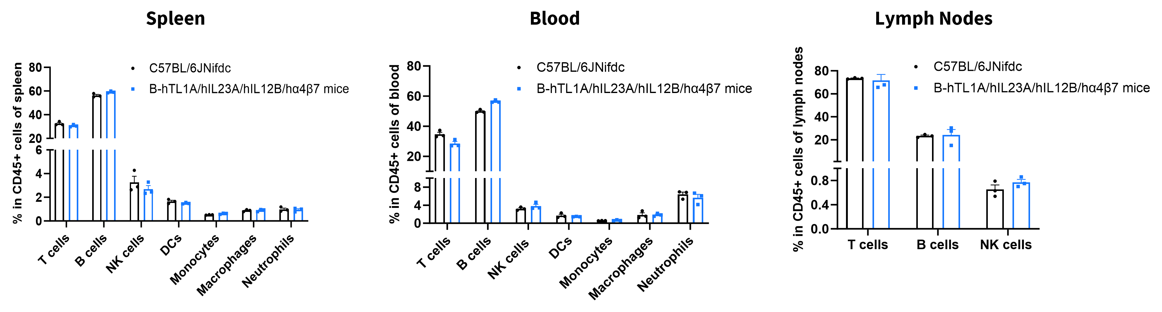
Analysis of leukocyte subpopulations by flow cytometry in immune organs and blood. Splenocytes, peripheral blood, and lymph nodes were isolated from female C57BL/6JNifdc and B-hTL1A/hIL23A/hIL12B/hα4β7 mice (female, 8-week-old, n = 3). Single live cells were gated on the CD45⁺ population and analyzed by flow cytometry as indicated. Values are expressed as mean ± SEM.
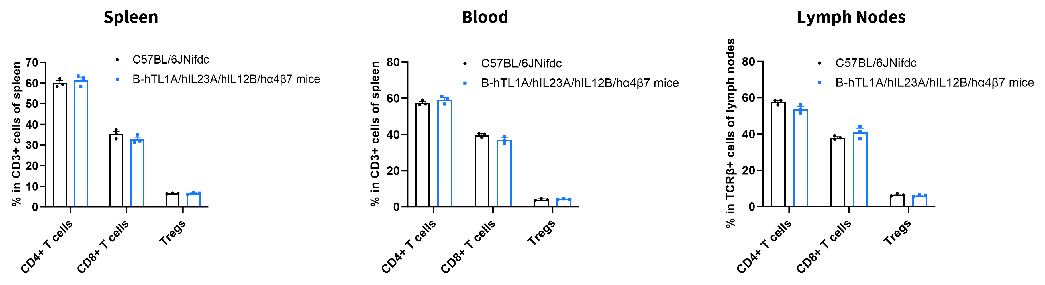
Analysis of T-cell subpopulations by flow cytometry in immune organs and blood. Splenocytes, peripheral blood, and lymph nodes were isolated from female C57BL/6JNifdc and B-hTL1A/hIL23A/hIL12B/hα4β7 mice (female, 8-week-old, n = 3). Single live cells were gated on the CD3⁺ T-cell population and analyzed by flow cytometry as indicated. Values are expressed as mean ± SEM.

Q1: What are TL1A/IL23A/IL12B/α4β7 humanized mice?
A1: TL1A/IL23A/IL12B/α4β7 humanized mice are genetically engineered mice expressing human TL1A, IL23A, IL12B, ITGA4, and ITGB7, enabling translational evaluation of inflammatory and gut-homing pathways in vivo.
Q2: What diseases can be studied using TL1A/IL23A/IL12B/α4β7 humanized mice?
A2: These mice are highly suitable for inflammatory bowel disease, Crohn’s disease, ulcerative colitis, autoimmune disease, and chronic inflammatory disease research.
Q3: Can this model be used for antibody validation?
A3: Yes. TL1A/IL23A/IL12B/α4β7 humanized mice support in vivo evaluation of anti-human TL1A, IL23, and α4β7 antibodies and biologics.
Q4: Does humanization alter immune cell development?
A4: No. Frequencies of major leukocyte and T cell populations in spleen, blood, and lymph nodes remain comparable to wild-type mice.
Q5: What makes this model unique?
A5: This model simultaneously humanizes multiple clinically relevant inflammatory pathways involved in intestinal inflammation and lymphocyte trafficking, enabling comprehensive translational drug evaluation.