Targeting Strategy
Gene targeting strategy for B-H11-hC3*R102G, mC3 KO mice.
The genome sequence of human C3 gene with R102G mutation encodes the whole molecule (ATG to STOP codon), including 3’UTR, were inserted into the Hipp11 (H11) locus in B-H11-hC3*R102G mice. The human C3 expression is driven by the human C3 promoter. Exons 2-40 of the mouse C3 gene were knocked out. As a result, the mouse C3 protein is not expressed anymore.
(R102G was an SNP named C3F. The C3F variant is associated with several diseases, including IgA nephropathy, C3G, and age-related macular degeneration.)
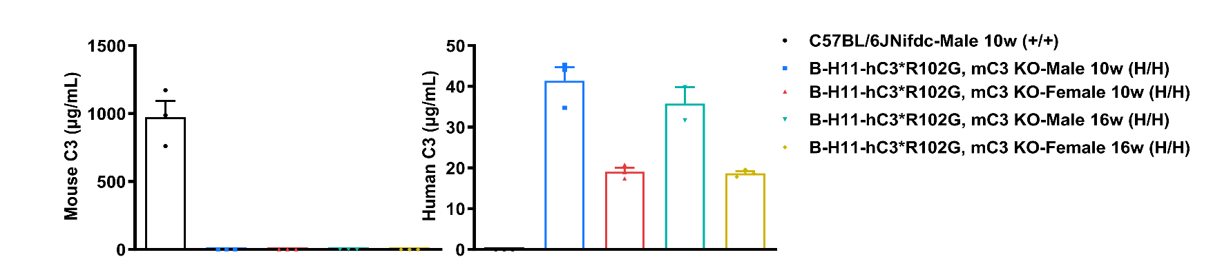
Protein Expression Analysis in Serum
Strain specific C3 expression analysis in wild-type C57BL/6JNifdc mice and homozygous B-H11-hC3*R102G, mC3 KO mice by ELISA. Serum was collected from wild-type C57BL/6JNifdc mice (+/+), and homozygous B-H11-hC3*R102G, mC3 KO mice (H/H, -/-) and analyzed by ELISA (mouse C3: ab157711, human C3: ab108823). Mouse C3 was only detectable in wild-type C57BL/6JNifdc mice. Human C3 was exclusively detectable in homozygous B-H11-hC3*R102G, mC3 KO mice. Values are expressed as mean ± SEM.
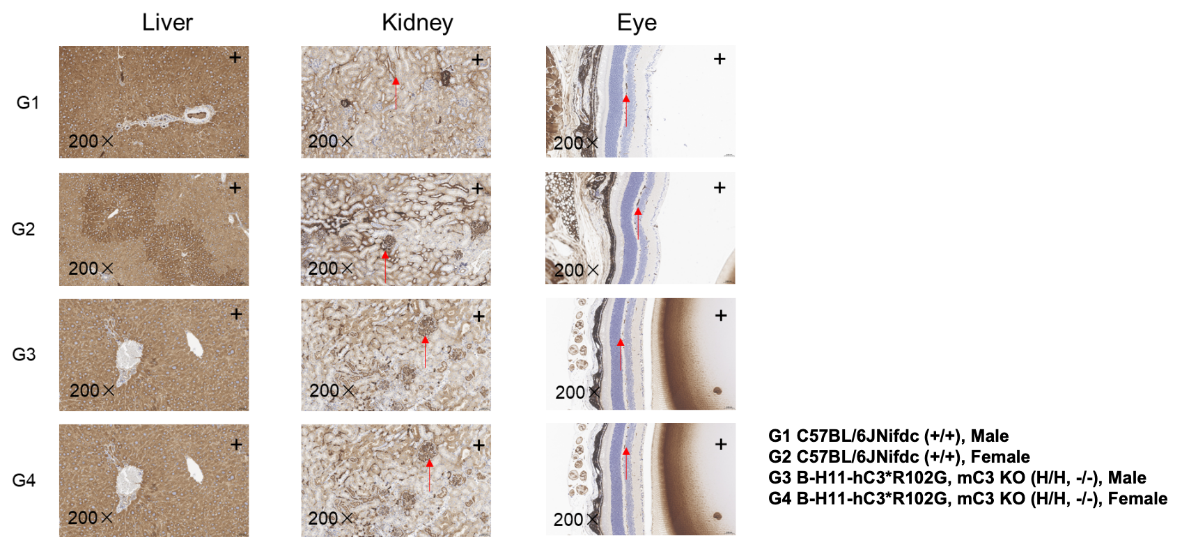
Immunohistochemistry (IHC) Staining Analysis
IHC Staining in B-H11-hC3*R102G, mC3 KO mice. The liver, kidney, and eye tissues of wild-type C57BL/6JNifdc mice (+/+) and homozygous B-H11-hC3*R102G, mC3 KO mice (H/H, -/-) were isolated at 16 weeks old and analyzed with IHC staining. C3 was detected in the liver, eye, and kidney of B-H11-hC3*R102G, mC3 KO mice and wild-type C57BL/6JNifdc mice, as the antibody cross-recognizes both human and mouse C3 (Abcam, ab200999). “+” indicate positive expression. Red arrows indicate a positive signal.
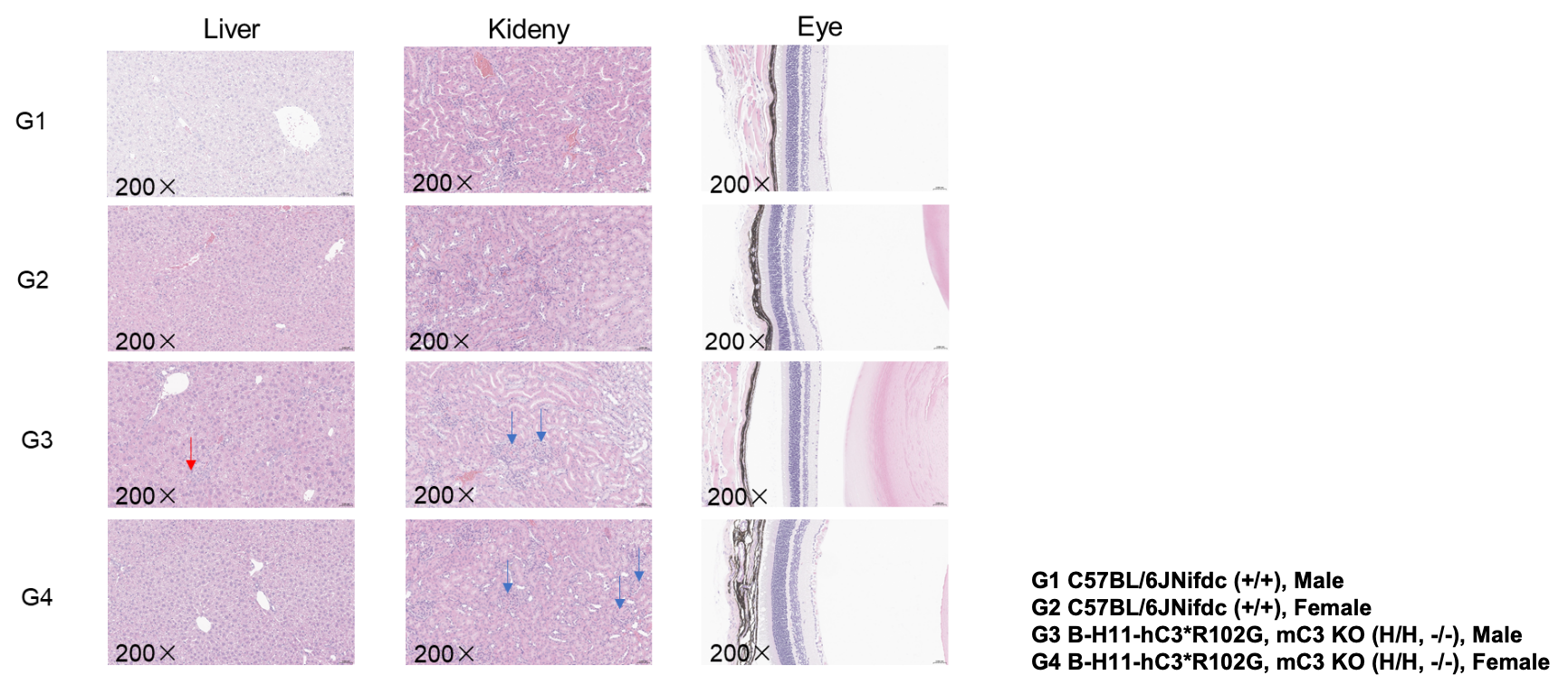
Histopathological Analysis
Histopathological analysis in B-H11-hC3*R102G, mC3 KO mice. The liver, kidney, and eye tissues of wild-type C57BL/6JNifdc mice (+/+) and homozygous B-H11-hC3*R102G, mC3 KO mice (H/H, -/-) were isolated at 16 weeks old and analyzed with IHC staining (male and female, n=6). The liver of male B-H11-hC3*R102G, mC3 KO mice (5/6) showed inflammatory cell infiltration. The kidneys of male (4/6) and female (5/6) B-H11-hC3*R102G, mC3 KO mice showed glomerular matrix proliferation. There were no obvious abnormalities in the eye of B-H11-hC3*R102G, mC3 KO mice. And there were no obvious abnormalities in wild-type C57BL/6JNifdc mice. Red arrows: inflammatory cell infiltration. Blue arrows: glomerular matrix proliferation.
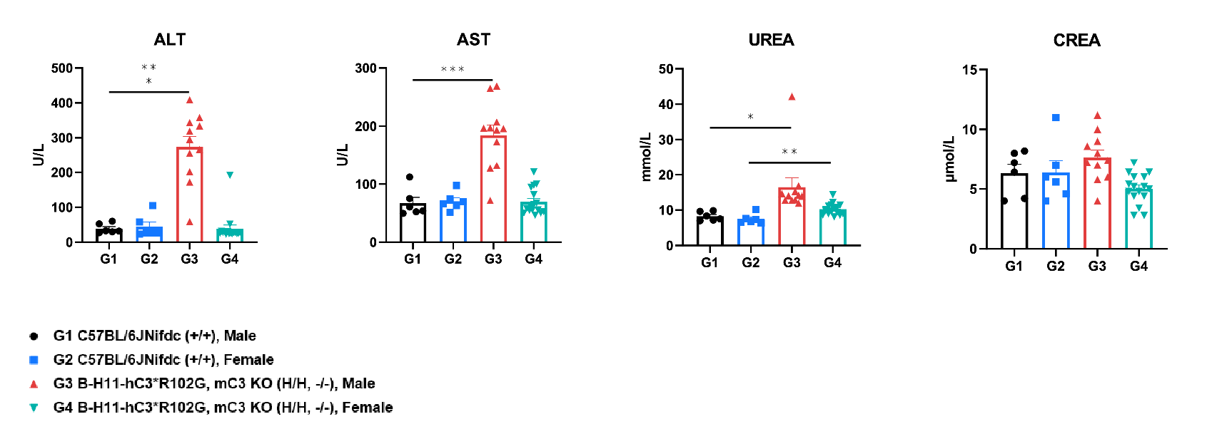
Biochemistry Analysis
Analysis of blood biochemicals in B-H11-hC3*R102G, mC3 KO mice and wild-type C57BL/6JNifdc mice. Serum was collected from wild-type C57BL/6JNifdc mice (+/+) and and homozygous B-H11-hC3*R102G, mC3 KO mice (H/H, -/-) (male n=11, female n=16, 16 weeks old) and analyzed for biochemistry. The ALT, AST, and UREA were increased in male B-H11-hC3*R102G, mC3 KO mice. Values are expressed as mean ± SEM. Significance was determined by t-test, *p<0.05,**p<0.01, ***p<0.001.
Survival Curve
Survival Curve of B-H11-hC3*R102G, mC3 KO mice. Graph showing the survival curve of homozygous B-H11-hC3*R102G, mC3 KO mice (H/H, -/-) (male, n=16; female, n=16). The survival rate of male B-H11-hC3*R102G, mC3 KO mice was around ~70% at 16 weeks old. And we didn’t observe that mice died in the female B-H11-hC3*R102G, mC3 KO mice until 16 weeks old.
* When publishing results obtained using this animal model, please acknowledge the source as follows: The animal model [B-H11-hC3*R102G/mC3 KO mice] (Cat# 114198) was purchased from Biocytogen.