Description
- The core function of MTARC1 is to act as a reductase, catalyzing the reduction of various N-hydroxylated compounds. It operates in concert with cytochrome b5 and cytochrome b5 reductase, forming an electron transfer chain that acquires electrons from NADH to facilitate this process. In recent years, the most notable discovery regarding MTARC1 has been its central role in hepatic lipid metabolism, particularly its strong association with MASLD. Genome-wide association studies (GWAS) have revealed that specific variants of the MTARC1 gene are significantly correlated with liver fat content and MASLD risk. A key variant, identified as the single nucleotide polymorphism (SNP) rs2642438, leads to a substitution of alanine with threonine at position 165 of the MTARC1 protein—known as the p.Ala165Thr (A165T) variant. This variant has been shown to have a protective effect: individuals carrying the A165T variant (even in a heterozygous state) exhibit lower liver fat content, a significantly reduced risk of developing MASLD/NAFLD, and a more favorable liver metabolic phenotype.
- Gene editing strategy: The exons 1-7 of mouse Mtarc1 gene that encode the whole molecule (ATG to STOP codon), including the promoter, 5’UTR, and 3’UTR, were replaced by the human counterparts in B-hMTARC1 mice. The human MTARC1 expression is driven by the human MTARC1 promoter, while mouse Mtarc1 gene transcription and translation will be disrupted.
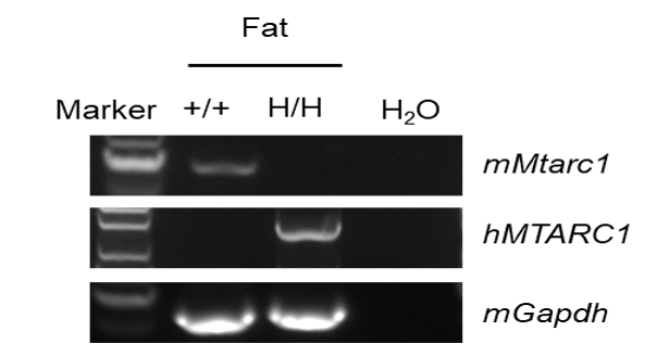
- Validation: Human MTARC1 mRNA was exclusively detectable in homozygous B-hMTARC1 mice but not in wild-type mice.
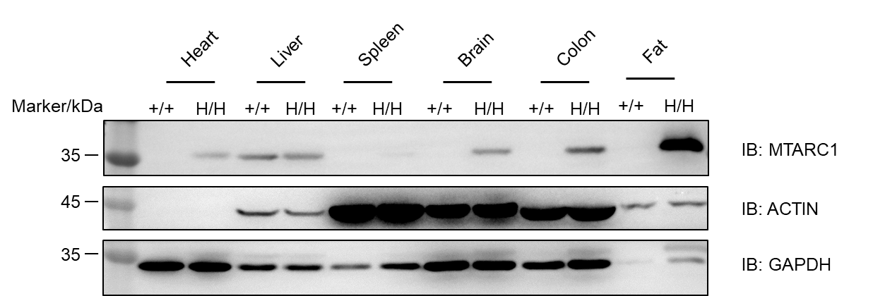
- MTARC1 protein was detected in the heart, liver, brain, colon, and fat of homozygous B-hMTARC1 mice, whereas in wild-type mice it was detected only in the liver.
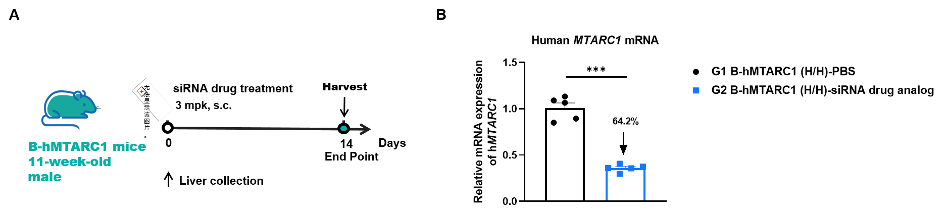
- The inhibitory efficiency of the small nucleic acid drugs: The human MTARC1 expression in the siRNA group was significantly reduced compared to the control group.
- Application: Applied to the efficacy evaluation and toxicity assessment of small nucleic acid drugs and inhibitors in the field of MASH.
Targeting Strategy
Gene targeting strategy for B-hMTARC1 mice. The exons 1-7 of mouse Mtarc1 gene that encode the whole molecule (ATG to STOP codon), including the promoter, 5’UTR and 3’UTR, were replaced by the human counterparts in B-hMTARC1 mice. The human MTARC1 expression is driven by the human MTARC1 promoter, while mouse Mtarc1 gene transcription and translation will be disrupted.
mRNA Expression Analysis
Strain specific analysis of MTARC1 mRNA expression in wild-type C57BL/6JNifdc mice and B-hMTARC1 mice by RT-PCR. Fat RNA was isolated from wild-type C57BL/6JNifdc mice (+/+) and homozygous B-hMTARC1 mice (H/H), then cDNA libraries were synthesized by reverse transcription, followed by PCR with mouse or human MTARC1 primers. Mouse Mtarc1 mRNA was only detectable in wild-type mice. Human MTARC1 mRNA was exclusively detectable in homozygous B-hMTARC1 mice but not in wild-type mice.

Strain specific analysis of MTARC1 mRNA expression in wild-type C57BL/6JNifdc mice and B-hMTARC1 mice by RT-qPCR. Liver and fat RNA were isolated from wild-type C57BL/6JNifdc mice (H/H) and homozygous B-hMTARC1 mice (H/H) (n=3, 8-week-old, male and female), then cDNA libraries were synthesized by reverse transcription, followed by PCR with mouse or human MTARC1 primers. (A) The mouse Mtarc1 mRNA expression of wild-type and homozygous mice by used mouse Mtarc1 primer. (B) The human MTARC1 mRNA expression of humanized wild-type and homozygous mice by used human MTARC1 primer. Values are expressed as mean ± SEM. Note: MTARC1 mRNA tissue-specific expression between mouse and human was different (doi: 10.1371/journal.pgen.1011179. eCollection 2024 Mar.).
Protein Expression Analysis
Western blot analysis of MTARC1 protein expression in homozygous B-hMTARC1 mice. Various tissue lysates were collected from wild-type C57BL/6JNifdc mice (+/+) and homozygous B-hMTARC1 mice (H/H), and then analyzed by western blot with cross reactive anti-MTARC1 antibody (Abcam, ab317262). 40 μg total proteins were loaded for western blotting analysis. MTARC1 was detected in the heart, liver, brain, colon and fat of homozygous B-hMTARC1 mice, whereas in wild-type mice it was detected only in the liver.
Note: MTARC1 tissue-specific expression between mouse and human was different (doi: 10.1371/journal.pgen.1011179. eCollection 2024 Mar.).
The Inhibitory Efficiency of the Nucleic Acid Drugs Against Human MTARC1
The inhibitory efficiency of the nucleic acid drugs against human MTARC1 in homozygous B-hMTARC1 mice. B-hMTARC1 mice (H/H) were randomly divided into two groups (male, 11-week-old, n=5/group). The human MTARC1 targeted nucleic acid drugs (synthesized according to patents), and PBS were administered to the mice individually. The nucleic acid drug was administered in the form of a PBS aqueous solution. The mice were sacrificed on day 14, and the liver tissue was collected to detect the expression level of human MTARC1 mRNA by qPCR. (A) The schematic diagram of experimental processing. (B) The expression of human MTARC1 mRNA in the liver tissues. Values are expressed as mean ± SEM. Significance was determined by t-test, *P<0.05, **P<0.01, ***P<0.001.
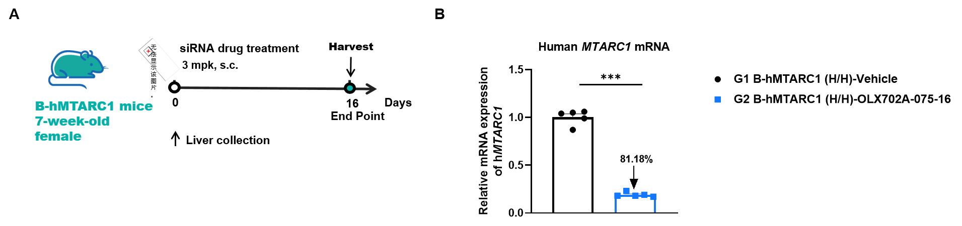
The inhibitory efficiency of the nucleic acid drugs against human MTARC1 in homozygous B-hMTARC1 mice. B-hMTARC1 mice (H/H) were randomly divided into two groups (female, 7-week-old, n=5/group). The human MTARC1 targeted nucleic acid drugs and the vehicle were administered to the mice individually. The mice were sacrificed on day 16, and the liver tissue was collected to detect the expression level of human MTARC1 mRNA by qPCR. (A) The schematic diagram of experimental processing. (B) The expression of human MTARC1 mRNA in the liver tissues. Values are expressed as mean ± SEM. Significance was determined by t-test, *P<0.05, **P<0.01, ***P<0.001. Note: Data are shared upon the client's approval.
* When publishing results obtained using this animal model, please acknowledge the source as follows: The animal model [B-hMTARC1 mice] (Cat# 113623) was purchased from Biocytogen.