Description
PD-1/PD-L1: An immune checkpoint and tumor-associated target for therapeutic intervention
- Gene Information: PD-1 is encoded by the PDCD1 gene on chromosome 2q37.3, while PD-L1 is encoded by the CD274 gene on chromosome 9p24.1. Both are type I transmembrane proteins; PD-1 belongs to the immunoglobulin superfamily and functions as an inhibitory receptor, whereas PD-L1 is a member of the B7 family of co-stimulatory molecules.
- Protein Expression: PD-1 is primarily expressed on activated T cells, B cells, and NK cells, whereas PD-L1 is expressed on antigen-presenting cells and many tumor cells, with expression often induced by IFN-γ.
- Signaling Pathway: Binding of PD-L1 to PD-1 recruits SHP2 phosphatase and suppresses TCR/CD28-mediated PI3K-AKT signaling, resulting in reduced T-cell proliferation, cytokine production, and cytotoxic activity.
- Therapeutic Inhibition: Anti-PD-1 and anti-PD-L1 antibodies block this inhibitory pathway, restoring T-cell function and enhancing anti-tumor immune responses.
VEGFA: A master regulator of angiogenesis and therapeutic intervention
- Gene Information: VEGFA is a protein-coding gene located on chromosome 6p21.1. It encodes a potent angiogenic growth factor that belongs to the PDGF/VEGF family, with multiple isoforms generated by alternative splicing.
- Protein Expression: VEGFA is secreted by tumor cells, cancer-associated fibroblasts, tumor-infiltrating macrophages, and endothelial cells themselves. Its expression is strongly upregulated by hypoxia, oncogenic mutations, and inflammatory mediators such as IL-6 and TGF-β, creating a pro-angiogenic gradient within the tumor microenvironment.
- Signaling Pathway: VEGFA binds with high affinity to receptor tyrosine kinases VEGFR-1 and VEGFR-2 on endothelial cells. This drives endothelial cell proliferation, migration, tube formation, vascular permeability, and survival. Additionally, VEGFA promotes the recruitment of immunosuppressive cells and upregulates PD-L1 on endothelial and tumor cells, directly linking angiogenesis to immune evasion.
- Therapeutic Inhibition: Anti-VEGFA monoclonal antibodies and small-molecule multikinase inhibitors targeting VEGFRs block ligand-receptor binding or downstream kinase activity.
GDF15: A stress-responsive cytokine and key regulator of energy homeostasis
- Gene Information: GDF15 is a protein-coding gene located on chromosome 19p13.11 and encodes a stress-responsive member of the TGF-β superfamily.
- Protein Expression: GDF15 is broadly expressed at low basal levels and is strongly induced by cellular stress, inflammation, tissue injury, and cancer. It is synthesized as a precursor and secreted as a mature homodimer. Circulating GDF15 levels are elevated in several pathological conditions and are closely associated with anorexia, body weight loss, and cancer cachexia.
- Signaling Pathway: GDF15 binds to GFRAL, which recruits the co-receptor RET to activate downstream ERK, AKT, and PLCγ signaling. The GDF15/GFRAL/RET axis plays a central role in regulating appetite, energy expenditure, and body weight.
- Therapeutic Inhibition: Targeting the GDF15/GFRAL/RET axis represents a potential therapeutic strategy for cancer cachexia, anorexia, and metabolic disorders. Blocking GDF15 or GFRAL signaling has demonstrated potential to alleviate anorexia and weight loss in preclinical models, while GDF15/GFRAL agonism is being investigated for metabolic disease applications.
Targeting Strategy
PD-1
- A chimeric CDS encoding the human PDCD1 extracellular domain and the mouse Pdcd1 transmembrane and cytoplasmic domains, followed by a WPRE-pA sequence, was inserted immediately after the initiation codon (ATG) to replace exon 1 of the mouse Pdcd1 gene in B-hPD-1 plus/hPD-L1/hVEGFA/hGDF15 mice.
- The endogenous mouse promoter and 5' UTR were retained. Chimeric PD-1 expression is driven by the endogenous mouse Pdcd1 promoter, while endogenous mouse Pdcd1 transcription and translation are disrupted.
PD-L1
- Exon 3 of the mouse Cd274 gene, which encodes the IgV domain, was replaced with human CD274 exon 3 in B-hPD-1 plus/hPD-L1/hVEGFA/hGDF15 mice.
- The endogenous mouse promoter, 5' UTR, 3' UTR, and genomic regions encoding the signal peptide, the non-IgV domain of the extracellular region, and the transmembrane and cytoplasmic domains were retained. Chimeric PD-L1 expression is driven by the endogenous mouse Cd274 promoter, while endogenous mouse Cd274 transcription and translation are disrupted.
VEGFA
- Exons 1-8 of the mouse Vegfa gene that encode the full-length protein were replaced by human VEGFA exons 1-8 in B-hPD-1 plus/hPD-L1/hVEGFA/hGDF15 mice.
- The promoter, 5' UTR, and 3' UTR regions of the endogenous mouse gene were retained, while endogenous mouse Vegfa transcription and translation are disrupted.
GDF15
- Exons 1-2 of the mouse Gdf15 gene that encode the full-length protein were replaced by human GDF15 exons 1-2 in B-hPD-1 plus/hPD-L1/hVEGFA/hGDF15 mice.
- The promoter, 5' UTR, and 3' UTR regions of the endogenous mouse gene were retained, while endogenous mouse Gdf15 transcription and translation are disrupted.
PD-1 Protein Expression Analysis in Spleen
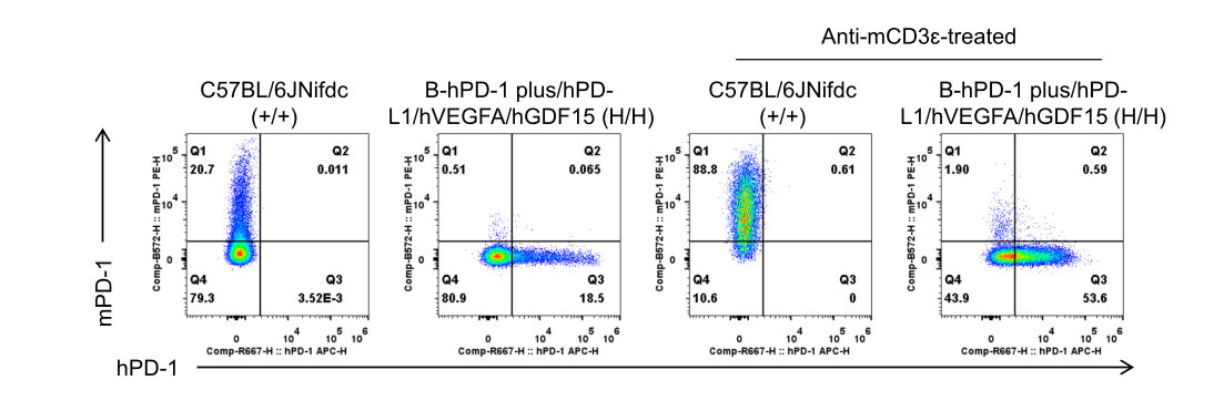
Strain-specific PD-1 expression analysis in wild-type C57BL/6JNifdc mice and homozygous B-hPD-1 plus/hPD-L1/hVEGFA/hGDF15 mice by flow cytometry. Splenocytes were collected from wild-type C57BL/6JNifdc mice (+/+) and homozygous B-hPD-1 plus/hPD-L1/hVEGFA/hGDF15 mice (H/H) (male, 6-week-old, n = 1/group) either left unstimulated or stimulated with an anti-mouse CD3ε antibody (7.5 μg, i.p.) in vivo for 24 hours. Protein expression was analyzed by flow cytometry using an anti-human PD-1 antibody (BioLegend, 329908) and an anti-mouse PD-1 antibody (BioLegend, 109104).
PD-L1 Protein Expression Analysis in Spleen
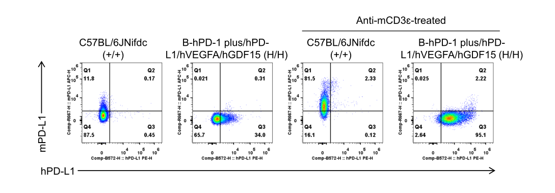
Strain-specific PD-L1 expression analysis in wild-type C57BL/6JNifdc mice and homozygous B-hPD-1 plus/hPD-L1/hVEGFA/hGDF15 mice by flow cytometry. Splenocytes were collected from wild-type C57BL/6JNifdc mice (+/+) and homozygous B-hPD-1 plus/hPD-L1/hVEGFA/hGDF15 mice (H/H) (male, 6-week-old, n = 1/group) either left unstimulated or stimulated with an anti-mouse CD3ε antibody (7.5 μg, i.p.) in vivo for 24 hours. Protein expression was analyzed by flow cytometry using an anti-human PD-L1 antibody (BioLegend, 329706) and an anti-mouse PD-L1 antibody (BioLegend, 124312).
VEGFA Protein Expression Analysis in Lung
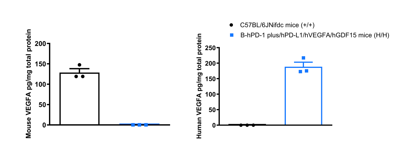
Strain-specific VEGFA expression analysis in wild-type C57BL/6JNifdc mice and homozygous B-hPD-1 plus/hPD-L1/hVEGFA/hGDF15 mice by ELISA. Lung tissue homogenate was collected from wild-type C57BL/6JNifdc mice (+/+) and homozygous B-hPD-1 plus/hPD-L1/hVEGFA/hGDF15 mice (H/H) (male, 6-week-old, n = 3). The expression levels of mouse and human VEGFA were analyzed by ELISA (anti-mouse VEGFA antibody: R&D Systems, MMV00; anti-human VEGFA antibody: R&D Systems, DVE00).
GDF15 Protein Expression Analysis in Serum
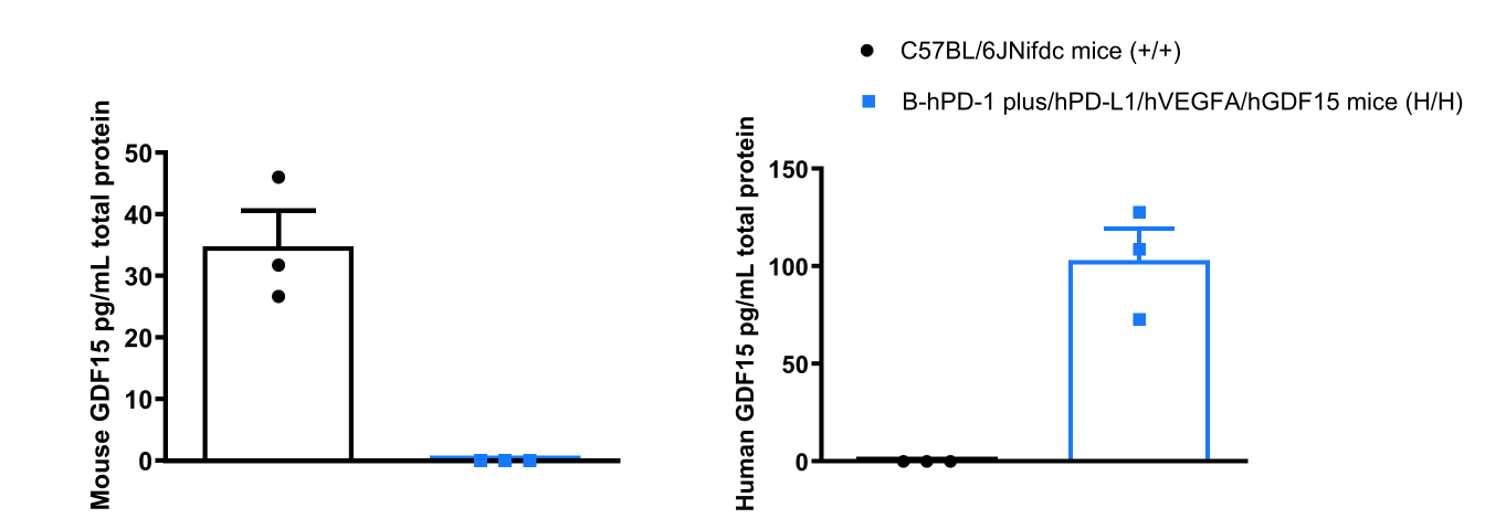
Strain-specific GDF15 expression analysis in wild-type C57BL/6JNifdc mice and homozygous B-hPD-1 plus/hPD-L1/hVEGFA/hGDF15 mice by ELISA. Serum was collected from wild-type C57BL/6JNifdc mice (+/+) and homozygous B-hPD-1 plus/hPD-L1/hVEGFA/hGDF15 mice (H/H) (male, 6-week-old, n = 3). The expression levels of mouse and human GDF15 were analyzed by ELISA (Mouse/Rat GDF-15 Quantikine ELISA Kit: R&D Systems, MGD150; Human GDF-15 Quantikine ELISA Kit: R&D Systems, DGD150).
* When publishing results obtained using this animal model, please acknowledge the source as follows: The animal model [B-hPD-1 plus/hPD-L1/hVEGFA/hGDF15 mice] (Cat# 114972) was purchased from Biocytogen.