Description
- Gene Information: Encoded by CD3E, D, G; part of the Ig superfamily. It forms the essential signaling backbone of the T-cell receptor (TCR) complex. CD28: serves as the primary and essential co-stimulatory receptor on T cells. Cooperation with TCR signaling leads to massive IL-2 production, metabolic reprogramming, robust clonal expansion, and enhanced T cell survival.
- Protein Expression: CD3: Constitutive and universal marker for all mature T cells (CD4+, CD8+). Always present on the cell surface. CD28: Constitutively expressed on the surface of most naive CD4+ T cells and a subset of CD8+ T cells in humans.
- Signaling Pathway: CD3 (Signal 1): Operates via ITAM phosphorylation and ZAP-70. It triggers the initial "on" switch, Ca2+ flux, and immediate cytotoxicity. CD28 (Signal 2): Ligand binding induces the phosphorylation of the cytoplasmic YMNM motif by Src-family kinases. This recruits and activates the PI3K/Akt/mTOR and MAPK/NF-kB pathways.
- Key Therapeutic: Traditional CD3 TCEs provide only the “first signal” which leads to rapid T-cell exhaustion and limited efficacy in solid tumors. By introducing the essential “second signal” (co-stimulation) and activating it via a tumor-dependent mechanism, can effectively breach immune-suppressive solid tumors without inducing systemic CRS.
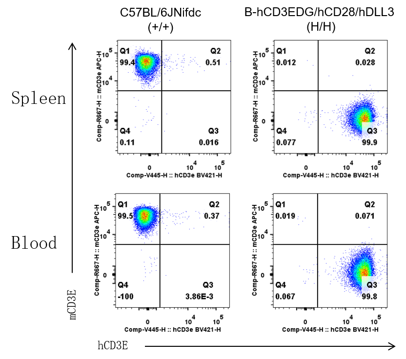
CD3E Protein Expression in Spleen and Blood
- Mouse CD3E was detected on T cells populations in wild-type C57BL/6JNifdc mice, but not in B-hCD3EDG/hCD28/hDLL3 mice.
- Human CD3E was detected on T cells populations in B-hCD3EDG/hCD28/hDLL3 mice, but not in wild-type C57BL/6JNifdc mice.
Mouse and human CD3E expression analysis in T cells of spleen and blood. Splenocytes and blood cells were collected from wild-type C57BL/6JNifdc mice (female, 6-week-old, n = 1) and homozygous B-hCD3EDG/hCD28/hDLL3 mice (female, 6-week-old, n = 1). CD3E expression on T cells was analyzed by flow cytometry using species-specific anti-CD3E antibodies (anti-human CD3E antibody, BD Horizon, 562426; anti-mouse CD3E antibody, Biolegend, 100312 ).
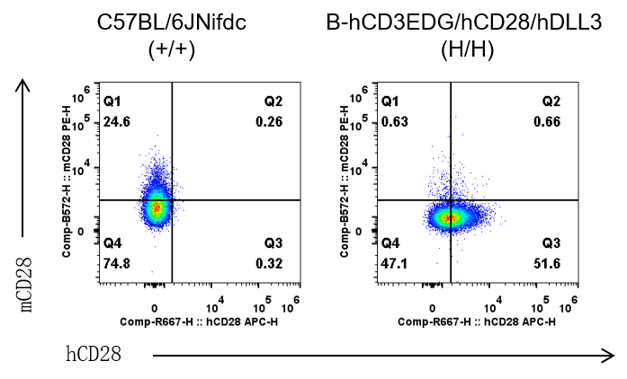
CD28 Protein Expression in Spleen
- Mouse CD28 was detected on T cells populations in wild-type C57BL/6JNifdc mice, but not in B-hCD3EDG/hCD28/hDLL3 mice.
- Human CD28 was detected on T cells populations in B-hCD3EDG/hCD28/hDLL3 mice, but not in wild-type C57BL/6JNifdc mice.
Mouse and human CD28 expression analysis in T cells of spleen. Splenocytes were collected from wild-type C57BL/6JNifdc mice (female, 6-week-old, n=1) and homozygous B-hCD3EDG/hCD28/hDLL3 mice (female, 6-week-old, n = 1). CD28 expression on T cells was analyzed by flow cytometry using species-specific anti-CD28 antibodies (anti-human CD28 antibody, Biolegend, 302911; anti-mouse CD28 antibody, Biolegend, 102105).
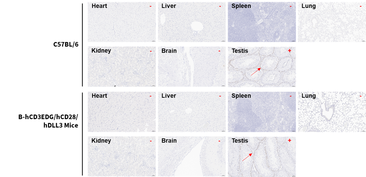
DLL3 Protein Expression Profile
- DLL3 expression was detected in the testis of C57BL/6 and homozygous B-hCD3EDG/hCD28/hDLL3 mice.
Immunohistochemical (IHC) analysis of DLL3 expression in homozygous B-hCD3EDG/hCD28/hDLL3 mice. The heart, liver, spleen, lung, kidney, brain, and testis were collected from C57BL/6 mice (+/+) (male, n=2) and homozygous B-hCD3EDG/hDLL3 mice (H/H) (male, n=2) , analyzed by IHC with anti-hDLL3 (abcam, ab229902). The arrow indicates tissue cells with positive DLL3 staining (brown). “+” indicates that the tissue is positive and “-” indicates that the tissue is negative.
* When publishing results obtained using this animal model, please acknowledge the source as follows: The animal model [B-hCD3EDG/hCD28/hDLL3 mice] (Cat# 114683) was purchased from Biocytogen.