

C57BL/6JNifdc-Cidebtm1(CIDEB)Bcgen/Bcgen • 112944

CIDEB: A novel important regulator in lipid metabolism

CIDEB

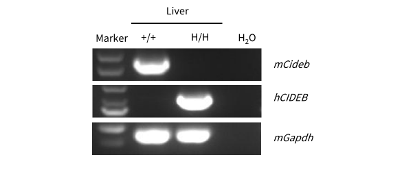
Strain-specific analysis of CIDEB mRNA expression in wild-type C57BL/6JNifdc mice and homozygous B-hCIDEB mice by RT-PCR. Liver RNA was isolated from wild-type C57BL/6JNifdc mice (+/+) and homozygous B-hCIDEB mice (H/H). cDNA libraries were synthesized by reverse transcription, followed by PCR with mouse- or human-specific CIDEB primers. Mouse Cideb mRNA was detectable exclusively in wild-type C57BL/6JNifdc mice. Human CIDEB mRNA was detectable exclusively in homozygous B-hCIDEB mice and was absent in wild-type mice.
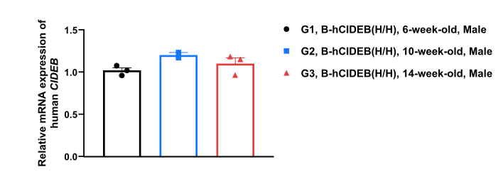
Strain specific analysis of CIDEB mRNA expression in B-hCIDEB mice by RT-PCR. Liver RNA were isolated from homozygous B-hCIDEB mice (H/H), then cDNA libraries were synthesized by reverse transcription, followed by PCR with human CIDEB primers. Human CIDEB mRNA expression remained stable in homozygous B-hCIDEB mice across different ages.

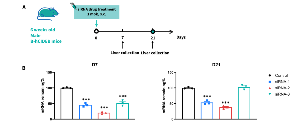
The inhibitory efficiency of the nucleic acid drugs against human CIDEB in homozygous B-hCIDEB mice. B-hCIDEB mice were randomly divided into four groups (male, 6 weeks old). The human CIDEB-targeted siRNA and control were administered to the mice individually. The mice were sacrificed on day 7 and day 21, and the liver tissues were collected to detect the expression level of human CIDEB mRNA by qPCR. (A) The schematic diagram of experimental processing. (B) The expression of human CIDEB mRNA in the liver. Data was shown as mean ± SEM, analyzed by one way-ANOVA (*p<0.05, p<0.01, ***p<0.001). The human CIDEB in the siRNA group was significantly reduced compared to the control group. (Data provided by a client.)
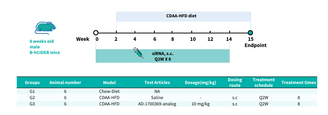
In vivo efficacy of the CDAA-HFD induced model in B-hCIDEB mice. B-hCIDEB mice were allowed to feed on a normal chow diet from week 0 for two weeks. After that, 6 mice were continually fed with the normal chow diet as the control, and the other mice were fed with the CDAA-HFD diet (60% energy from fat, 0.1% methionine, and deficient in choline, XTMRCD60). The mice in siRNA treatment groups were injected subcutaneously with the saline or AD-1700369-analog (synthesized according to patents) from week 0 once every 2 weeks for 14 weeks.
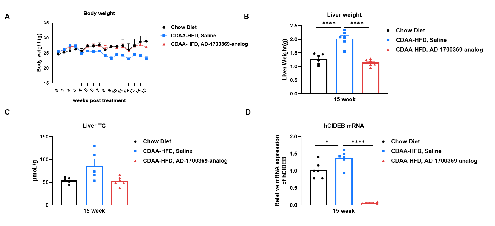
In vivo efficacy of the CDAA-HFD induced model in B-hCIDEB mice. (A) Body weight of different groups after treatment. (B) Liver weight of different groups after treatment. (C) The liver TG of different groups after treatment. (D) The expression level of human CIDEB mRNA by qPCR. Data was shown as mean ± SEM, analyzed by one way-ANOVA (*p<0.05, **p<0.01, ***p<0.001, ****p<0.0001).

In vivo efficacy of the CDAA-HFD induced model in B-hCIDEB mice. (A), Representative pictures of H&E staining showing degree of NASH. (B) NAFLD activity score (NAS) in different treatment groups. (C) Representative pictures of Sirius Red Staining showing the degree of liver fibrosis. (D) Quantitively positive area of Sirius Red Staining. Data were shown as mean ± SEM, analyzed by one way-ANOVA (*p<0.05, **p<0.01, ***p<0.001, ****p<0.0001). Scare bar:200μm.
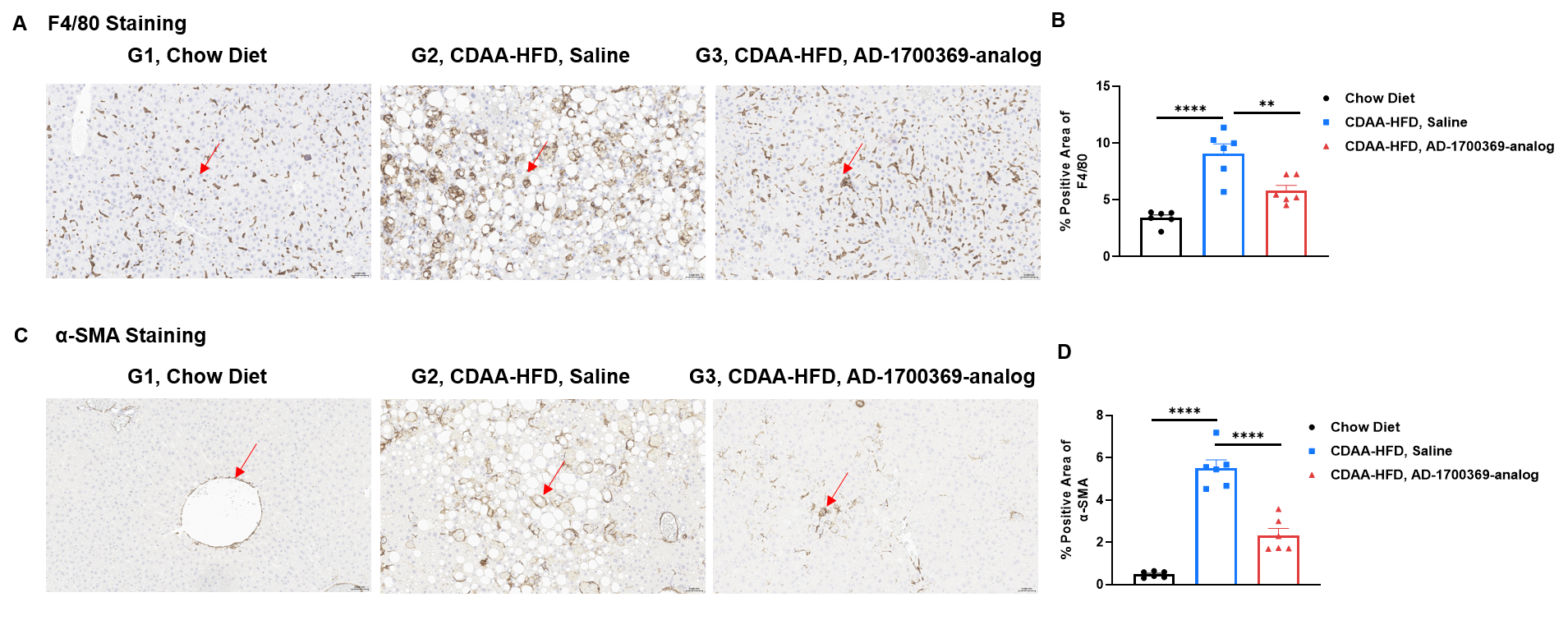
In vivo efficacy of the CDAA-HFD induced model in B-hCIDEB mice. (A), Representative pictures of IHC staining showing F4/80 expression (B) Quantitively positive area of F4/80 expression. (C) Representative pictures of IHC staining showing α-smooth muscle actin (α-SMA) expression (D) Quantitively positive area of α-SMA expression. Data were shown as mean ± SEM, analyzed by one way-ANOVA (*p<0.05, **p<0.01, ***p<0.001, ****p<0.0001). Scare bar:200μm.
