

C57BL/6N-Cxcl13tm1(CXCL13)Bcgen/Cxcr5tm1(CXCR5)Bcgen/Bcgen • 111189

CXCL13
Exons 1-4 of mouse Cxcl13 gene that encode the whole molecule (ATG to STOP codon) were replaced by human counterparts in B-hCXCL13/hCXCR5 mice. The promoter, 5’UTR and 3’UTR region of the mouse gene were retained. The human CXCL13 expression was driven by endogenous mouse Cxcl13 promoter, while mouse Cxcl13 gene transcription and translation will be disrupted.
CXCR5
Exons 1-2 of mouse Cxcr5 gene that encode the entire protein (from ATG to stop codon) were replaced by human counterparts in B-hCXCL13/hCXCR5 mice. The promoter, 5’UTR and 3’UTR region of the mouse gene were retained. The human CXCR5 expression was driven by endogenous mouse Cxcr5 promoter, while mouse Cxcr5 gene transcription and translation will be disrupted. B-hCXCL13/hCXCR5 mice (111189) were obtained by mating B-hCXCL13 mice (111136) and B-hCXCR5 mice (110970).

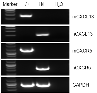
Strain specific analysis of CXCL13 and CXCR5 gene expression in WT and homozygous B-hCXCL13/hCXCR5 mice by RT-PCR. Mouse Cxcl13 and Cxcr5 mRNA were detectable in splenocytes of wild-type (+/+) mice. Human CXCL13 and CXCR5 mRNA were detectable only in homozygous B-hCXCL13/hCXCR5 mice , but not in wild-type (+/+) mice.
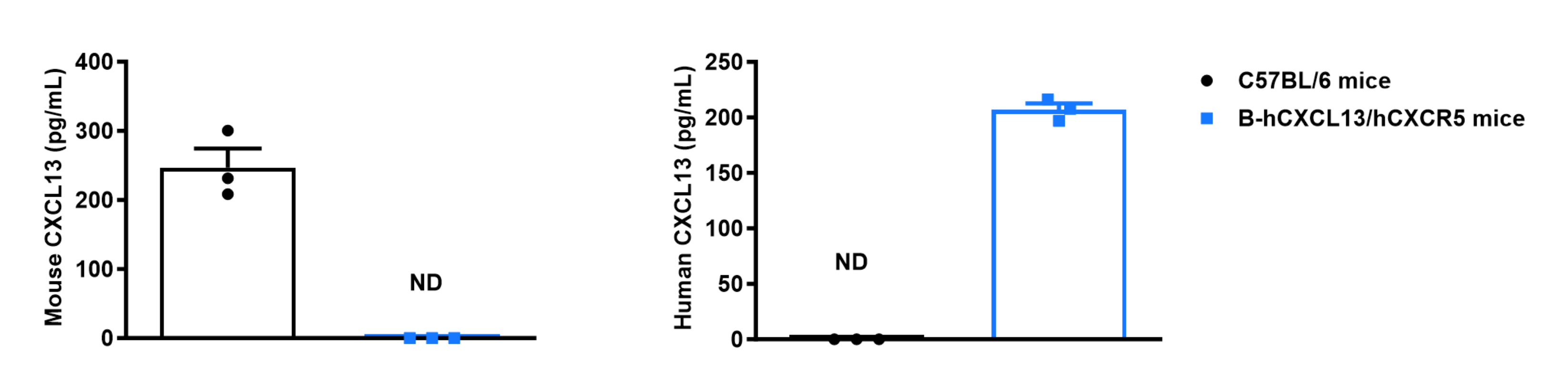
Strain specific CXCL13 expression analysis in wild-type C57BL/6 mice and homozygous humanized B-hCXCL13/hCXCR5 mice by ELISA. Serum was collected from wild-type C57BL/6 mice and homozygous B-hCXCL13/hCXCR5 mice (female, 7-week-old, n = 3). Expression level of mouse and human CXCL13 were analyzed by ELISA (anti-mouse CXCL13: R&D, MCX130; anti-human CXCL13: R&D, DCX130). Values are expressed as mean ± SEM. ND: not detectable.
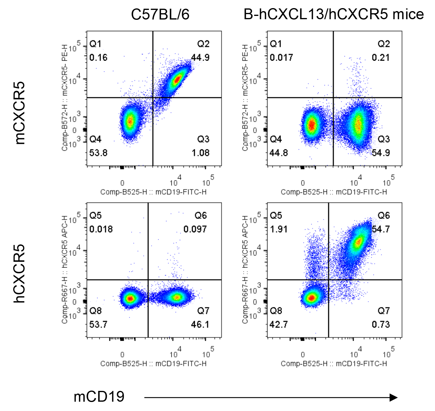
Strain specific CXCR5 expression analysis in wild-type C57BL/6 mice and homozygous humanized B-hCXCL13/hCXCR5 mice by flow cytometry. Splenocytes were collected from wild-type C57BL/6 mice and homozygous B-hCXCL13/hCXCR5 mice. Protein expression was analyzed with anti-mouse CXCR5 antibody (Biolegend, 145503) and anti-human CXCR5 antibody (Biolegend, 356907) by flow cytometry.
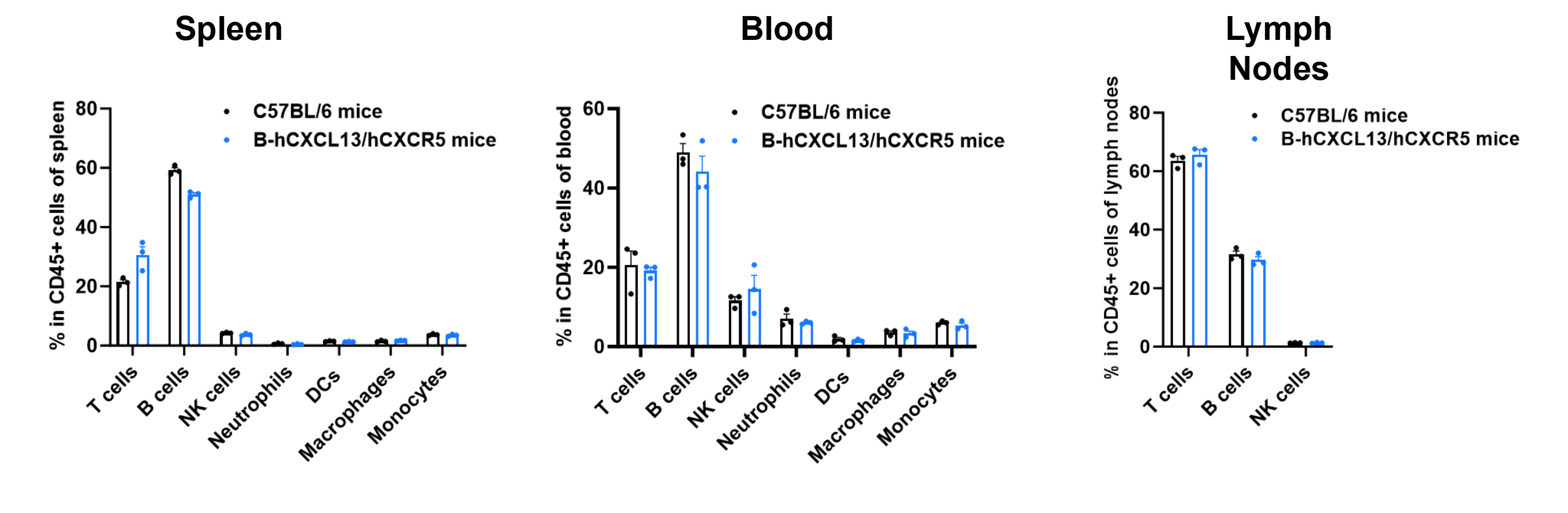
Analysis of T-cell subpopulations by flow cytometry in immune organs and blood. Splenocytes, peripheral blood, and lymph nodes were isolated from C57BL/6 mice and homozygous B-hCXCL13/hCXCR5 mice (female, 6-week-old, n = 3). Single live cells were gated on the TCRβ⁺ T-cell population and analyzed by flow cytometry as indicated. Values are expressed as mean ± SEM.
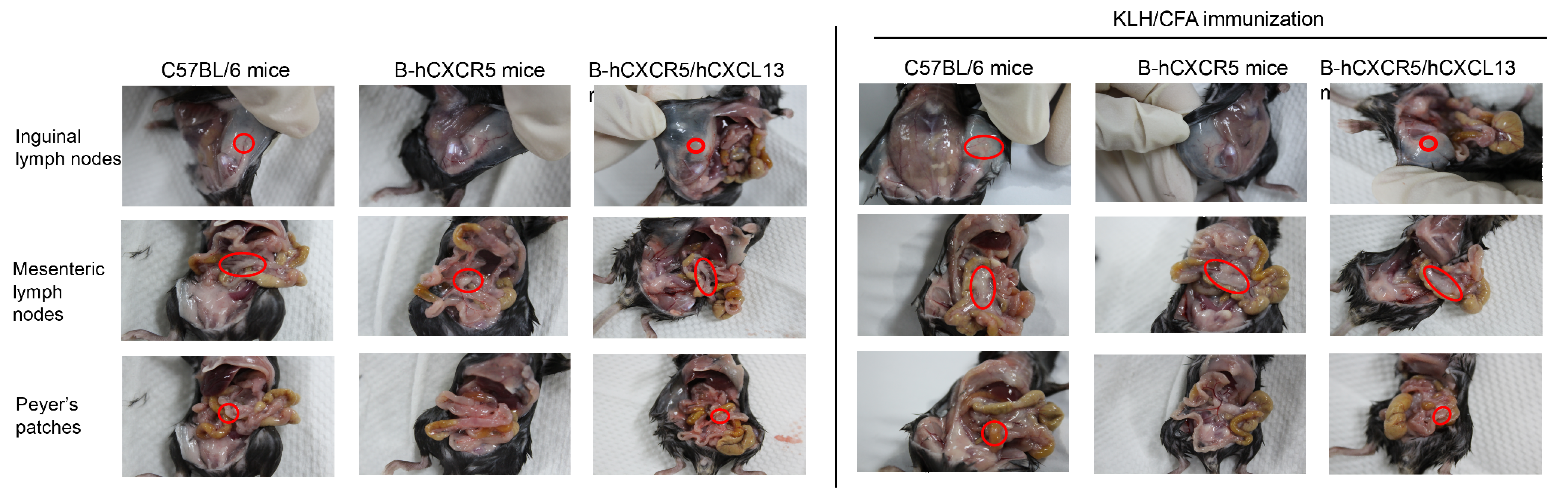
C57BL/6 mice, B-hCXCR5 mice and B-hCXCR5/hCXCL13 mice (female, 8-week-old, n=3) were immunized intraperitoneally with 200 μg keyhole limpet hemocyanin (KLH) protein emulsified in complete Freund's adjuvant (CFA). After 14 days of immunization, the lymph nodes were observed and the spleen was analyzed by hematologic and epithelial analysis (H&E). Note: CXCR5 homozygous null mutants lack inguinal lymph nodes, have a few abnormal or no Peyer's patches, morphologically altered primary lymphoid follicles and no functional germinal centers in their spleen.(https://www.informatics.jax.org/marker/MGI:103567)
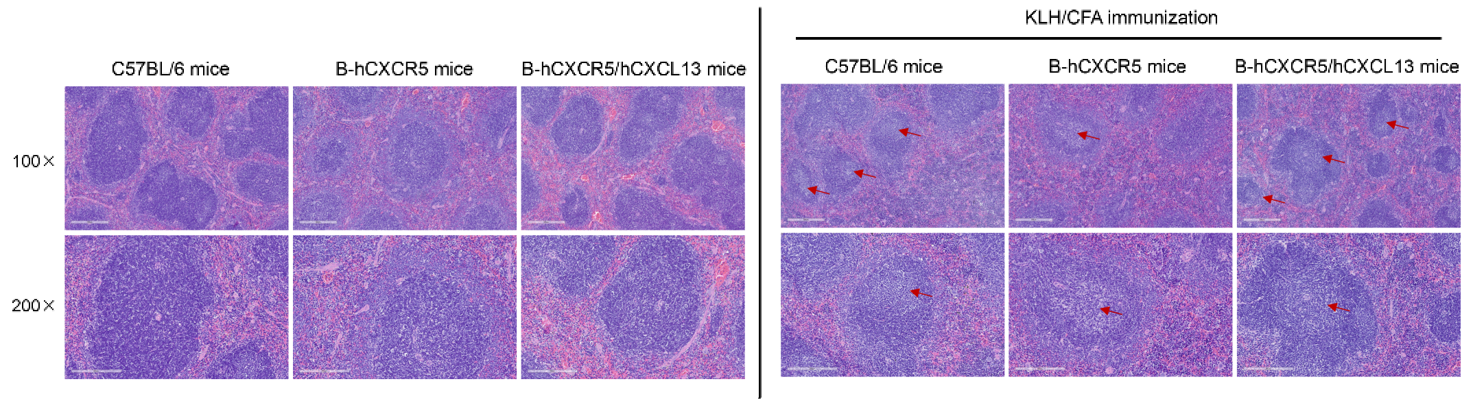
C57BL/6 mice, B-hCXCR5 mice and B-hCXCR5/hCXCL13 mice (female, 8-week-old, n=3) were immunized intraperitoneally with 200 μg keyhole limpet hemocyanin (KLH) protein emulsified in complete Freund's adjuvant (CFA). After 14 days of immunization, the lymph nodes were observed and the spleen was analyzed by hematologic and epithelial analysis (H&E). Note: CXCR5 homozygous null mutants lack inguinal lymph nodes, have a few abnormal or no Peyer's patches, morphologically altered primary lymphoid follicles and no functional germinal centers in their spleen.(https://www.informatics.jax.org/marker/MGI:103567)
