


IL6/IL6R: A key cytokine signaling axis in inflammation and its therapeutic intervention

IL6
IL6R

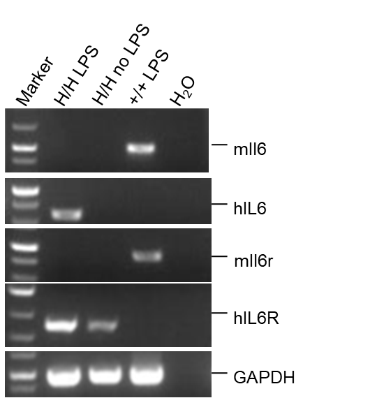
Strain specific analysis of IL6 and IL6R gene expression in B-hIL6/hIL6R mice by RT-PCR. Mouse Il6 and Il6r mRNA were detectable in spleen of wild-type mice (+/+). Human IL6 was detectable only in homozygous B-hIL6/hIL6R mice (H/H; H/H) after stimulated with LPS for 3h. Human IL6R mRNA was detectable in homozygous B-hIL6/hIL6R mice, but not in wild-type mice with or without LPS stimulation.

Strain specific IL6 expression analysis in wild-type C57BL/6JNifdc mice and homozygous B-hIL6/hIL6R mice by ELISA. Serum was collected from wild-type C57BL/6JNifdc mice (+/+) (female, n=3, 8-week-old), homozygous B-hIL6/hIL6R mice(H/H ; H/H) (female, n=3, 8-week-old), the production of IL6 in serum were assessed after 3h of stimulation with LPS (20 μg/200uL, i.p.) in vivo. Expression level of human IL6 were analyzed by ELISA (Human IL-6 ELISA MAX TM Deluxe Set (430508)). Human IL6 was only detectable in homozygous homozygous B-hIL6/hIL6R mice (n=3). Values are expressed as mean ± SEM.
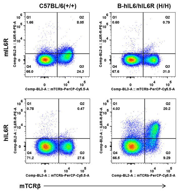
Strain specific IL6R expression analysis in wild-type C57BL/6 mice, homozygous B-hIL6/hIL6R mice by flow cytometry. Splenocytes were collected wild-type C57BL/6 mice(+/+) and homozygous B-hIL6/hIL6R mice (H/H;H/H). The protein expression was analyzed with anti-mouse CD126 (IL-6Rα chain) Antibody (Biolegend, 115805) and anti-human CD126 (IL-6Rα) Antibody (Biolegend, 352803) by flow cytometry. Mouse IL6R was detectable in T cells of C57BL/6 mice. Human IL6R was detectable in T cells of homozygous B-hIL6/hIL6R mice.
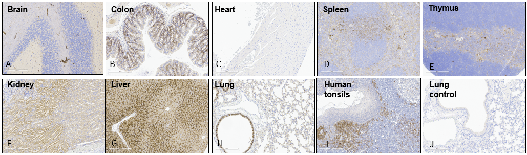
Representative human IL6R expression in different tissues of B-hIL6/IL6R mice by IHC. Tissues were collected from homozygous B-hIL6/hIL6R mice and stained with antibodies for human IL6R (A-I) or anti-IgG antibodies (J). Human tonsils as positive control (I); Mouse lung stained with anti-IgG antibodies as a negative control (J); As shown in the figure, human IL6R was detected in brain, colon, kidney, liver, lung, spleen and thymus, but not in heart. Original magnification ×200. Abbreviations: IHC, immunohistochemistry.
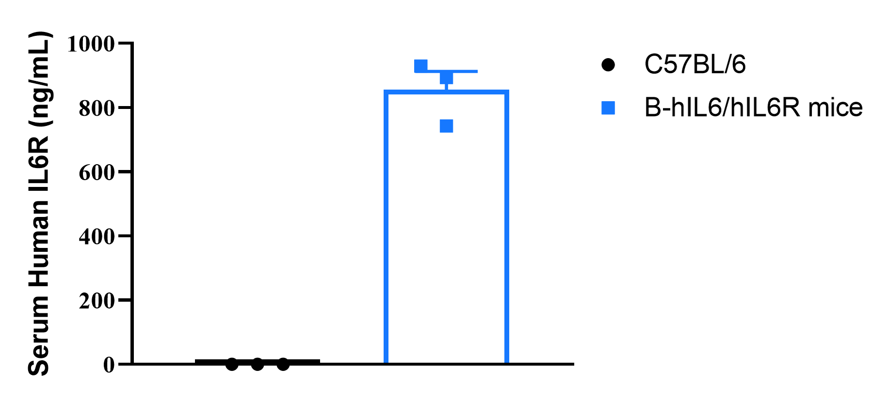
Strain specific IL6R expression analysis in wild-type C57BL/6 mice and homozygous humanized B-hIL6/IL6R mice by ELISA. Plasma (EDTA) was collected from wild-type C57BL/6 mice (female, 6-week-old, n=3) and homozygous B-hIL6/IL6R mice (female, 6-week-old, n=3). Expression level of human IL6R were analyzed by ELISA (human IL6R ELISA kit: R&D, DR600). Human IL6R was exclusively detectable in homozygous B-hIL6/IL6R mice. Values are expressed as mean ± SEM.
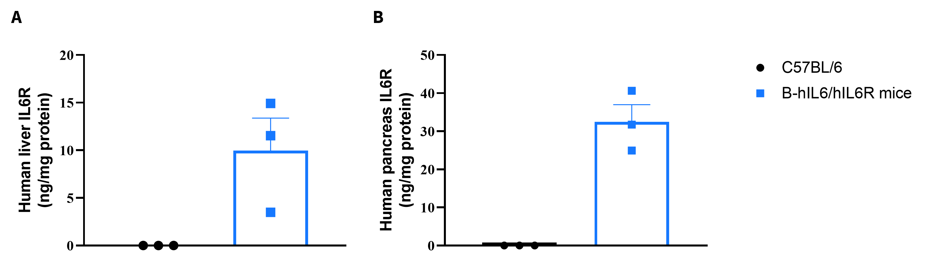
Soluble human IL6R expression analysis in homozygous B-hIL6/hIL6R mice by ELISA. Liver and pancreas were isolated from wild-type C57BL/6 mice (female, 7-week-old, n=3) and homozygous B-hIL6/hIL6R mice (female, 7-week-old, n=3), and analyzed by ELISA with species-specific anti-human IL6R ELISA kit (human IL6R ELISA kit: R&D, DR600). Soluble human IL6R was detectable in liver (A) and pancreas (B) of homozygous B-hIL6/hIL6R mice but not in wild-type mice.
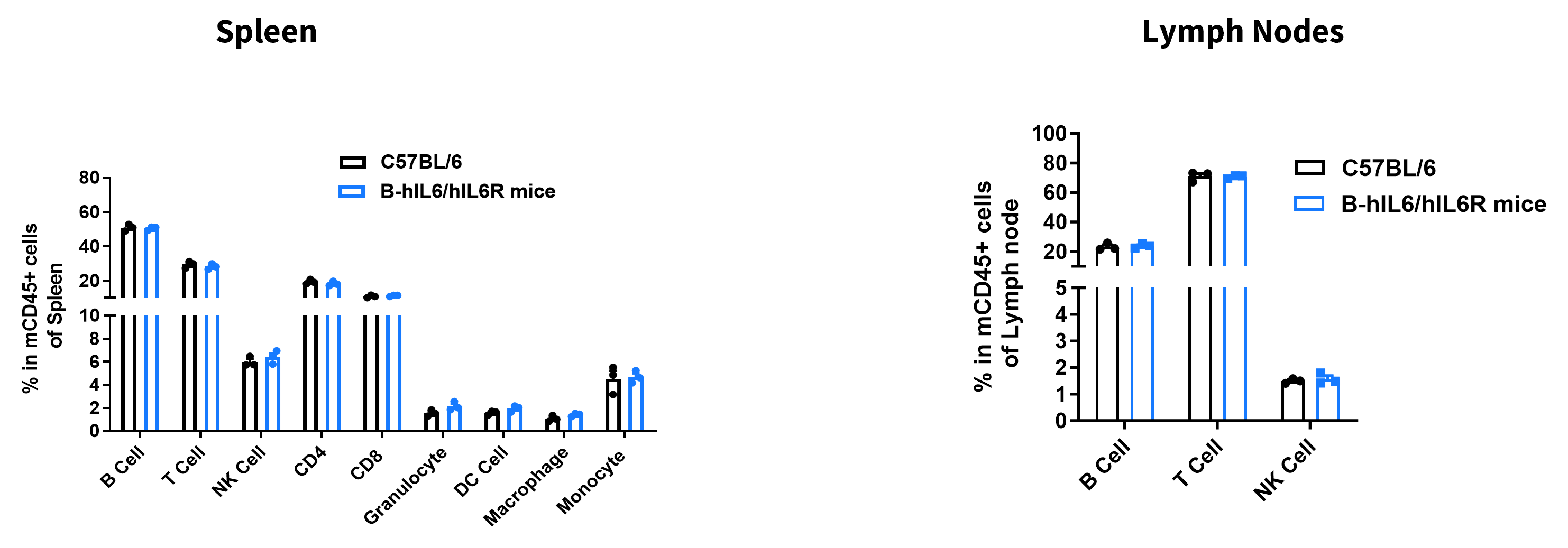
Analysis of leukocyte subpopulations by flow cytometry in spleen and lymph node. Splenocytes and lymph nodes were isolated from female C57BL/6 and B-hIL6/hIL6R mice (female, 9-week-old, n = 3). Single live cells were gated on the CD45⁺ population and analyzed by flow cytometry as indicated. Values are expressed as mean ± SEM.
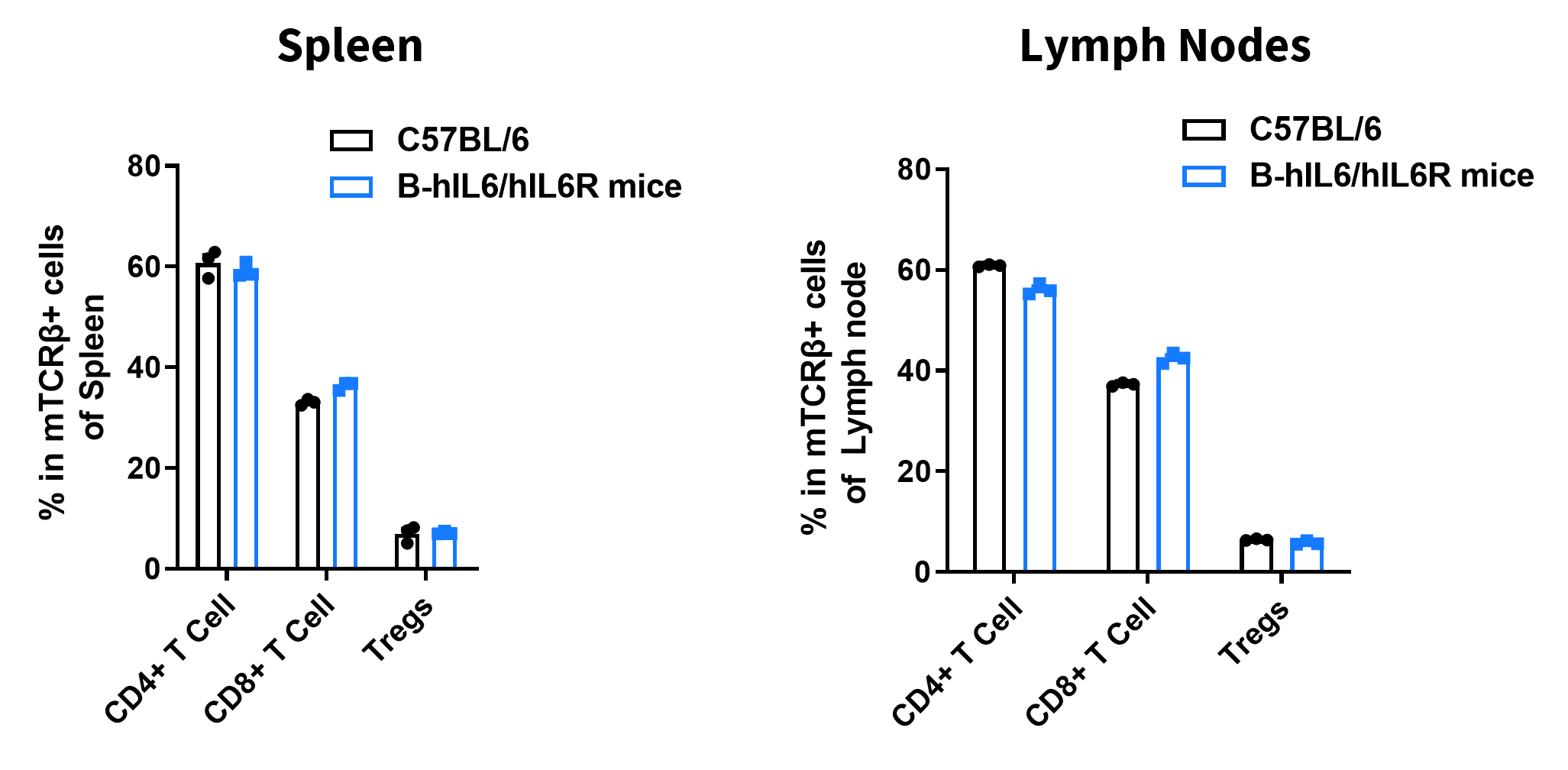
Analysis of T-cell subpopulations by flow cytometry in spleen and lymph node. Splenocytes and lymph nodes were isolated from female C57BL/6 and B-hIL6/hIL6R mice (female, 9-week-old, n = 3). Single live cells were gated on the TCRβ⁺ T-cell population and analyzed by flow cytometry as indicated. Values are expressed as mean ± SEM.
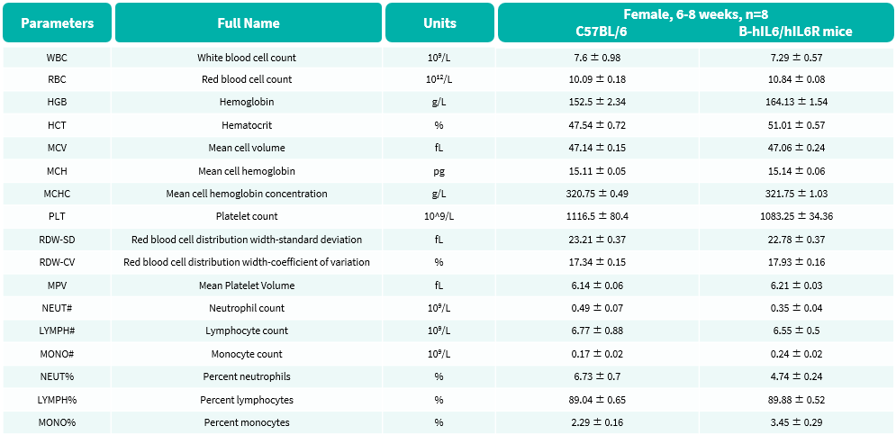
Complete blood count (CBC) of B-hIL6/hIL6R mice. Values are expressed as mean ± SD.
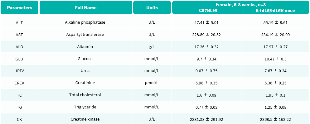
Blood biochemical parameters of B-hIL6/hIL6R mice are shown. Values are expressed as mean ± SD.

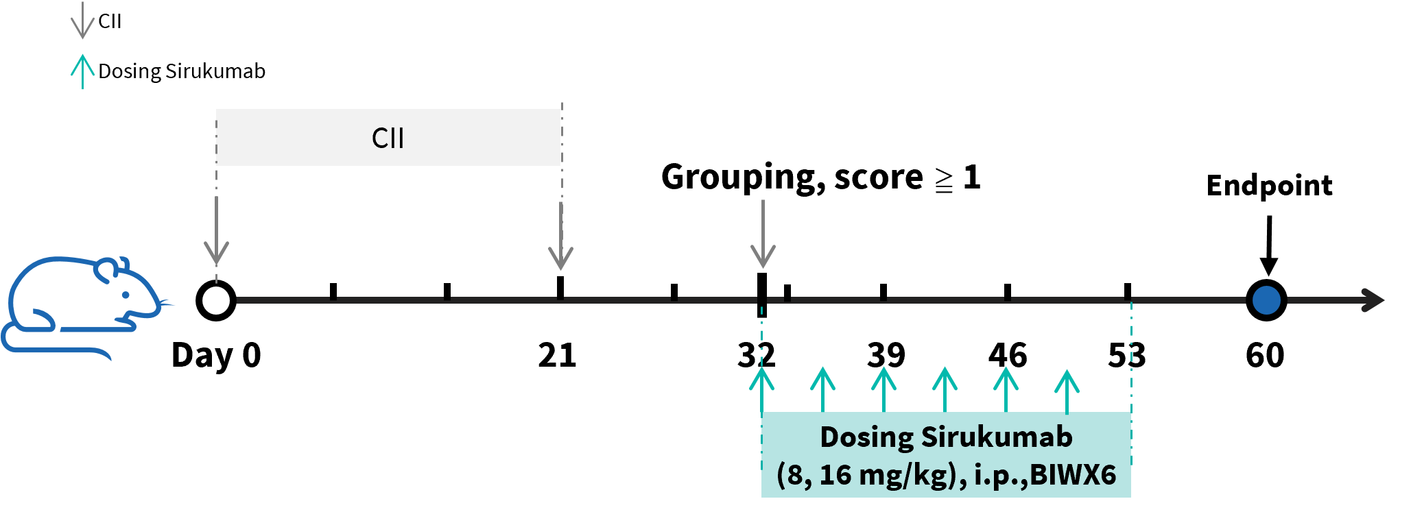
Efficacy of anti-human IL6 antibodies in B-hIL6/hIL6R mice with collagen induced arthritis (CIA) model. Arthritis was induced by the subcutaneous injection of CII emulsion into B-hIL6/hIL6R mice on Day 0 and Day 21 (male, n=6 in each group). The development of arthritis was monitored and the arthritis score was evaluated every day. The mice were divided into groups at day 32 (Clinical score ≧ 1). The treatment group was intraperitoneally injected with different doses of anti-human IL6 antibody sirukumab analog (in house).
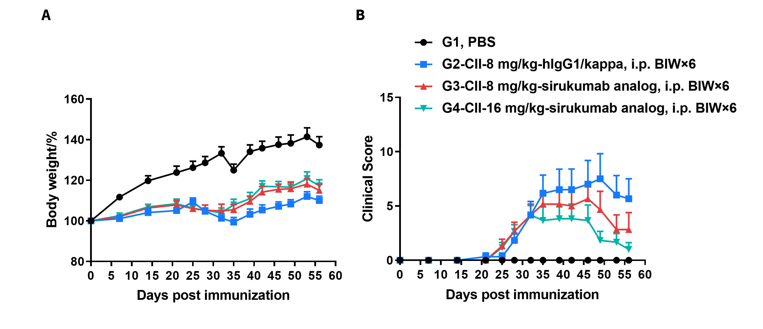
Efficacy of anti-human IL6 antibodies in B-hIL6/hIL6R mice with collagen induced arthritis (CIA) model. Mice in each group were treated with sirukumab analog (in house). Body weight change(A) and clinical score (B) were evaluated during treatment twice a week. There was no significant change in body weight, while total clinical score increased in the groups except control during days 21 to 28. It indicated that the arthritis mouse model had been constructed successfully. Clinical scores decreased in the two groups treated with sirukumab analog and dose-dependent. The results indicated the B-hIL6/hIL6R mice provide a powerful preclinical model for in vivo evaluation of anti-human IL6 antibodies.
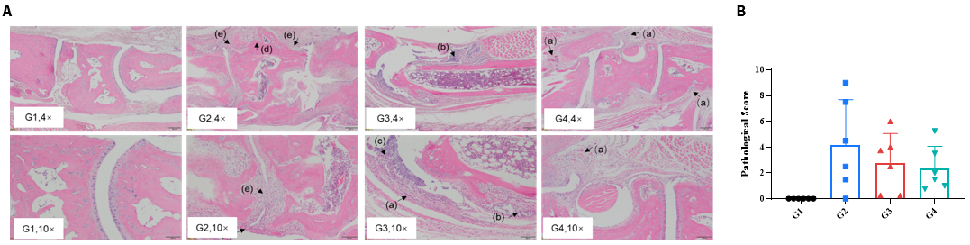
Efficacy of anti-human IL6 antibody in B-hIL6/hIL6R mice with collagen induced arthritis (CIA) model. Histopathological examination was performed on the joints of the extremities at endpoint. (A) Hematoxylin and eosin (H&E) staining. (B) Score of arthritis histology. G1 showed no significant abnormal changes. G2 showed bone structure damage(d), articular cavity or periarticular space disappeared (e), and pannus were observed (a), suggesting that the CIA model was successfully established. Compared with the G2 group, the low-dose group (G3) showed slight inflammatory cell infiltration (b) and pannus (a) and synovial hyperplasia (c). However, in the high dose group (G4), there was only partial pannus, the arthrosis disappeared and the articular cavity was obvious.
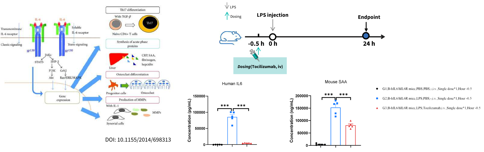
Human IL6 and mouse SAA expression analysis in homozygous humanized B-hIL6/hIL6R mice by ELISA. Mice received LPS injection (i.p.) 30 min following PBS or tocilizumab (i.v.,provided by client) treatment. Serum was collected from homozygous B-hIL6/hIL6R mice twenty four hours after LPS injection. Expression level of human IL6 and mouse SAA were analyzedby ELISA (ELISA MAX™ Deluxe Set Human IL6, Biolegend, 430507; Mouse SAA SimpleStep ELISA® Kit, abcam, ab215090). Pre-treatment of tocilizumab significantly decreased human IL6 andmouse SAA expression at twenty four hours after LPS injection, indicating a successful inhibition of the downstream activation caused by hIL6R. Values are expressed as mean ± SEM.Significance was determined by one-way ANOVA test . ***p < 0.001.
