B-hPD-1 plus/hIL2/hIL2RA/hIL2RB/hIL2RG mice

C57BL/6-Pdcd1tm3(PDCD1)BcgenIl2tm1(IL2)Bcgen Il2ratm1(IL2RA)Bcgen ll2rbtm2(IL2RB)Bcgen ll2rgtm2(IL2RG)Bcgen/Bcgen • 112742

B-hPD-1 plus/hIL2/hIL2RA/hIL2RB/hIL2RG mice

Product nameB-hPD-1 plus/hIL2/hIL2RA/hIL2RB/hIL2RG mice
Catalog number112742
Strain nameC57BL/6-Pdcd1tm3(PDCD1)BcgenIl2tm1(IL2)Bcgen Il2ratm1(IL2RA)Bcgen ll2rbtm2(IL2RB)Bcgen ll2rgtm2(IL2RG)Bcgen/Bcgen
Strain backgroundC57BL/6
NCBI gene ID5133,3558,3559,3560,3561 (Human)
AliasesPD1, PD-1, CD279, SLEB2, hPD-1, hPD-l, hSLE1, ADMIO4, AIMTBS;IL-2, TCGF, lymphokine;P55, CD25, IL2R, IMD41, TCGFR, IDDM10;CD122, IMD63, IL15RB, P70-75;P64, CIDX, IMD4, CD132, SCIDX, IL-2RG, SCIDX1

이 페이지에서

  • Description
  • Targeting strategy
  • Phenotypic analysis

포스터

모두 보기

    출판물

      Description
      • Programmed cell death protein 1 (PDCD1), also known as PD-1, functions as an inhibitory receptor on activated T cells and helps regulate immune responses. It modulates various T-cell activities, such as limiting the effector functions of CD8+ T cells and fostering the development of regulatory T cells (Tregs) from CD4+ T cell precursors. The interleukin-2 (IL2) protein, encoded by the IL2 gene, is a critical cytokine secreted by activated CD4+ and CD8+ T lymphocytes, playing a pivotal role in the proliferation of T and B lymphocytes. The expression of IL2 in mature thymocytes is characterized by monoallelic regulation, representing a distinctive mechanism for the precise control of gene expression. The high-affinity IL2 receptor is composed of the interleukin 2 receptor alpha (IL2RA) and beta (IL2RB) chains, in conjunction with the common gamma chain (IL2RG). Homodimeric configurations of the alpha chains (IL2RA) yield a low-affinity receptor, while the beta chains (IL2RB) in a homodimeric state produce a receptor with medium affinity. Typically, IL2RA is an integral membrane protein; however, a soluble form has been identified and is known to arise from extracellular proteolytic processes.
      • Human PD-1 gene encoding the extracellular region and mouse PD-1 gene encoding the transmembrane and cytoplasmic region were inserted after the initiation codon ATG of mouse PD-1 gene. The mouse Il2 gene that encodes the full coding sequence was replaced by human IL2 full coding sequence. The mouse Il2ra gene that encodes the extracellular domain was replaced by human IL2RA counterpart gene sequences. The mouse Il2rb gene that encodes the extracellular domain was replaced by human IL2RB counterpart gene sequences. The mouse Il2rg gene that encodes the full coding region sequences was replaced by human IL2RG counterpart gene sequences.
      • Human PD-1, IL2RA, IL2RB, and IL2RG were exclusively detectable in homozygous B-hPD-1 plus/hIL2/hIL2RA/hIL2RB/hIL2RG mice, but not in wild-type C57BL/6JNifdc mice.
      • B-hPD-1 plus/hIL2/hIL2RA/hIL2RB/hIL2RG mice provide a powerful preclinical model for in vivo evaluation of anti-tumor antibodies.
      • Application: For example, this product is used for pharmacodynamics and safety evaluation of antibodies for cancers.
      Targeting Strategy

      Gene targeting strategy for B-hPD-1 plus/hIL2/hIL2RA/hIL2RB/hIL2RG mice.

      • Human PD-1 gene encoding the extracellular region and mouse PD-1 gene encoding the transmembrane and cytoplasmic region were inserted after the initiation codon ATG of mouse PD-1 gene in B-hPD-1 plus/hIL2/hIL2RA/hIL2RB/hIL2RG mice.
      • The exons 1-4 of mouse Il2 gene that encode the whole molecule (ATG to STOP codon) were replaced by human counterparts in B-hPD-1 plus/hIL2/hIL2RA/hIL2RB/hIL2RG mice. The promoter, 5’UTR and 3’UTR region of the mouse gene are retained. The human IL2 expression is driven by endogenous mouse Il2 promoter, while mouse Il2 gene transcription and translation will be disrupted.
      • The exons 2-6 of mouse Il2ra gene that encode extracellular domain were replaced by human counterparts in B-hPD-1 plus/hIL2/hIL2RA/hIL2RB/hIL2RG mice. The genomic region of mouse Il2ra gene that encodes cytoplasmic portion is retained. The promoter, 5’UTR and 3’UTR region of the mouse gene are also retained. The chimeric IL2RA expression is driven by endogenous mouse Il2ra promoter, while mouse Il2ra gene transcription and translation will be disrupted.
      • The exons 2-8 of mouse Il2rb gene that encode extracellular domain were replaced by human counterparts in B-hPD-1 plus/hIL2/hIL2RA/hIL2RB/hIL2RG mice. The genomic region of mouse Il2rb gene that encodes cytoplasmic portion is retained. The promoter, 5’UTR and 3’UTR region of the mouse gene are also retained. The chimeric IL2RB expression is driven by endogenous mouse Il2rb promoter, while mouse Il2rb gene transcription and translation will be disrupted.
      • The exons 1-7 of mouse Il2rg gene that encode signal peptide, extracellular domain, transmembrane domain, cytoplasmic region  were replaced by human counterparts in B-hPD-1 plus/hIL2/hIL2RA/hIL2RB/hIL2RG mice. The promoter and 5’UTR region of the mouse gene are retained. The human IL2RG expression is driven by endogenous mouse Il2rg promoter, while mouse Il2rg gene transcription and translation will be disrupted.
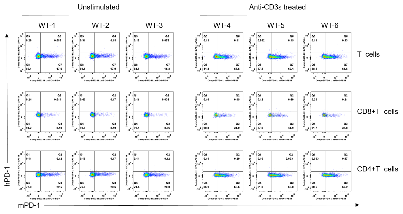
      The PD-1 Expression in Wild-type(WT) Mice

      Strain specific PD-1 expression analysis in wild-type C57BL/6JNifdc mice by flow cytometry. Splenocytes were collected from wild-type C57BL/6JNifdc mice (male, 6-week-old, n=3/group) stimulated with or without anti-CD3ε antibody (7.5 μg/mice, i.p.) in vivo for 24 h,  and analyzed by flow cytometry with species-specific anti-mouse PD-1 antibody (Biolegend, 109104) and anti-human PD-1 antibody (Biolegend, 329908). Mouse PD-1 was only detectable in wild-type mice.

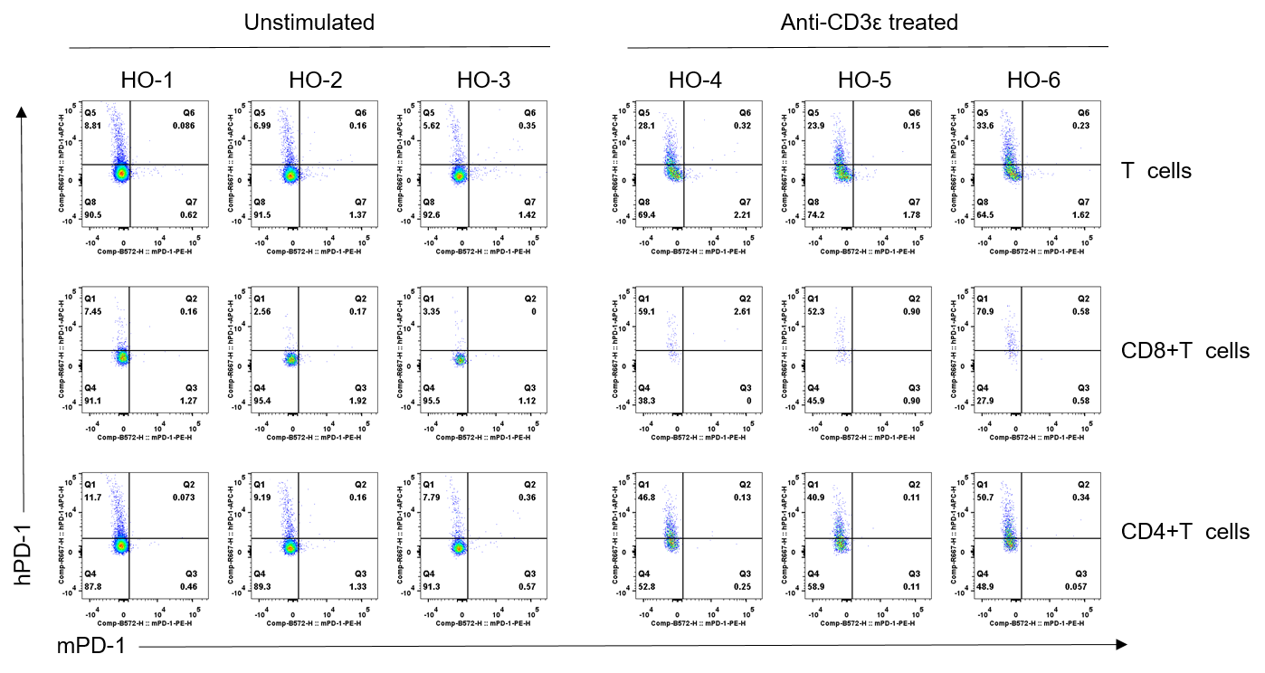
      The PD-1 Expression in B-hPD-1 plus/hIL2RA/hIL2RB/hIL2RG Mice

      Strain specific PD-1 expression analysis in homozygous B-hPD-1 plus/hIL2RA/hIL2RB/hIL2RG mice by flow cytometry. Splenocytes were collected from homozygous B-hPD-1 plus/hIL2RA/hIL2RB/hIL2RG mice (male, 6-week-old, n=3/group) stimulated with or without anti-CD3ε antibody (7.5 μg/mice, i.p.) in vivo for 24 h,  and analyzed by flow cytometry with species-specific anti-mouse PD-1 antibody (Biolegend, 109104) and anti-human PD-1 antibody (Biolegend, 329908). Human PD-1 was only detectable in homozygous B-hPD-1 plus/hIL2RA/hIL2RB/hIL2RG mice.

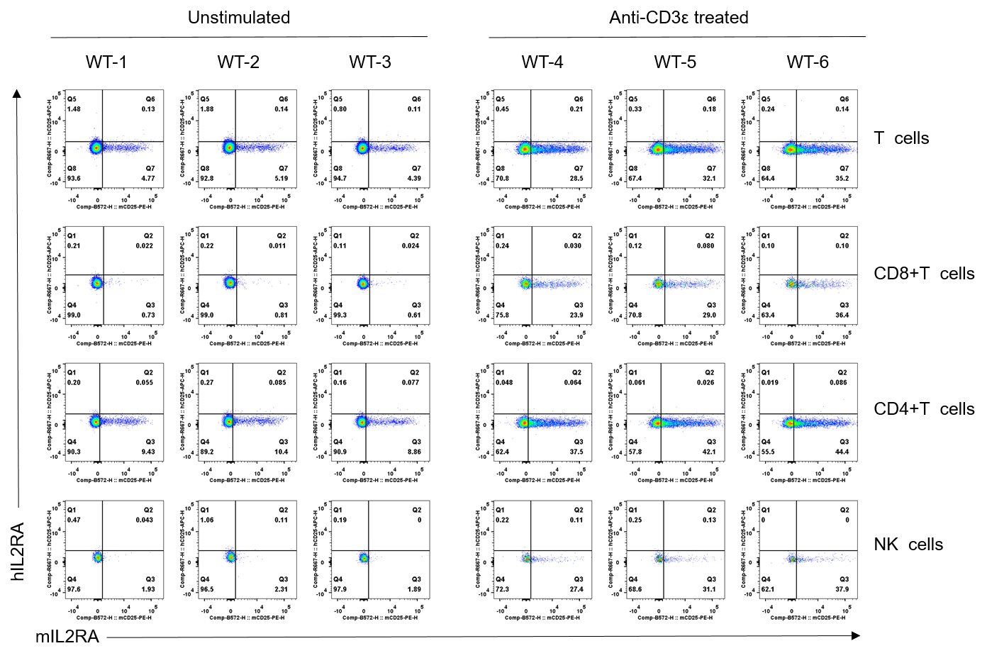
      The IL2RA Expression in Wild-type(WT) Mice

      Strain specific IL2RA expression analysis in wild-type C57BL/6JNifdc mice by flow cytometry. Splenocytes were collected from wild-type C57BL/6JNifdc mice (male, 6-week-old, n=3/group) stimulated with or without anti-CD3ε antibody (7.5 μg/mice, i.p.) in vivo for 24 h,  and analyzed by flow cytometry with species-specific anti-mouse IL2RA antibody (Biolegend, 102008) and anti-human IL2RA antibody (Biolegend, 302610). Mouse IL2RA was only detectable in wild-type mice.

      The IL2RA Expression in B-hPD-1 plus/hIL2RA/hIL2RB/hIL2RG Mice

      Strain specific IL2RA expression analysis in homozygous B-hPD-1 plus/hIL2RA/hIL2RB/hIL2RG mice by flow cytometry. Splenocytes were collected from homozygous B-hPD-1 plus/hIL2RA/hIL2RB/hIL2RG mice (male, 6-week-old, n=3/group) stimulated with or without anti-CD3ε antibody (7.5 μg/mice, i.p.) in vivo for 24 h,  and analyzed by flow cytometry with species-specific anti-mouse IL2RA antibody (Biolegend, 102008) and anti-human IL2RA antibody (Biolegend, 302610). Human IL2RA was only detectable in homozygous B-hPD-1 plus/hIL2RA/hIL2RB/hIL2RG mice.

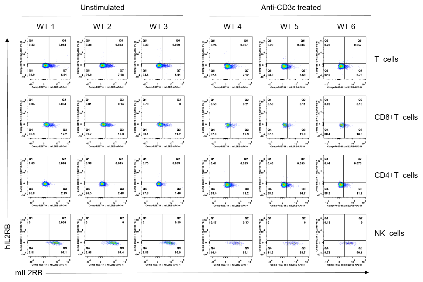
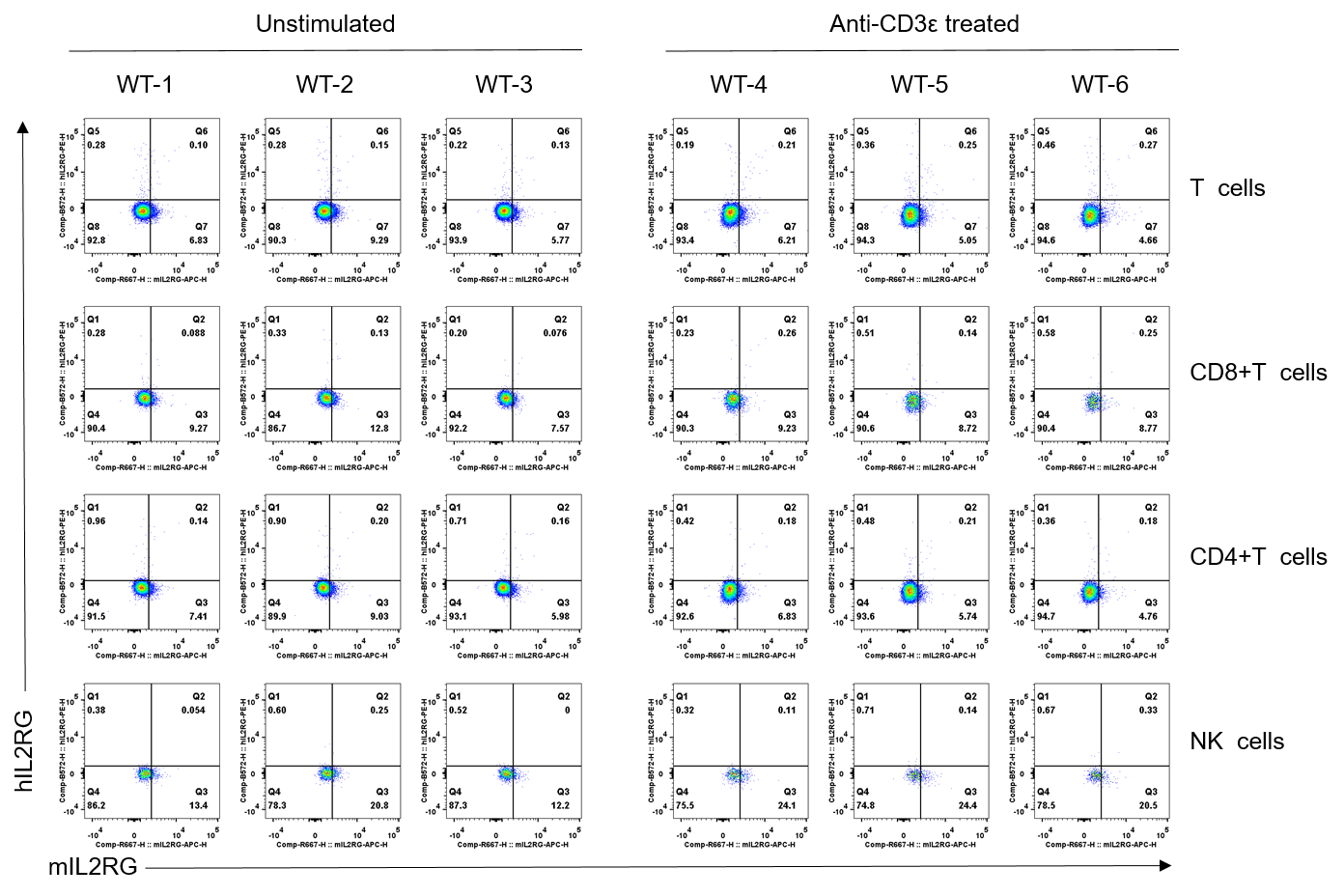
      The IL2RB Expression in Wild-type(WT) Mice

      Strain specific IL2RB expression analysis in wild-type C57BL/6JNifdc mice by flow cytometry. Splenocytes were collected from wild-type C57BL/6JNifdc mice (male, 6-week-old, n=3/group) stimulated with or without anti-CD3ε antibody (7.5 μg/mice, i.p.) in vivo for 24 h,  and analyzed by flow cytometry with species-specific anti-mouse IL2RB antibody (Biolegend, 105912) and anti-human IL2RB antibody (Biolegend, 339005). Mouse IL2RB was only detectable in wild-type mice.

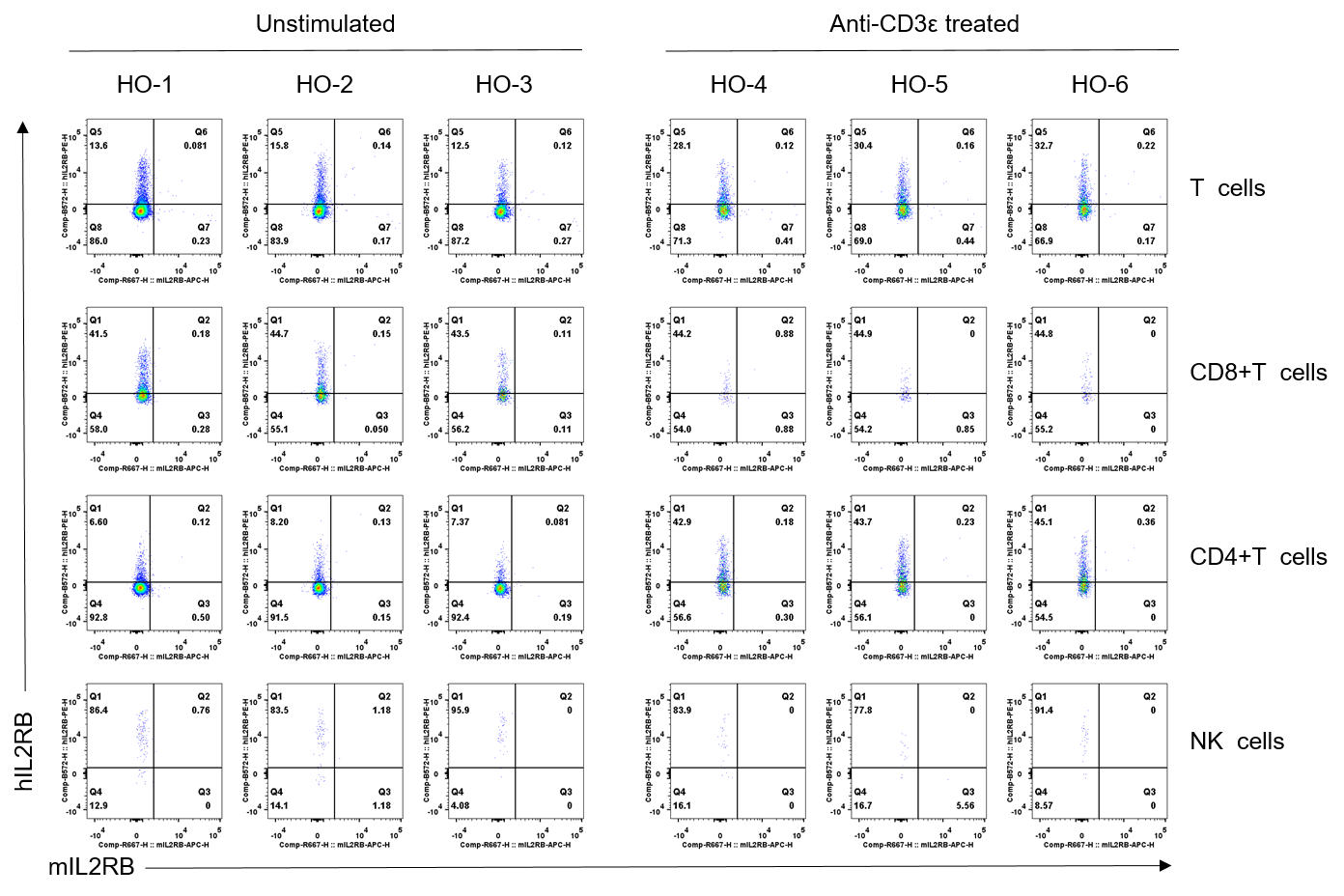
      The IL2RB Expression in B-hPD-1 plus/hIL2RA/hIL2RB/hIL2RG Mice

      Strain specific IL2RB expression analysis in homozygous B-hPD-1 plus/hIL2RA/hIL2RB/hIL2RG mice by flow cytometry. Splenocytes were collected from homozygous B-hPD-1 plus/hIL2RA/hIL2RB/hIL2RG mice (male, 6-week-old, n=3/group) stimulated with or without anti-CD3ε antibody (7.5 μg/mice, i.p.) in vivo for 24 h,  and analyzed by flow cytometry with species-specific anti-mouse IL2RB antibody (Biolegend, 105912) and anti-human IL2RB antibody (Biolegend, 339005). Human IL2RB was only detectable in homozygous B-hPD-1 plus/hIL2RA/hIL2RB/hIL2RG mice.

      The IL2RG Expression in Wild-type(WT) Mice

      Strain specific IL2RG expression analysis in wild-type C57BL/6JNifdc mice by flow cytometry. Splenocytes were collected from wild-type C57BL/6JNifdc mice (male, 6-week-old, n=3/group) stimulated with or without anti-CD3ε antibody (7.5 μg/mice, i.p.) in vivo for 24 h,  and analyzed by flow cytometry with species-specific anti-mouse IL2RG antibody (Biolegend, 132307) and anti-human IL2RB antibody (Biolegend, 338605). Mouse IL2RB was only detectable in wild-type mice.

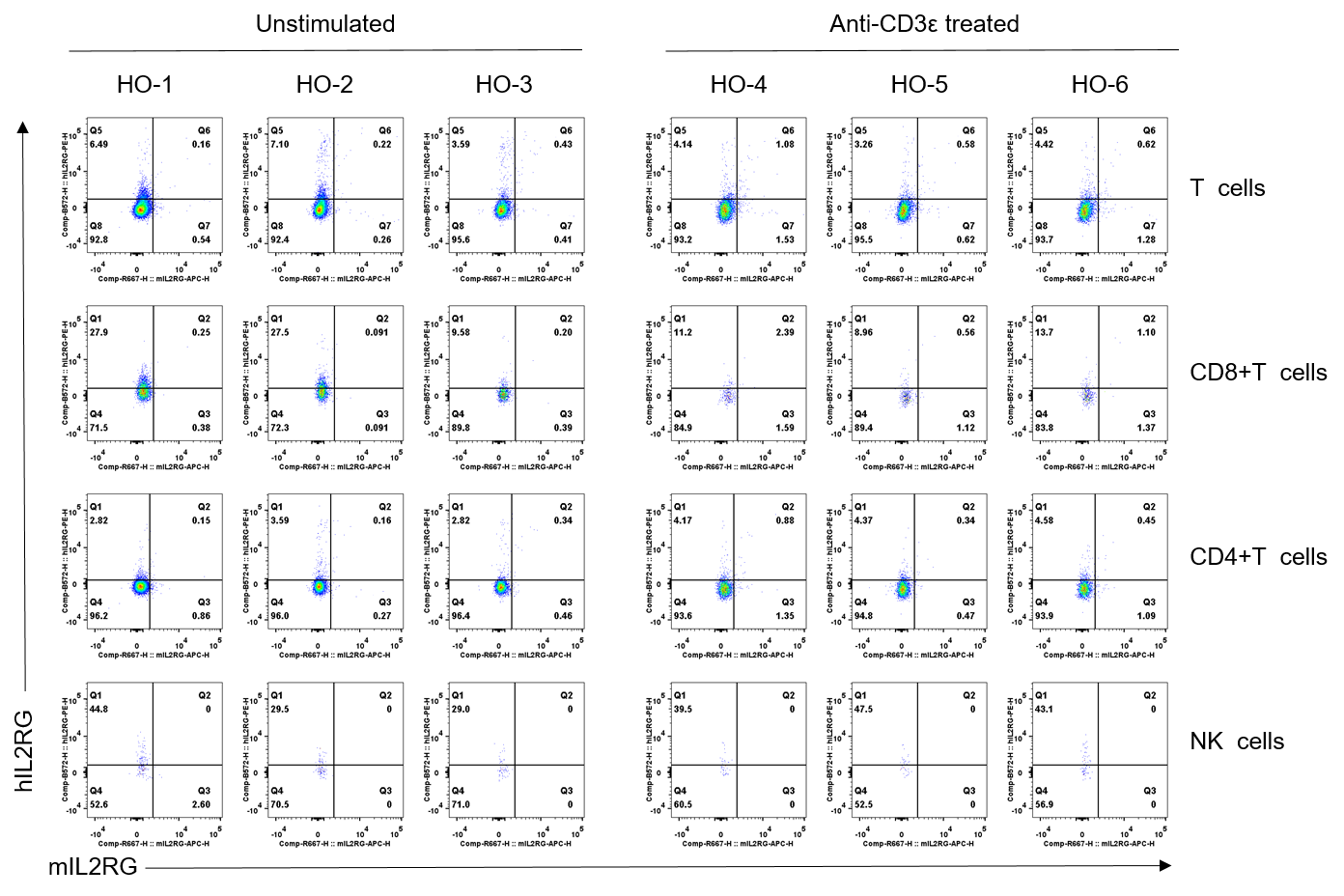
      The IL2RG Expression in B-hPD-1 plus/hIL2RA/hIL2RB/hIL2RG Mice

      Strain specific IL2RG expression analysis in homozygous B-hPD-1 plus/hIL2RA/hIL2RB/hIL2RG mice by flow cytometry. Splenocytes were collected from homozygous B-hPD-1 plus/hIL2RA/hIL2RB/hIL2RG mice (male, 6-week-old, n=3/group) stimulated with or without anti-CD3ε antibody (7.5 μg/mice, i.p.) in vivo for 24 h,  and analyzed by flow cytometry with species-specific anti-mouse IL2RG antibody (Biolegend, 132307) and anti-human IL2RG antibody (Biolegend, 338605). Human IL2RG was only detectable in homozygous B-hPD-1 plus/hIL2RA/hIL2RB/hIL2RG mice.

      * When publishing results obtained using this animal model, please acknowledge the source as follows: The animal model [B-hPD-1 plus/hIL2/hIL2RA/hIL2RB/hIL2RG mice] (Cat# 112742) was purchased from Biocytogen.