

C57BL/6N-Tnfsf15tm3(TNFSF15)Bcgen Tnfrsf25tm3(TNFRSF25)Bcgen/Bcgen • 114943

TL1A/DR3: A key inflammation signaling pathway in chronic intestinal inflammation and fibrosis-related diseases
Gene Information: TNF Superfamily Member 15 (TNFSF15, also known as TL1A) is a protein-coding gene located on chromosome 9q32. This cytokine is a ligand for receptor TNFRSF25 (also known as DR3) and TNFRSF6B (also known as DcR3).
Protein Expression: TL1A is expressed in various immune cells (such as monocytes, macrophages, dendritic cells, and T cells) as well as in non-immune cells (such as synovial fibroblasts and endothelial cells). TL1A is a type II transmembrane protein that exists in either membrane-bound (mTL1A) or soluble (sTL1A) forms.
Signaling Pathway: TL1A competes with Death Receptor 3 (DR3) for binding, providing stimulus signals for downstream signaling pathways, thereby regulating the proliferation, activation, apoptosis of effector cells, and the production of cytokines and chemokines.

TL1A
DR3
The exons 1-10 of mouse DR3 gene that encode the whole molecule (ATG to STOP codon), including promoter, 5’ UTR and 3’ UTR were replaced by human counterparts in B-hTL1A plus/hDR3 mice.
B-hTL1A plus/hDR3 mice were obtained by mating B-hTL1A mice plus (112949) and B-hDR3 mice (113926).

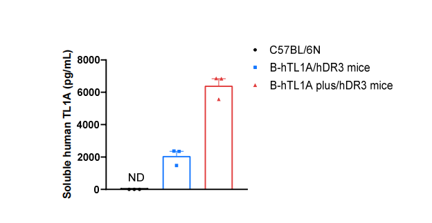
Soluble TL1A expression analysis in B-hTL1A plus/hDR3 mice by ELISA. Bone marrow derived dendritic cells (BMDC) were produced by culturing the bone marrow from wild-type C57BL/6N mice (+/+), homozygous B-hTL1A/hDR3 mice (H/H; H/H), and homozygous B-hTL1A plus/hDR3 mice (H/H; H/H), which were stimulated with LPS in vitro. After stimulation, the supernatants were collected and the level of soluble TL1A was analyzed by ELISA. Soluble human TL1A was exclusively detectable in homozygous B-hTL1A/hDR3 mice and B-hTL1A plus/hDR3 mice, but not in wild-type C57BL/6N mice. Values are expressed as mean ± SEM. ND: not detectable.
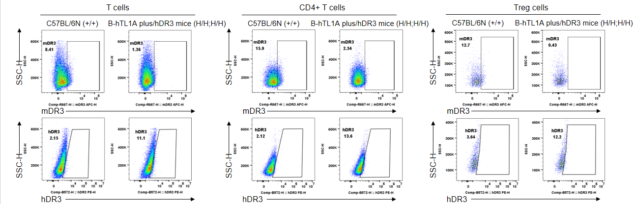
Strain specific DR3 expression analysis in wild-type C57BL/6N mice and homozygous B-hTL1A plus/hDR3 mice by flow cytometry. Splenocytes were collected from wild-type C57BL/6N mice (+/+) and homozygous B-hTL1A plus/hDR3 mice (H/H;H/H), stimulated with anti-mouse CD3ε antibody and anti-mouse CD28 antibody in vivo for 24 h. Protein expression was analyzed with anti-mouse DR3 antibody (Biolegend, 144407) and anti-human DR3 antibody (Biolegend, 307105) by flow cytometry. Mouse DR3 was detectable in wild-type C57BL/6N mice, human DR3 was detectable in homozygous B-hTL1A plus/hDR3 mice.
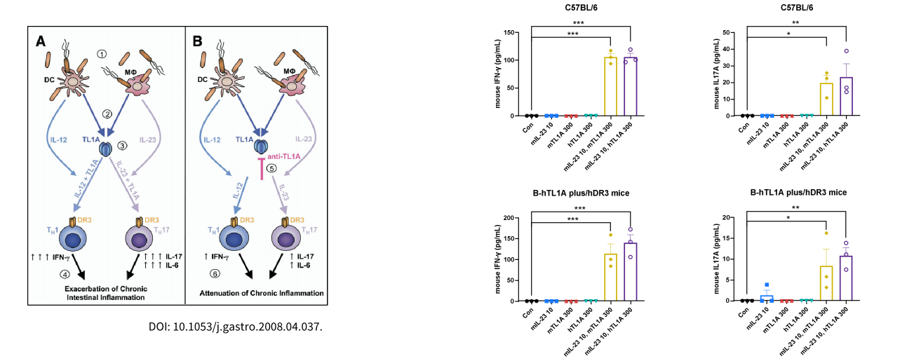
Ex vivo functional analysis in B-hTL1A plus/hDR3 mice. Splenocytes were collected from wild type C57BL/6 mice (+/+) and homozygous B hTL1A plus/hDR3 mice (H/H;H/H), then the production of mouse IFN γ and mouse IL 17A in supernatants were assessed by ELISA after 72 h of incubation with mIL23 (10 ng/mL) and m/hTL1A (300 ng/mL) in vitro.
