Description
CD30L: A crucial co-stimulatory driver that amplifies T-cell activation
- Gene Information: TNF Superfamily Member 8 (TNFSF8, also known as CD30L) is a protein-coding gene located on chromosome 9q32-q33.1. This cytokine is a ligand for receptor TNFRSF8 (also known as CD30).
- Protein Expression: CD30L is rarely expressed under resting conditions and is predominantly localized on the surface of stimulated and activated immune cells. Activated T cells serve as the primary source of CD30L expression, though it is also expressed on activated Bcells, monocytes, macrophages, and dendritic cells.
- Signaling Pathway: The binding of CD30 and CD30L forms a trimer,which can generate signal transduction for binding with TRAF members,induce the activation of NF-κB and MAPK pathways, and participate inthe processes of tumor cell proliferation and apoptosis, as well as theactivation and proliferation of T and B cells.
- Therapeutic Inhibition: Blocking the interaction between CD30 and CD30L can abrogate the co-stimulatory signals driving immune cell hyperactivation, thereby ameliorating autoimmune inflammatory responses.
RAG2: An essential chromatin-sensing cofactorin V(D)J recombination
- Gene Information: Recombination Activating 2 (RAG2) is a protein-coding gene located on chromosome 11p12, which is involved in the initiation of V(D)J recombination during B and T cell development.
- Protein Expression: RAG2 exists exclusively in T and B cells at specific early developmental stages within the bone marrow and thymus. It is expressed and survives only during the G0/G1 phase, and the Rag2 protein is rapidly degraded as soon as the cell undergoes DNA replication.
- Signaling Pathway: RAG2 forms a DNA-cleaving complex with RAG1. In this complex, RAG1 provides the catalytic activity, RAG2 acts as a structural scaffold: its N-terminus binds tightly to DNA, and its C-terminal PHD finger anchors the complex to chromatin via trimethylated histone H3 (H3K4me3), which is the core component in the immune system that controls the assembly of diverse immune receptors.
- Therapeutic Inhibition: Complete deficiency of RAG2 leads to severe immunodeficiency characterized by a near-total absence of mature T and B cells, thereby precluding the development of autoimmunity. RAG2 mutations can cause Omenn syndrome, a severe combined immunodeficiency associated with autoimmune-like symptoms.
Targeting Strategy
CD30L
- The exons 1 4 of mouse Cd30l gene that encode extracellular domain were replaced by human counterparts in B hCD30L, Rag2 KO mice.
- The genomic region of mouse Cd30l gene that encodes transmembrane domain and cytoplasmic portion was retained. The promoter and 5’UTR region of the mousegene were also retained. The 3’UTR region of the mouse gene are replaced by human counterparts. The CD30L expression was driven by endogenous mouse Cd30lpromoter, while mouse Cd30l gene transcription and translation will be disrupted.
Rag2
- The exon 3 and 3’UTR region of mouse Rag2 were knocked out in B hCD30L, Rag2 KO mice, resulting in a disruption of the Rag2 gene.
CD30L Protein Expression Analysis in Spleen
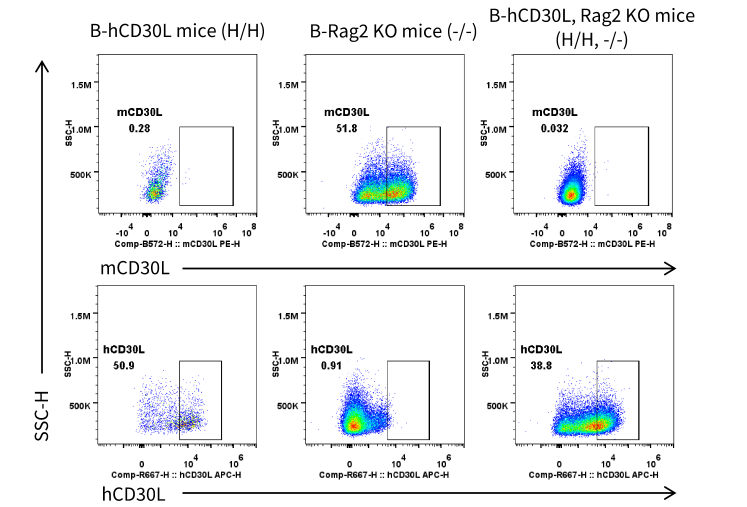
- Mouse CD30L was exclusively detectable in DCs of wild-type B-Rag2 KO mice. 、
- Human CD30L was exclusively detectable in DCs of homozygous B-hCD30L mice and B-hCD30L, Rag2 KO mice.
Strain specific CD30L expression analysis in homozygous B-hCD30L, Rag2 KO mice by flow cytometry. Splenocytes were collected from wild-type B-Rag2 KO mice (-/-),homozygous B-hCD30L mice (H/H), and homozygous B-hCD30L, Rag2 KO mice (H/H;-/-). Splenocytes are incubated in a medium containing Cell Activation Cocktail (withoutBrefeldin A) (Biolegend, 423303) before analysis of CD30L surface expression on gated DCs. Protein expression was analyzed with anti-mouse CD30L antibody (Biolegend,106405) and anti-human CD30L antibody (RD, FAB1028A) by flow cytometry.
Analysis of Leukocyte Subpopulations
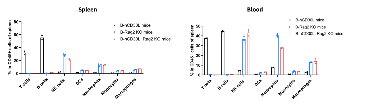
- The percentages of T cells, B cells, NK cells, DCs, monocytes, macrophages, and neutrophils in homozygous B-hCD30L, Rag2 KO micewere similar to those in B-Rag2 KO mice, and B-hCD30L, Rag2 KO mice presented a near-total absence of T and B cells.
- Humanization of CD30L does not affect normal immune cell development or splenic distribution.
Analysis of leukocyte subpopulations by flow cytometry in immune organs and blood. Splenocytes and peripheral blood were isolated from wild-type B-Rag2 KO mice, homozygous B-hCD30L mice, and homozygous B-hCD30L, Rag2 KO mice (female, n=3, 8-week-old). Single live cells were gated on the CD45⁺ population and analyzed by flow cytometry as indicated. Values are expressed as mean ± SEM.
Analysis of T Cell Subpopulations
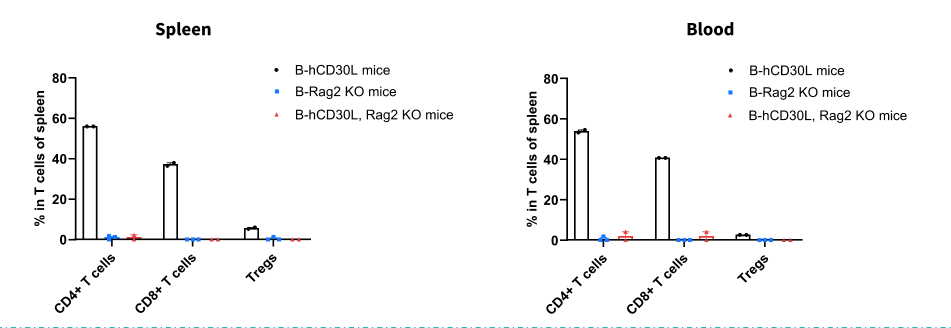
- The proportions of CD4⁺ T cells, CD8⁺ T cells, and Tregs in homozygous B-hCD30L, Rag2 KO mice were comparable to those in B-Rag2 KO mice, and B-hCD30L, Rag2 KO mice presented a near-total absence of CD4+ T cells, CD8+ T cells, and Tregs.
- Humanization of CD30L does not affect normal T cell development, differentiation, or splenic distribution.
Analysis of T-cell subpopulations by flow cytometry in immune organs and blood. Splenocytes and peripheral blood were isolated from wild-type B-Rag2KO mice, homozygous B-hCD30L mice, and homozygous B-hCD30L, Rag2 KO mice (female, n=3, 8-week-old). Single live cells were gated on the TCRβ⁺ T-cellpopulation and analyzed by flow cytometry as indicated. Values are expressed as mean ± SEM.
* When publishing results obtained using this animal model, please acknowledge the source as follows: The animal model [B-hCD30L, Rag2 KO mice] (Cat# 114032) was purchased from Biocytogen.