mRNA expression analysis
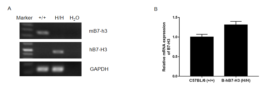
Strain specific analysis of B7-H3 gene expression in C57BL/6 and B-hB7-H3 mice. (A) Mouse B7-h3 mRNA was detectable in splenocytes of wild-type C57BL/6 mice (+/+). Human B7-H3 mRNA was detectable only in homozygous B-hB7-H3 mice (H/H), but not in wild-type mice. (B) Bone marrow were collected from wild-type C57BL/6 mice (+/+) and homozygous B-hB7-H3 mice (H/H), and then stimulated with GM-CSF and IL4 for 7 days. After that, the cells were stimulated with 10 μg/mL LPS for 24 h.
Protein expression analysis
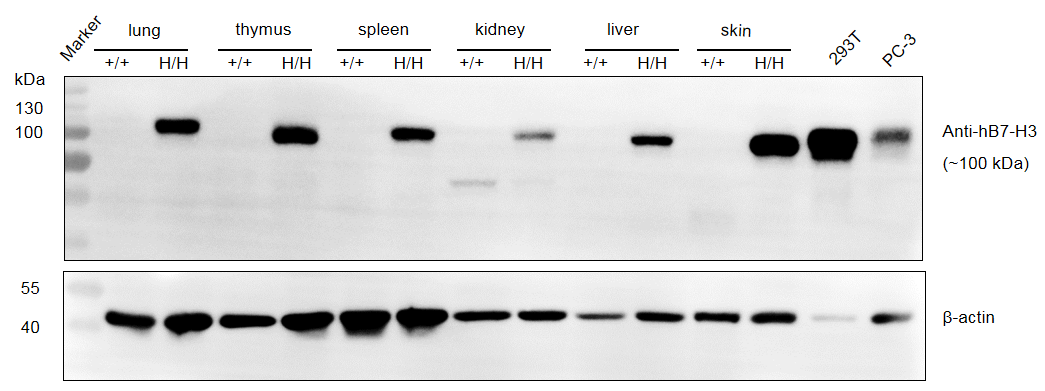
Strain specific B7-H3 expression analysis in homozygous B-hB7-H3 mice by western blot. Tissues were collected from wild-type C57BL/6 mice (+/+) and homozygous B-hB7-H3 mice (H/H), and analyzed by western blot with species-specific anti-B7-H3 antibody (Abcam, ab219648).
Protein Expression by IHC
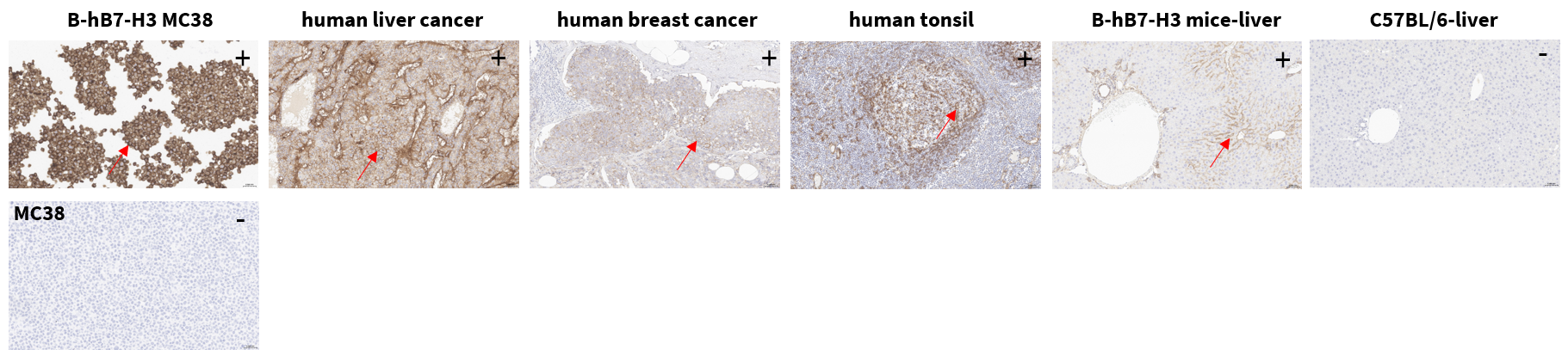
B7-H3 protein expression by immunohistochemistry (IHC). B-hB7-H3 MC38 cells, human liver cancer, human breast cancer, human tonsil, B-hB7-H3 mice tissue, C57BL/6 liver, and wild-type MC38 cells were stained using anti-B7-H3 antibody (Abcam, ab219648). Positive staining was observed in B-hB7-H3 MC38 cells, human positive-control tissues, and B-hB7-H3 mice tissue, while C57BL/6 liver and wild-type MC38 cells were negative. Images were captured at 200× magnification. “+” indicates positive staining; “−” indicates negative staining; red arrows indicate positive cells.
Protein Expression Profile in Normal Tissues by IHC
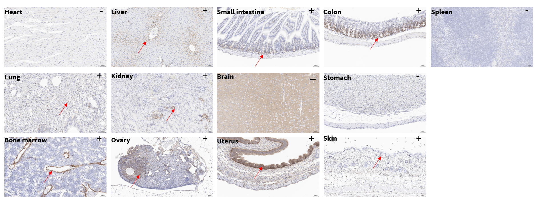
Human B7-H3 protein expression profile in normal tissues of homozygous B-hB7-H3 mice by IHC. Tissues were stained with anti-B7-H3 antibody (Abcam, ab219648). Human B7-H3 was detected in liver, lung, kidney, skin, colon, small intestine, bone marrow, ovary, and uterus, mainly in epithelial, stromal, or perivascular compartments. Heart, spleen, and stomach were negative. Brain exhibited diffuse staining and was classified as suspected positive/weak positive due to the lack of clear cell-type-specific localization. Images: 200×. “+” positive; “−” negative; “±” suspected positive/weak positive; red arrows indicate positive regions.
B7-H3 IHC Specificity Control in Wild-Type C57BL/6 Tissues
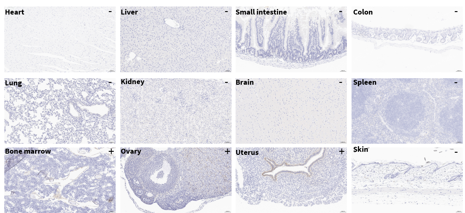
IHC specificity control in wild-type C57BL/6 tissues. Representative tissues from wild-type C57BL/6 mice were stained with anti-B7-H3 antibody (Abcam, ab219648). No obvious staining was observed in most tissues, while weak or localized brown staining was observed in bone marrow, ovary, and uterus. These signals may be associated with endogenous mouse B7-H3/Cd276 expression and indicate potential weak cross-reactivity of the antibody with mouse B7-H3 under the tested IHC conditions. Overall, the staining intensity and distribution in wild-type tissues were much lower than those observed in B-hB7-H3 mice, supporting preferential detection of human B7-H3 in the humanized model. Images: 200×. “+” positive; “−” negative.
Spleen Leukocyte Subpopulation Analysis
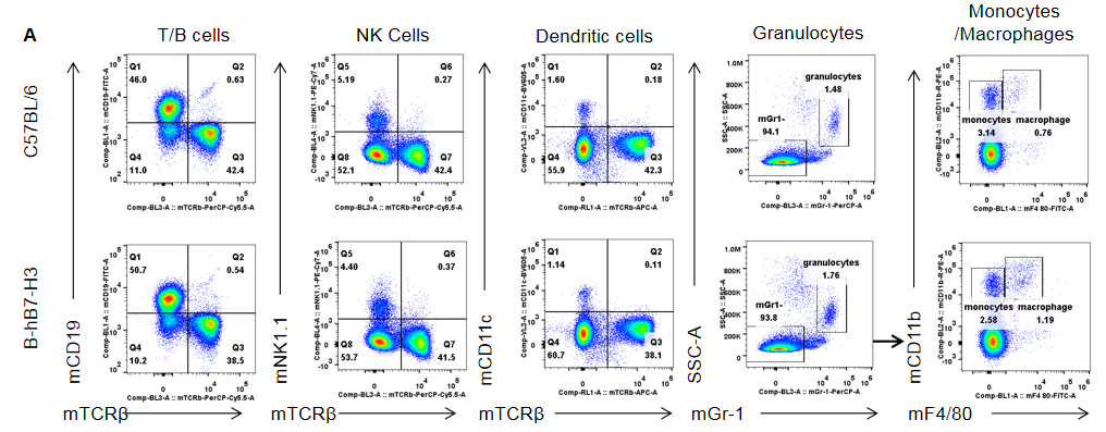
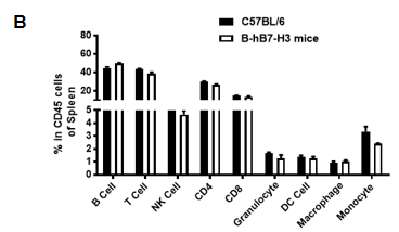
Analysis of spleen leukocyte subpopulations by flow cytometry. Splenocytes were isolated from female C57BL/6 mice and B-hB7-H3 mice (6-week-old, n = 3). (A) Representative FACS plots gated on single live CD45+ cells. (B) Frequencies of T cells, B cells, NK cells, granulocytes, monocytes/macrophages and dendritic cells. Results indicate that B7-H3 humanization does not alter the overall development, differentiation or splenic distribution of these leukocyte populations. Values are expressed as mean ± SEM.
Spleen T-Cell Subpopulation Analysis
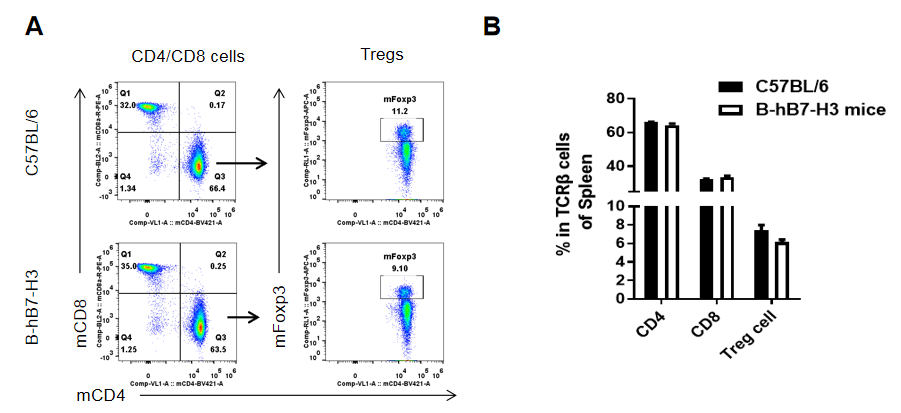
Analysis of spleen T-cell subpopulations by flow cytometry. Splenocytes were isolated from female C57BL/6 and B-hB7-H3 mice (6-week-old, n = 3). (A) Representative FACS plots gated on TCRβ+ T cells. (B) Frequencies of CD4+ T cells, CD8+ T cells and Tregs. Results show that B7-H3 humanization does not affect normal splenic T-cell development, differentiation or distribution. Values are expressed as mean ± SEM.
Lymph Node Leukocyte Subpopulation Analysis
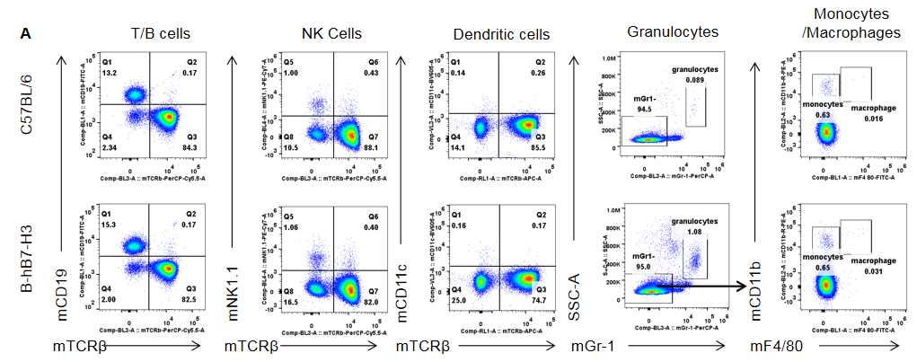
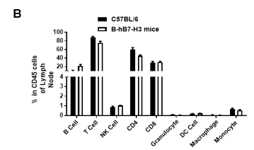
Analysis of lymph node leukocyte subpopulations by flow cytometry. Leukocytes were isolated from female C57BL/6 mice and B-hB7-H3 mice (6-week-old, n = 3). (A) Representative FACS plots gated on single live CD45+ cells. (B) Frequencies of T cells, B cells, NK cells, granulocytes, monocytes/macrophages and dendritic cells. Results indicate that B7-H3 humanization does not alter the overall development, differentiation or distribution of these leukocyte populations. Values are expressed as mean ± SEM.
Lymph Node T-Cell Subpopulation Analysis
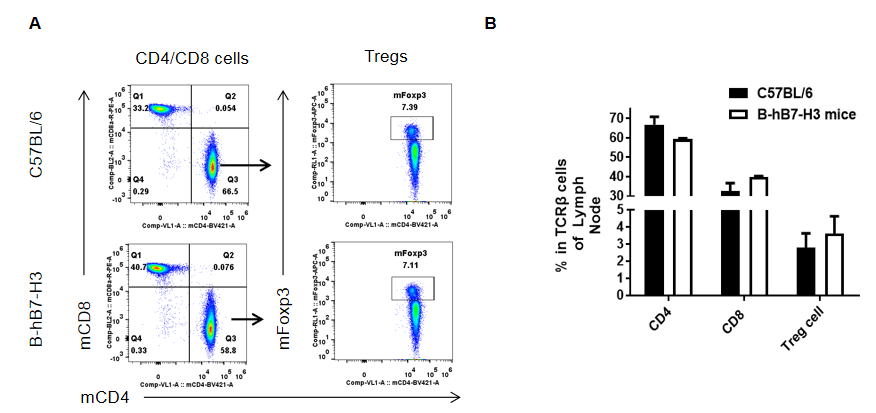
Analysis of lymph node T-cell subpopulations by flow cytometry. Leukocytes were isolated from female C57BL/6 and B-hB7-H3 mice (6-week-old, n = 3). (A) Representative FACS plots gated on TCRβ+ T cells. (B) Frequencies of CD4+ T cells, CD8+ T cells and Tregs. Results show that B7-H3 humanization does not affect normal lymph node T-cell development, differentiation or distribution. Values are expressed as mean ± SEM.
Peripheral Blood Leukocyte Subpopulation Analysis
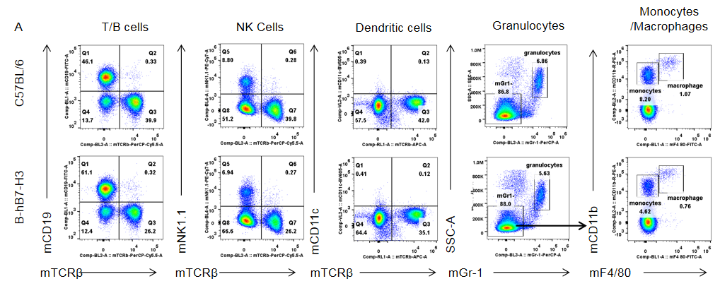
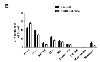
Analysis of peripheral blood leukocyte subpopulations by flow cytometry. Blood was collected from female C57BL/6 and B-hB7-H3 mice (6-week-old, n = 3). (A) Representative FACS plots gated on single live CD45+ cells. (B) Frequencies of T cells, B cells, NK cells, granulocytes, monocytes/macrophages and dendritic cells. Results indicate that B7-H3 humanization does not alter peripheral leukocyte distribution. Values are expressed as mean ± SEM.
Peripheral Blood T-Cell Subpopulation Analysis
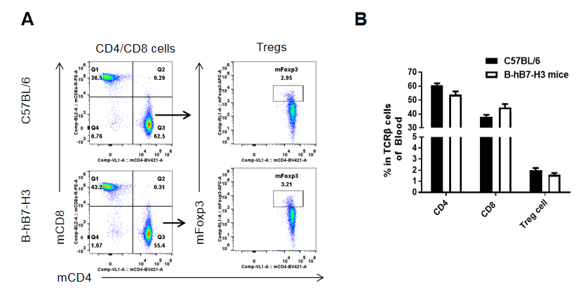
Analysis of peripheral blood T-cell subpopulations by flow cytometry. Blood was collected from female C57BL/6 and B-hB7-H3 mice (6-week-old, n = 3). (A) Representative FACS plots gated on TCRβ+ T cells. (B) Frequencies of CD4+ T cells, CD8+ T cells and Tregs. Results show that B7-H3 humanization does not affect peripheral T-cell development, differentiation or distribution. Values are expressed as mean ± SEM
Hematology Analysis
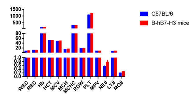
Complete blood count (CBC) analysis of B-hB7-H3 mice. Blood was collected from female C57BL/6 and B-hB7-H3 mice (6-week-old, n = 5) and analyzed for CBC parameters. B-hB7-H3 mice showed blood cell composition and morphology comparable to C57BL/6 controls, indicating no obvious hematological abnormalities associated with B7-H3 humanization. Values are expressed as mean ± SEM.
Blood Chemistry Analysis
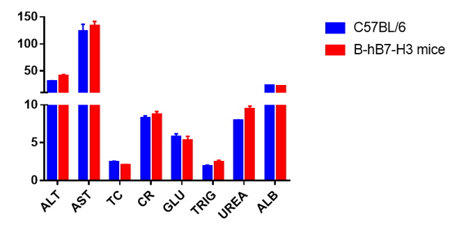
Blood chemistry analysis of B-hB7-H3 mice. Serum was collected from female C57BL/6 and B-hB7-H3 mice (6-week-old, n = 5) and analyzed for ALT, AST and other clinical chemistry parameters. No obvious differences were observed between B-hB7-H3 mice and C57BL/6 controls, indicating no apparent liver or systemic clinical chemistry abnormalities associated with B7-H3 humanization. Values are expressed as mean ± SEM.
Anti-Human B7-H3 Antibody Binding Assay
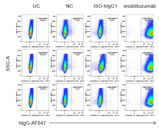
Anti-human B7-H3 antibody binding assay by flow cytometry. B-hB7-H3 MC38 cells were stained with enoblituzumab analog (in-house) or isotype control and analyzed by flow cytometry. Single live cells were gated for analysis. B-hB7-H3 MC38 cells bound enoblituzumab analog, confirming surface human B7-H3 expression and antibody target engagement in vitro.
Enoblituzumab Analog Efficacy in B-hB7-H3 MC38 Tumor Model
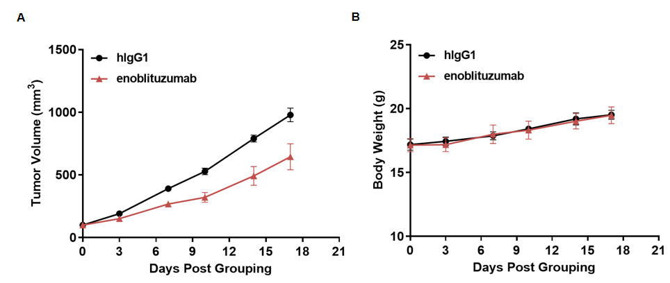
Antitumor activity of enoblituzumab analog in the B-hB7-H3 MC38 syngeneic tumor model. B-hB7-H3 MC38 cells were subcutaneously implanted into homozygous B-hB7-H3 mice (female, 6–7 weeks old, n = 5). Mice were randomized when tumor volume reached approximately 100 mm3 and treated with enoblituzumab analog (in-house). (A) Tumor volume. (B) Body weight. Enoblituzumab analog inhibited tumor growth, supporting B-hB7-H3 mice as a model for in vivo evaluation of anti-human B7-H3 antibodies. Values are expressed as mean ± SEM.
Anti-Human B7-H3 Antibody Efficacy in B-hB7-H3 MC38 Model
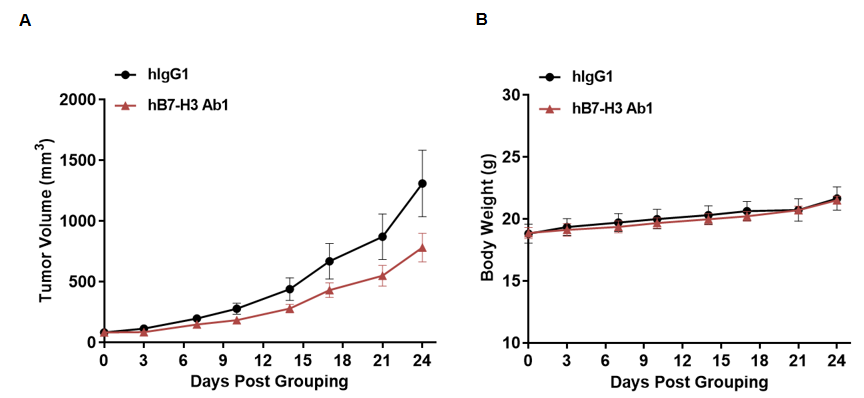
Antitumor activity of anti-human B7-H3 antibody in the B-hB7-H3 MC38 syngeneic tumor model. B-hB7-H3 MC38 cells were subcutaneously implanted into homozygous B-hB7-H3 mice (female, 6–7 weeks old, n = 6). Mice were randomized when tumor volume reached approximately 80 mm3 and treated with anti-human B7-H3 antibody (in-house). (A) Tumor volume. (B) Body weight. Anti-human B7-H3 antibody inhibited tumor growth, supporting this model for anti-human B7-H3 efficacy evaluation. Values are expressed as mean ± SEM.
Anti-Human B7-H3 Antibody Efficacy in B-hB7-H3 EL4 Model
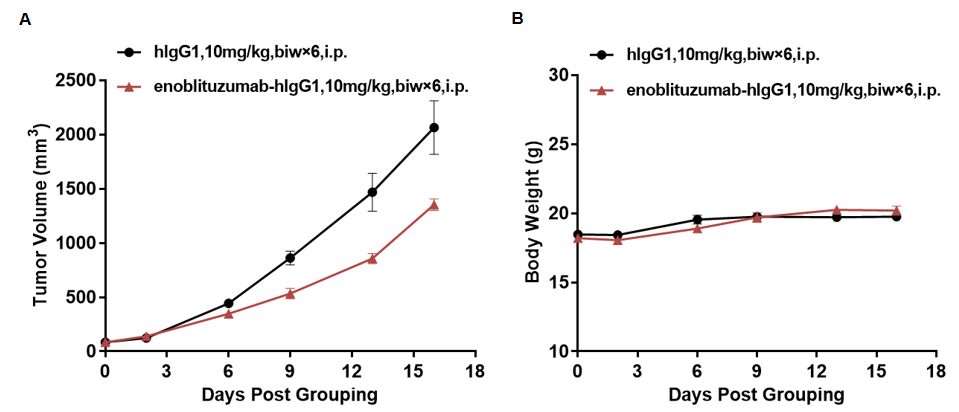
Antitumor activity of anti-human B7-H3 antibodies in the B-hB7-H3 EL4 syngeneic tumor model. B-hB7-H3 EL4 cells were subcutaneously implanted into homozygous B-hB7-H3 mice (female, 6–7 weeks old, n = 6). Mice were randomized when tumor volume reached approximately 80 mm3 and treated with anti-human B7-H3 antibody using the indicated dosing schedules. (A) Tumor volume. (B) Body weight. Anti-human B7-H3 antibody inhibited B-hB7-H3 EL4 tumor growth, demonstrating utility of B-hB7-H3 mice for in vivo efficacy evaluation. Values are expressed as mean ± SEM.
Combination Therapy with Enoblituzumab Analog and Anti-mouse PD-1
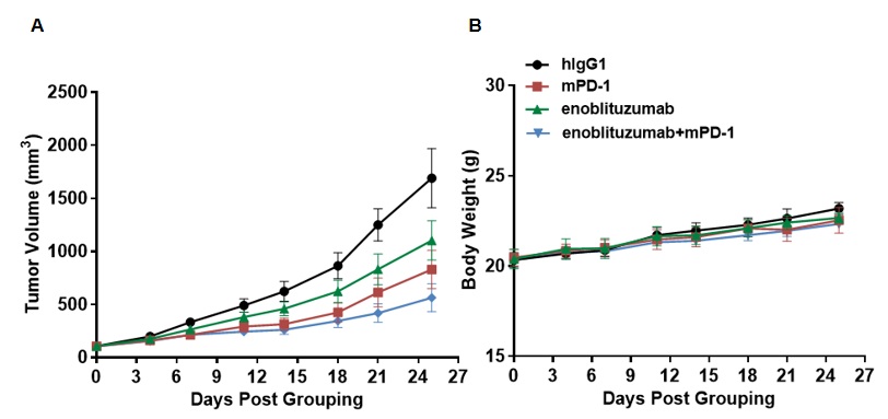
Antitumor activity of enoblituzumab analog combined with anti-mouse PD-1 antibody in B-hB7-H3 mice. B-hB7-H3 MC38 cells were subcutaneously implanted into homozygous B-hB7-H3 mice (female, 7–8 weeks old, n = 6). Mice were randomized when tumor volume reached approximately 100 mm3 and treated with enoblituzumab analog, anti-mouse PD-1, or the combination. (A) Tumor volume. (B) Body weight. Combination therapy showed greater tumor-growth inhibition than individual treatments, supporting evaluation of anti-human B7-H3 antibody combinations in B-hB7-H3 mice. Values are expressed as mean ± SEM.
* When publishing results obtained using this animal model, please acknowledge the source as follows: The animal model [B-hB7-H3 mice] (Cat# 110028) was purchased from Biocytogen.