


IL-17A/IL17F: Core pro-inflammatory cytokines driving autoimmune skin and joint diseases

IL17A
IL17F

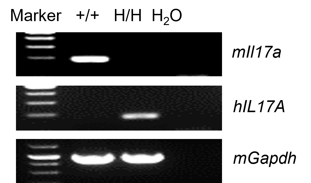
Strain specific analysis of IL17A gene expression in wild type (WT) mice and B-hIL17A/hIL17F mice by RT-PCR. Small intestine RNA were isolated from wild-type C57BL/6JNifdc mice (+/+) and homozygous B-hIL17A/hIL17F mice (H/H, H/H), then cDNA libraries were synthesized by reverse transcription, followed by PCR with mouse or human IL17A primers. Mouse Il17a mRNA were detectable in wild-type mice. Human IL17A mRNA was detectable in homozygous B-hIL17A/hIL17F mice but not in wild-type mice.
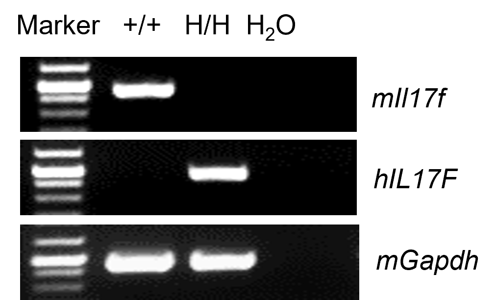
Strain specific analysis of IL17A gene expression in wild type (WT) mice and B-hIL17A/hIL17F mice by RT-PCR. Kidney RNA were isolated from wild-type C57BL/6JNifdc mice (+/+) and homozygous B-hIL17A/hIL17F mice (H/H, H/H), then cDNA libraries were synthesized by reverse transcription, followed by PCR with mouse or human IL17F primers. Mouse Il17f mRNA were detectable in wild-type mice. Human IL17F mRNA was detectable in homozygous B-hIL17A/hIL17F mice but not in wild-type mice.

Strain specific IL17A expression analysis in homozygous B-hIL17A/hIL17F mice by ELISA. Serum were collected from WT mice and homozygous B-hIL17A/hIL17F mice (H/H, H/H) stimulated with anti-mCD3ε and anti-mCD28 antibody in vivo, and analyzed by ELISA with mouse IL-17A ELISA Kit (Biolegend, 432507) and human IL-17A ELISA Kit (Biolegend, 433917). Mouse IL17A was detectable in WT mice. Human IL17A was detectable in homozygous B-hIL17A/hIL17F mice (H/H). ND: not detectable.

Strain specific IL17F expression analysis in homozygous B-hIL17A/hIL17F mice by ELISA. Naïve CD4+ T cells were sorted from splenocytes of wild type (WT) mice and homozygous B-hIL17A/hIL17F mice (H/H, H/H), and induced into Th17 cells. Th17 cells were stimulated by PMA and lonomycin. The Th17 cells culture supernatants were collected and analyzed by ELISA with with mouse IL-17F ELISA Kit (Biolegend, 436107) and human IL-17F ELISA Kit (Biolegend, 435107). Mouse IL17F was detectable in WT mice. Human IL17F was exclusively detectable in homozygous B-hIL17A/hIL17F mice (H/H, H/H). ND: not detectable.
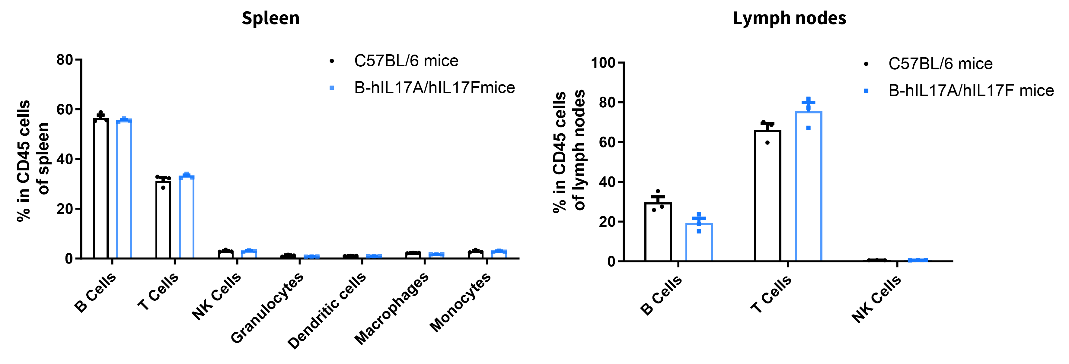
Analysis of leukocyte subpopulations by FACS. Splenocytes and lymph nodes were isolated from female C57BL/6 and B-hIL17A/hIL17F mice(n=3, 7-week-old). Flow cytometry analysis of the splenocytes and lymph nodes was performed to assess leukocyte subpopulations. Percent of T, B, NK, DC, granulocyte, monocyte, and macrophage cells in heterozygous B-hIL17A/hIL17F mice was similar to those in the C57BL/6 mice, demonstrating that introduction of B-hIL17A/hIL17F in place of its mouse counterpart does not change the overall development, differentiation or distribution of these cell types in spleen.
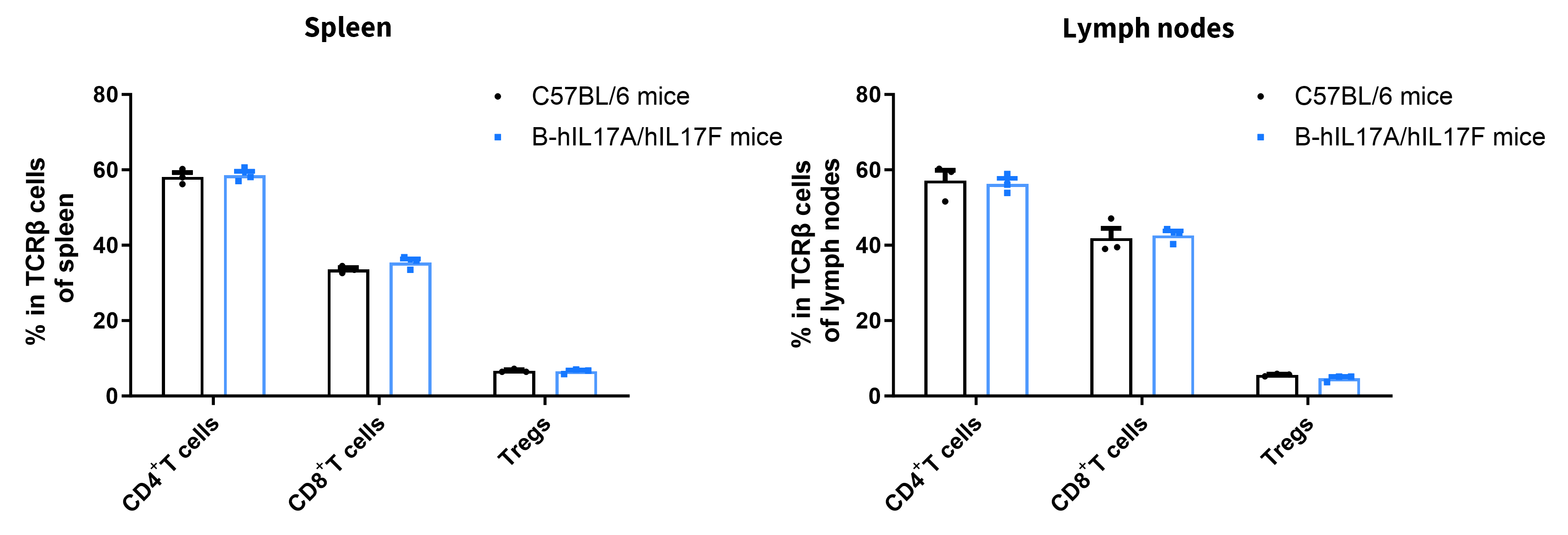
Analysis of spleen T cell subpopulations by FACS. Splenocytes and lymph nodes were isolated from female C57BL/6 and B-hIL17A/hIL17F mice (n=3, 7-week-old). Flow cytometry analysis of the splenocytes and lymph nodes was performed to assess leukocyte subpopulations. Percent of CD8+ T cells, CD4+ T cells, and Treg cells in homozygous B-hIL17A/hIL17F mice was similar to those in the C57BL/6 mice, demonstrating that introduction of B-hIL17A/hIL17F mice in place of its mouse counterpart does not change the overall development, differentiation or distribution of these T cell subtypes in lymph node. Values are expressed as mean ± SEM.
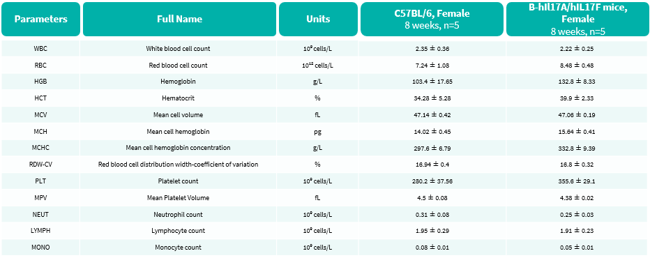
Complete blood count (CBC) of B-hIL17A/hIL17F mice. Values are expressed as mean ± SD.
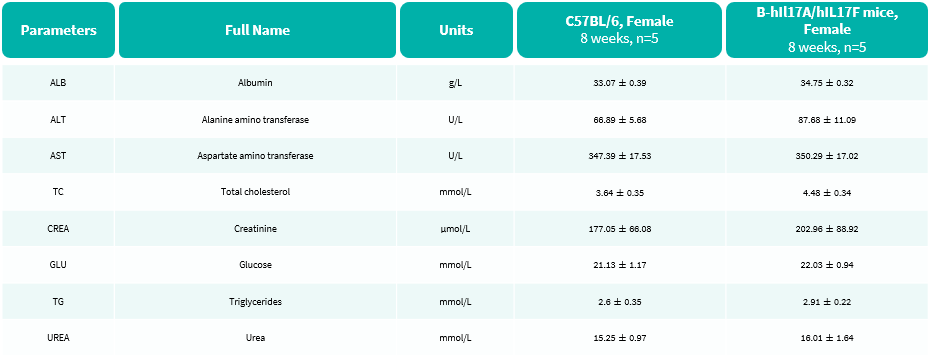
Blood biochemical parameters of B-hIL17A/hIL17F mice are shown. Values are expressed as mean ± SD.

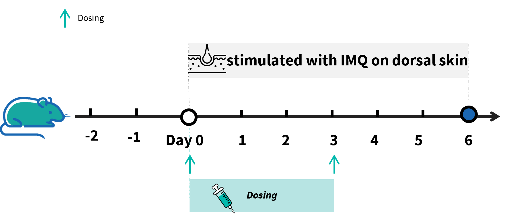
Experimental schedule for induction of IMQ-induced psoriasis model in B-hIL17A/hIL17F mice. Mice at 10 week-old of age received a daily topical of commercially available IMQ cream on the shaved back for 6 consecutive days to induce IMQ-induced psoriasis model. Control mice were treated similarly with Vaseline cream. Severity of skin inflammation was daily scored and back skin was collected at the endpoint. IMQ: imiquimod.
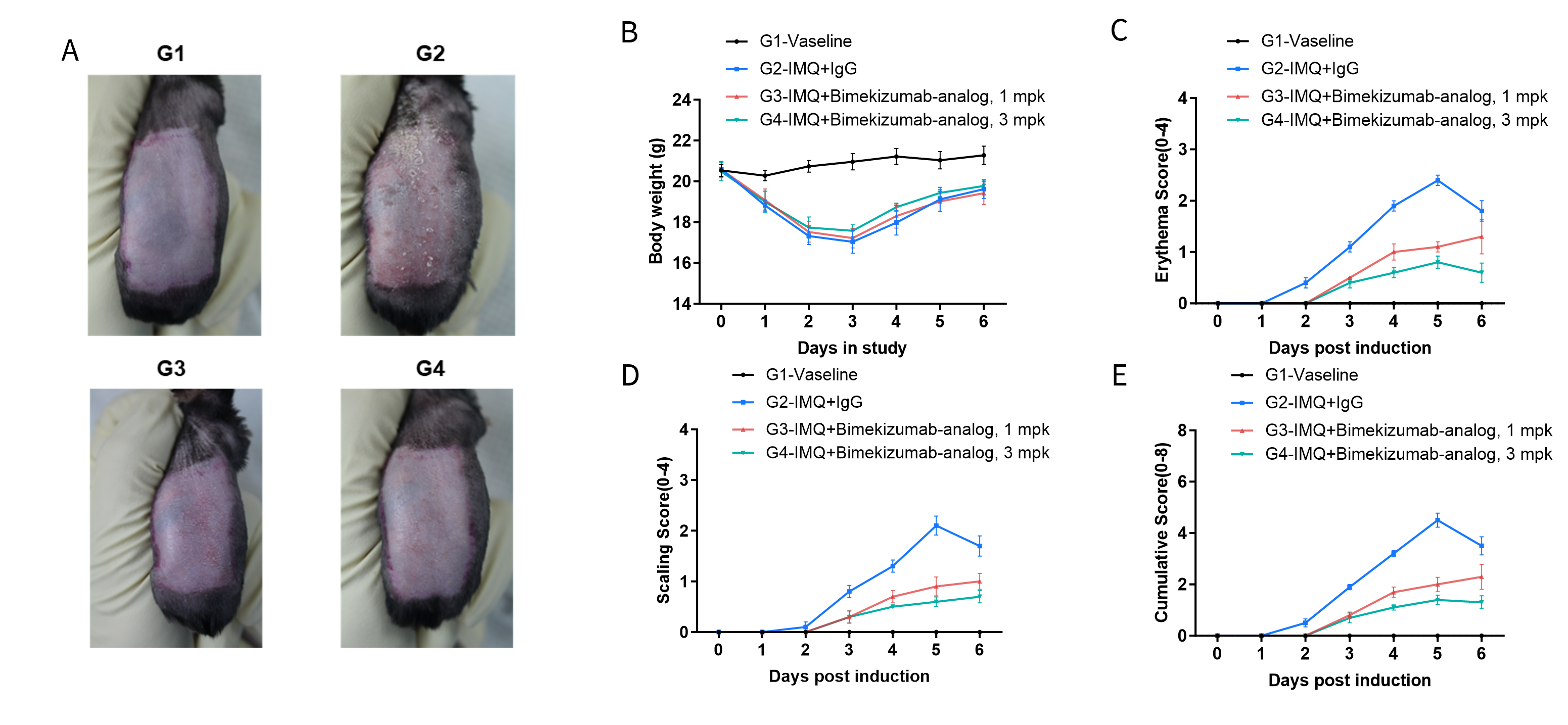
IMQ-induced psoriasis model in B-hIL17A/hIL17F mice. Mice (female, 10 week-old, n=5) were scored daily for up to 6 days for body weight and clinical signs of IMQ-induced psoriasis model following treatment with imiquimod (IMQ) cream. Mice in each group were treated with different dose of bimekizumab-analog, produced in house. Doses are shown in legend. (A) Phenotypical presentation of mouse back skin after 6 days of treatment. (B) Body weight changes during treatment. (C-D) Erythema and scaling score of the back was scored daily on a scale from 0 to 4. Additionally, the cumulative score (erythema plus scaling) is depicted. Values are expressed as mean ± SEM.
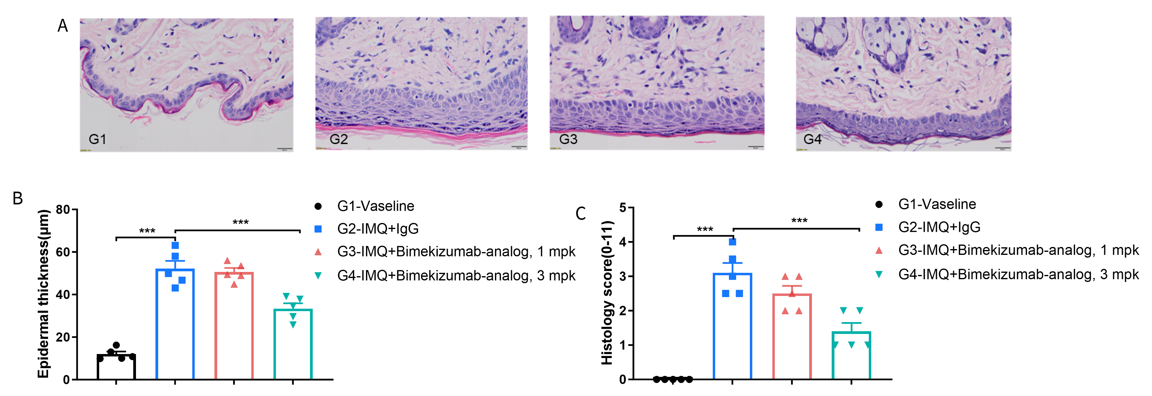
Dose dependent effects of antibody on keratinocyte proliferation and inflammatory cell infiltration in IMQ-induced psoriasis model in B-hIL17A/hIL17F mice. Back skin was collected at the endpoint and stained with Hematoxylin and eosin (H&E). (A) H&E staining of the back skin of mice. (B) Epidermal thickness of the mice. (C) Histological changes were scored on a scale from 0 to 11. Results indicated that bimekizumab-analog (in house) significantly reduced psoriasis-like skin lesions in B-hIL17A/hIL17F mice, confirming that B-hIL17A/hIL17F mice is a powerful model for in vivo evaluation of anti-human IL17A and IL17F antibody.
