FcRn MOA: pH-Dependent Binding of IgG to FcRn
In acidic environments such as endosomes, IgG binds strongly to FcRn, facilitating its transport and recycling.
Upon reaching the plasma membrane, the pH shifts to near-neutral, triggering the release of IgG from FcRn into the extracellular fluid.

CD98HC
FcRn
B-hCD98HC/hFcRn mice are generated by mating B-hCD98HC mice (110983) with B-hFcRn mice (110001).

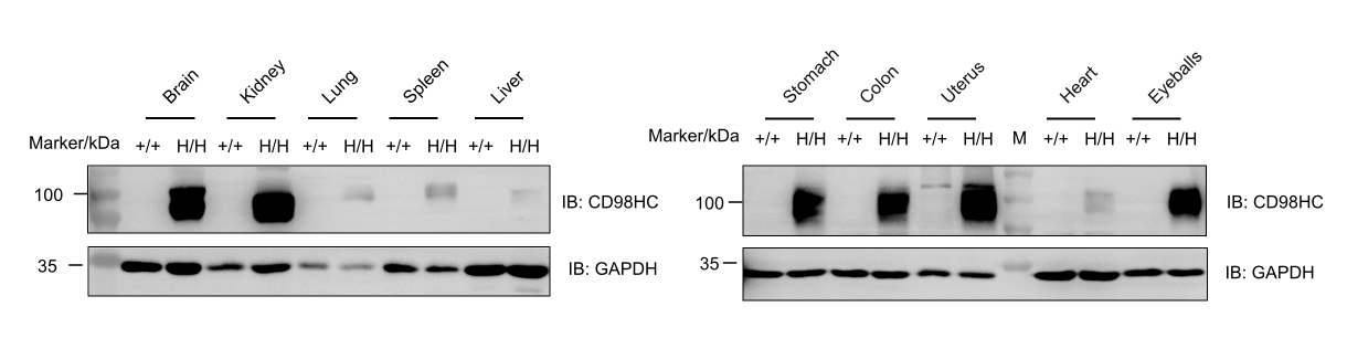
Protein expression analysis of CD98HC in homozygous B-hCD98HC/hFcRn mice. Various tissue lysates were collected from wild-type C57BL/6JNifdc mice (+/+) and homozygous B-hCD98HC/hFcRn mice (H/H), and then analyzed by western blot with anti-CD98HC antibody (Proteintech, 15193-1-AP). 40 μg total protein was loaded for western blotting analysis. Human CD98HC was exclusively detected in heart, liver, spleen, lung, kidney, brain, stomach, colon, uterus and eyeballs from homozygous B-hCD98HC/hFcRn mice.
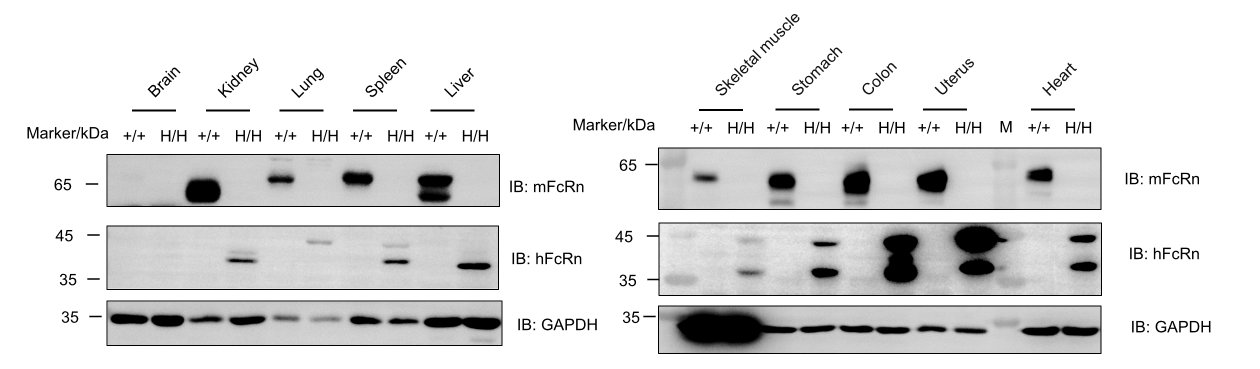
Protein expression analysis of FcRn in homozygous B-hCD98HC/hFcRn mice. Various tissue lysates were collected from wild-type C57BL/6JNifdc mice (+/+) and homozygous B-hCD98HC/hFcRn mice (H/H), and then analyzed by western blot with anti-FcRn antibody (human FcRn: NOVUS, NBP1-89128; mouse FcRn: R&D, AF6775). 40 μg total protein was loaded for western blotting analysis. mFcRn was only detected in heart, liver, spleen, lung, kidney, skeletal muscle, stomach, colon and uterus from wild-type C57BL/6JNifdc mice. hFcRn was exclusively detected in heart, liver, spleen, lung, kidney, skeletal muscle, stomach, colon and uterus from homozygous B-hCD98HC/hFcRn mice.
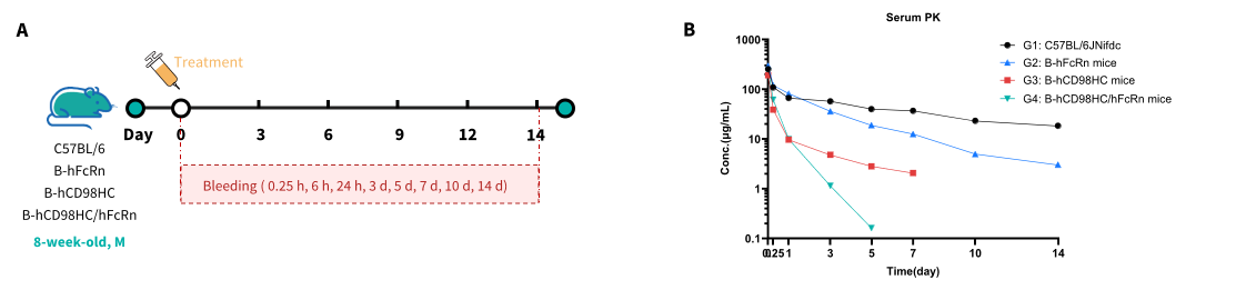
Serum pharmacokinetic (PK) analysis of anti-CD98HC antibody in wild-type and humanized TFR1/FcRn mouse strains. (A) Scheme design. Male mice (n = 9 per group) from wild-type C57BL/6 mice (G1), B-hFcRn mice (G2), B-hCD98HC mice (G3) and B-hCD98HC/hFcRn mice (G4) received a single intravenous (i.v.) dose of 10 mg/kg anti-human CD98HC antibody (monovalent). Blood samples were collected at 0.25 h, 6 h, 24 h, and on days 3, 5, 7, 10 and 14 post-administration. (B) Serum concentrations of anti-CD98HC antibody were measured by ELISA. Mice lacking human CD98hc (G1, G2) displayed typical slow IgG clearance. Accelerated antibody elimination was observed in humanized CD98HC mice (G3 and G4) due to target-mediated drug disposition (TMDD). Binding of the mAb to cell-surface human CD98hc triggers receptor internalization and lysosomal degradation of antibody-antigen complexes. G4 showed the most rapid clearance due to combined human CD98hc-dependent TMDD and human FcRn background.
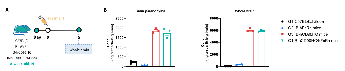
Brain pharmacokinetic (PK) analysis of anti-CD98HC antibody in wild-type and humanized TFR1/FcRn mouse strains. (A) Scheme design. Male mice (n = 9 per group) from wild-type C57BL/6 mice (G1), B-hFcRn mice (G2), B-hCD98HC mice (G3) and B-hCD98HC/hFcRn mice (G4) received a single intravenous (i.v.) dose of 10 mg/kg anti-human CD98HC antibody (monovalent). Brain tissues were collected on day 5 post-administration. (B) Brain parenchyma and whole brain concentrations of anti-CD98HC antibody were measured by ELISA. The test antibody specifically recognizes human CD98HC and does not cross-react with endogenous murine CD98HC, therefore brain penetration is not detectable in wild-type mice (G1, G2). Data are shown as mean ± SEM.