
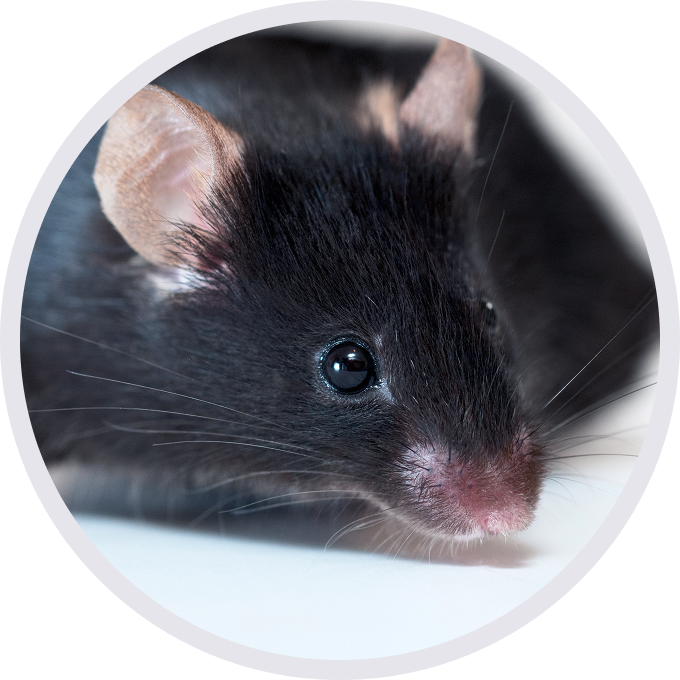

ICOS: ICOS Costimulates T-Cell Activation and Enhances Humoral Immune Responses.

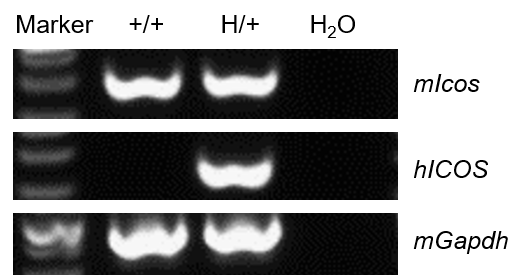
Species specific analysis of ICOS gene expression in wild-type C57BL/6 mice and heterozygous ICOS humanized mice by RT-PCR.Thymus RNA was isolated from wild-type C57BL/6 mice (+/+) and heterozygous B-hICOS mice (H/+), and then cDNA libraries were synthesized by reverse transcription, followed by PCR with mouse Icos primers and human ICOS primers. The positive band was confirmed to be correct by sequencing.
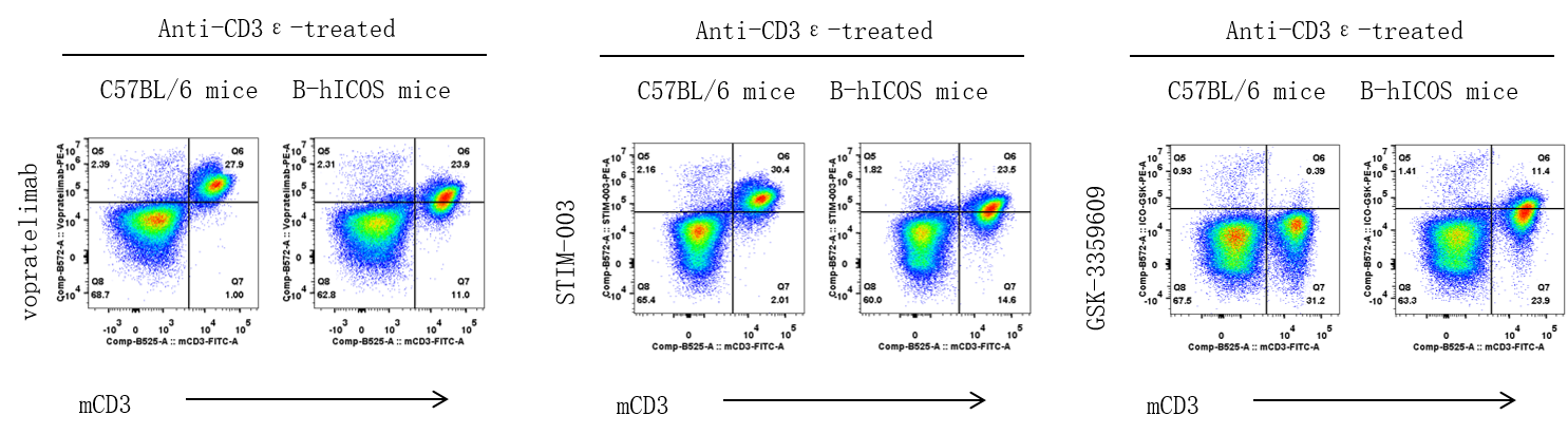
Strain specific ICOS expression analysis in wild-type C57BL/6 mice and homozygous ICOS humanized mice by flow cytometry.Splenocytes were collected from wild-type C57BL/6 mice and homozygous B-hICOS mice stimulated with anti-mouse CD3ε antibody in vivo for 24 hours. Protein expression was analyzed with anti-human ICOS antibody vopratelimab (in house), STIM-003 (in house) and GSK-3359609 (in house) by flow cytometry.

Mouse spleen CD4/CD8 ICOS expression and proliferation in wild-type C57BL/6 mice 、heterozygous ICOS humanized mice and homozygous ICOS humanized mice by flow cytometry.Splenocytes from wild-type (WT) mice (+/+), heterozygous B-hICOS mice (H/+) and homozygous B-hICOS mice (H/H) were stained by CellTrace Violet staining solution, and incubated for 72h in the presence of anti-mCD3ε, or anti-mCD3ε and anti-mCD28, or anti-mCD3ε and human ICOSL recombinant protein. The expression and proliferation of CD4+ T cells and CD8+ T cells were measured by flow cytometry.
Note: This experiment was performed by the client using B-hICOS mice. All the other materials were provided by the client.

Mouse spleen CD4/CD8 ICOS expression and proliferation in wild-type C57BL/6 mice 、heterozygous ICOS humanized mice and homozygous ICOS humanized mice by flow cytometry.Splenocytes from wild-type (WT) mice (+/+), heterozygous B-hICOS mice (H/+) and homozygous B-hICOS mice (H/H) were stained by CellTrace Violet staining solution, and incubated for 72h in the presence of anti-mCD3ε, or anti-mCD3ε and anti-mCD28, or anti-mCD3ε and human ICOSL recombinant protein. The expression and proliferation of CD4+ T cells and CD8+ T cells were measured by flow cytometry.
Note: This experiment was performed by the client using B-hICOS mice. All the other materials were provided by the client.

Mouse spleen CD4/CD8 ICOS expression and proliferation in wild-type C57BL/6 mice、heterozygous ICOS humanized mice and homozygous humanized B-hICOS mice by flow cytometry.Splenocytes from wild-type (WT) mice (+/+), heterozygous B-hICOS mice (H/+) and homozygous B-hICOS mice (H/H) were stained by CellTrace Violet staining solution, and incubated for 72 hours in the presence of anti-mCD3ε antibody, or anti-mCD3ε antibody and anti-mCD28 antibody, or anti-mCD3ε antibody and human ICOSL recombinant protein. The expression and proliferation of CD4+ T cells and CD8+ T cells were measured by flow cytometry.
Note: This experiment was performed by the client using B-hICOS mice. All the other materials were provided by the client.
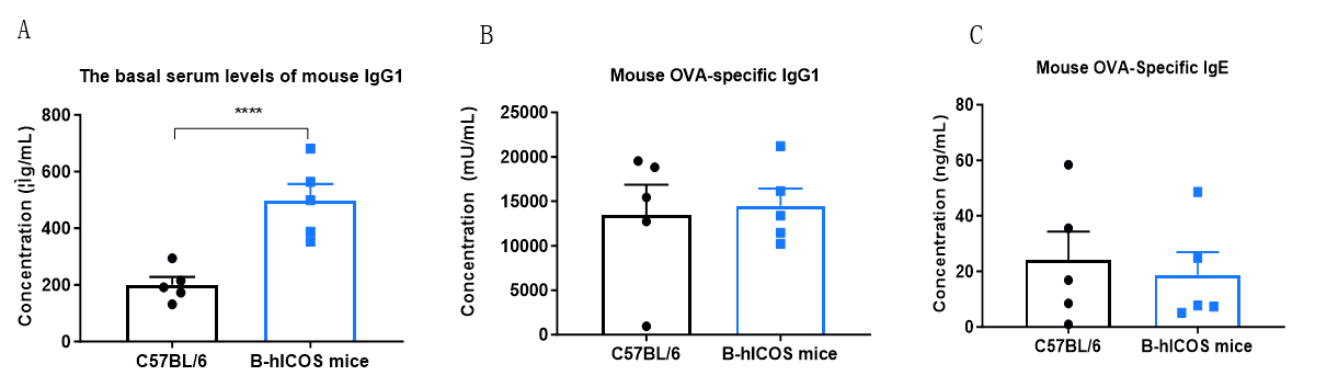
ICOS delivers a co-stimulatory signal that is essential both for efficient interaction between T and B cells and for normal antibody responses to T-cell-dependent antigens. Mice homozygous for disruptions in the ICOS gene show reduced basal IgG1 levels and impaired interactions between T and B cells. Immunoglobulin class switching, including production of allergy-mediating IgE, is defective1. To determine the physiological function of ICOS/ICOSL, the basal serum levels of IgG1 in wild type (WT) mice and homozygous B-hICOS mice (8-week-old, n=5) were measured (A). Serum from WT mice and homozygous B-hICOS mice challenged with the T-cell-dependent antigen ovalbumin (OVA) was further analyzed, and the presence of OVA-specific IgG1 and IgE was determined by ELISA (B, C).
1.Dong C, Juedes AE, Temann UA, Shresta S, Allison JP, Ruddle NH, Flavell RA. ICOS co-stimulatory receptor is essential for T-cell activation and function. Nature. 2001 Jan 4;409(6816):97-101. doi: 10.1038/35051100. PMID: 11343121.