Beta-2 microglobulin (B2M) encodes a serum protein that associates with major histocompatibility complex (MHC) class I heavy chains on the surface of nearly all nucleated cells. Structurally, B2M exhibits a predominantly β-pleated sheet conformation and can form amyloid fibrils under certain pathological conditions. HLA-A is a member of the human MHC class I heavy chain paralogues and is anchored in the cell membrane. MHC class I molecules play a fundamental role in adaptive immunity by presenting intracellularly derived peptides to cytotoxic T cells and are ubiquitously expressed in most cell types.
In HLA-A11.1 humanized mice (B-HLA-A11.1), exons 1–3 of the mouse B2m gene were replaced with a genetic construct containing the human B2M coding sequence (CDS), together with the leader sequence and α1/α2 domains of the human HLA-A*1101 allele, ligated to a fragment of the murine H-2Dᵇ gene encoding the α3 domain, transmembrane region, and cytoplasmic tail.
Validation data confirm species-specific expression of human B2M and HLA-A*1101 exclusively in homozygous HLA-A11.1 humanized mice (B-HLA-A11.1).
Key Advantages
Validation
Application

In HLA-A11.1 humanized mice (B-HLA-A11.1), exons 1–3 of the mouse B2m gene, which encode β2-microglobulin essential for MHC class I surface expression, were replaced with a sequence comprising the human B2M coding sequence together with the HLA-A*1101 gene. The HLA-A*1101 construct includes the leader sequence and the α1 and α2 domains of human HLA-A*1101, which are fused to a fragment of the murine H-2Dᵇ gene encoding the α3 domain, transmembrane region, and cytoplasmic tail. This design enables stable expression of functional human HLA-A11.1 molecules within a murine physiological context while disrupting endogenous mouse B2m expression.

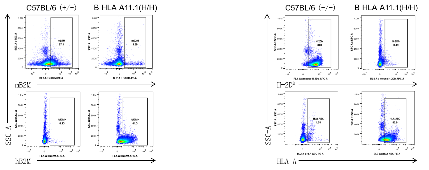
Strain specific B2M and HLA expression analysis in homozygous HLA-A11.1 humanized mice by flow cytometry. Splenocytes from both wild-type C57BL/6 (+/+) and homozygous HLA-A11.1 humanized mice (H/H) were analyzed by flow cytometry. B2M protein expression was analyzed by flow cytometry using species-specific anti-mouse B2M antibody (Biolegend, 154503) and anti-human B2M (Biolegend, 316305). HLA expression was analyzed by flow cytometry using species-specific anti-H-2Db antibody (Biolegend, 114607) and anti-HLA-A,B,C antibody (Biolegend, 311406).
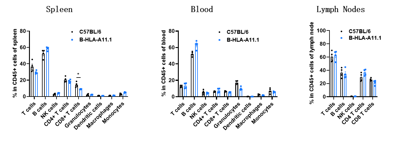
Analysis of leukocyte subpopulations by flow cytometry in immune organs and blood of HLA-A11.1 humanized mice. Splenocytes, peripheral blood, and lymph nodes were isolated from female C57BL/6 and HLA-A11.1 humanized mice (female, 8-week-old, n = 3). Single live cells were gated on the CD45⁺ population and analyzed by flow cytometry as indicated. Values are expressed as mean ± SEM.
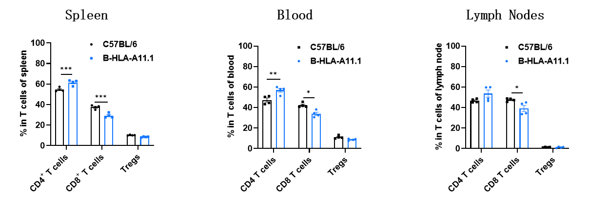
Analysis of T-cell subpopulations by flow cytometry in immune organs and blood of HLA-A11.1 humanized mice. Splenocytes, peripheral blood, and lymph nodes were isolated from female C57BL/6 and HLA-A11.1 humanized mice (female, 8-week-old, n = 3). Single live cells were gated on the CD3⁺ T-cell population and analyzed by flow cytometry as indicated. Values are expressed as mean ± SEM.
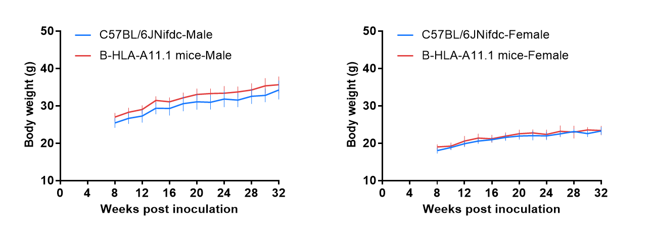
Growth curve of wild-type C57BL/6JNifdc and HLA-A11.1 humanized mice. Eight-week-old wild-type C57BL/6JNifdc and HLA-A11.1 humanized mice were grouped by sex (10 males and 10 females). Body weight was measured on the same day of every two weeks, until 32 weeks. The minimum and maximum body weights shown in the table were calculated as the mean ± SD. The growth curve of HLA-A11.1 humanized mice was similar to that of C57BL/6JNifdc mice.
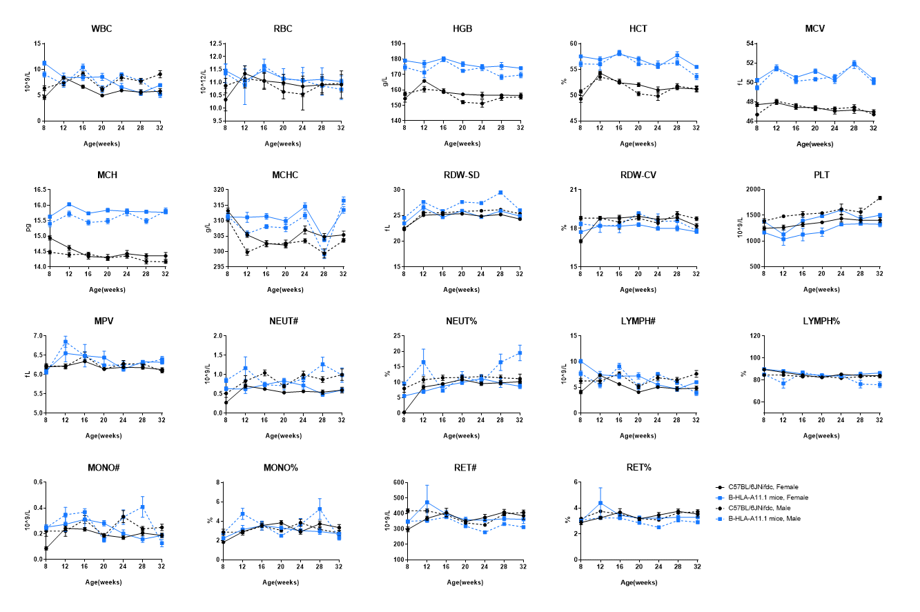
Complete blood count (CBC) of HLA-A11.1 humanized mice. Values are expressed as mean ± SD.
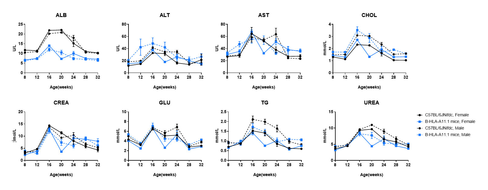
Blood biochemical parameters of HLA-A11.1 humanized mice are shown. Values are expressed as mean ± SD.
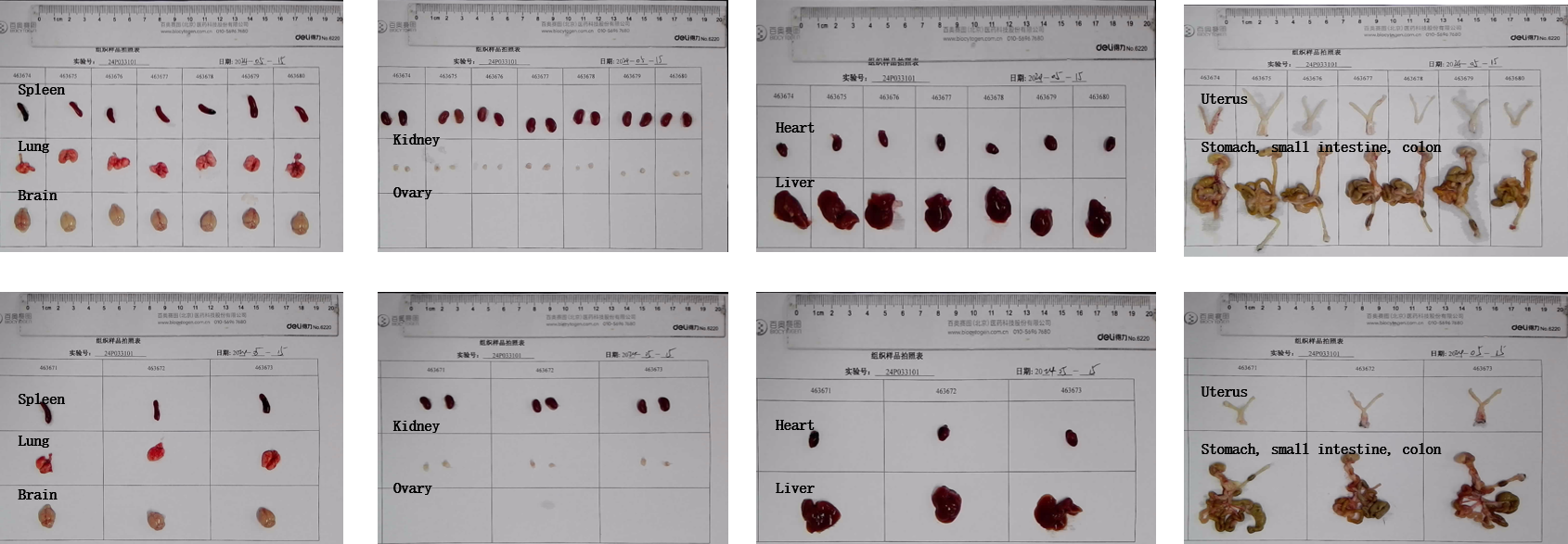
Organs of female HLA-A11.1 humanized mice (8-week-old, n = 10).
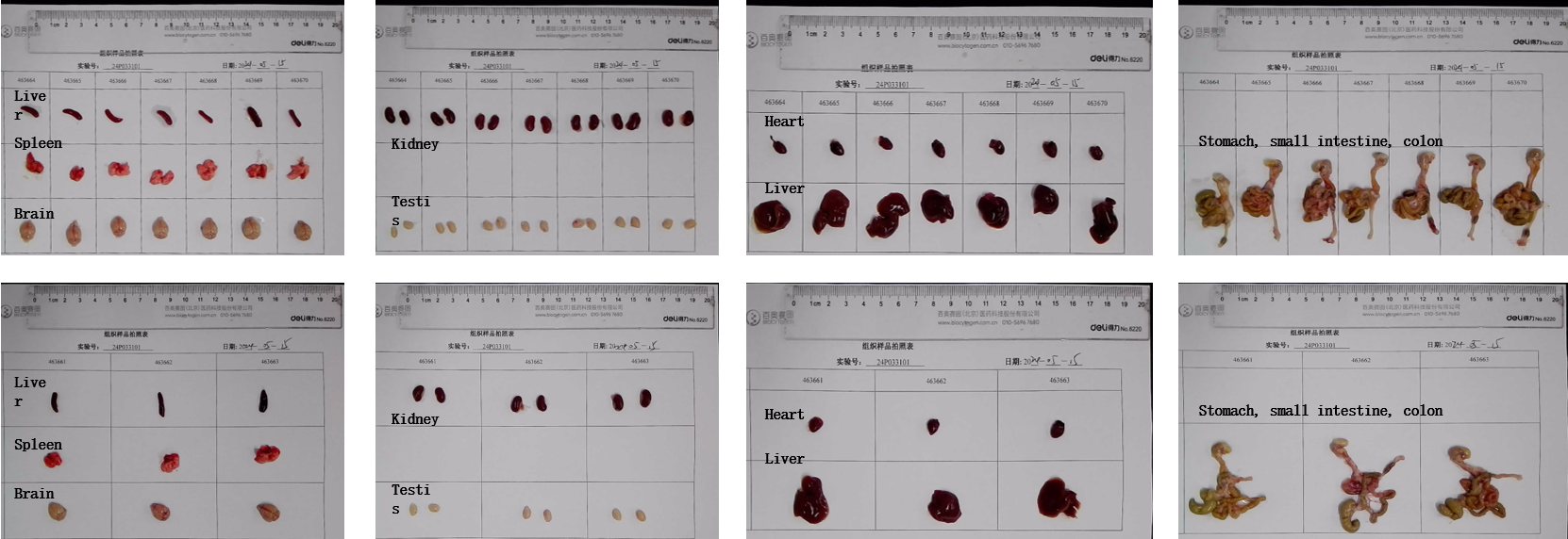
Organs of male HLA-A11.1 humanized mice (8-week-old, n = 10).
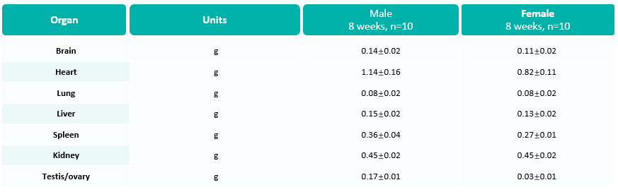
Average weights of major organs in HLA-A11.1 humanized mice. Values are expressed as mean ± SD.
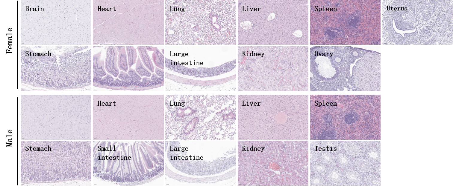
Histopathological analysis of organs in HLA-A11.1 humanized mice. Major organs from HLA-A11.1 humanized mice were collected at 32 weeks of age and analyzed by H&E staining (male, n = 10; female, n = 10).
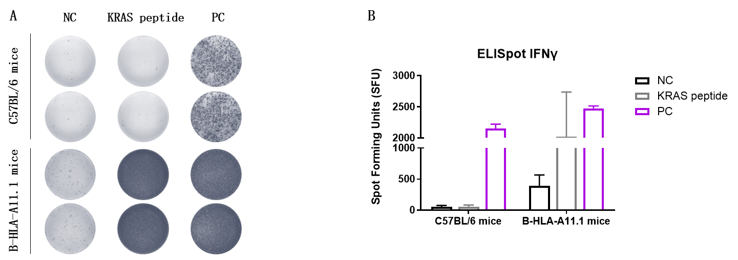
Detection of vaccine-induced immune responses in HLA-A11.1 humanized mice by IFN-γ ELISpot assay. Female HLA-A11.1 humanized mice at the age of 7-8 weeks were divided into PBS group, Group 2 and Group 3 (n = 2), and then inoculated PBS or vaccines at the inside muscle of both legs. Three weeks after the last immunization, mice were sacrificed. The splenocytes were extracted and stimulated with individual peptides or target-unrelated polypeptide as negative control (NC) or anti-CD3 antibody as positive control. Then IFN-γ-secreting cells were subsequently measured using an ELISpot assay. No significant differences in body weight were observed among groups (Data was not shown). (A) Representative results showing stimulation of splenocytes harvested from immunized mice with negative control, or peptide vaccines, or positive control in duplicates. (B) ELISpot Quantification: Statistical summary of IFN-γ-secreting cells.
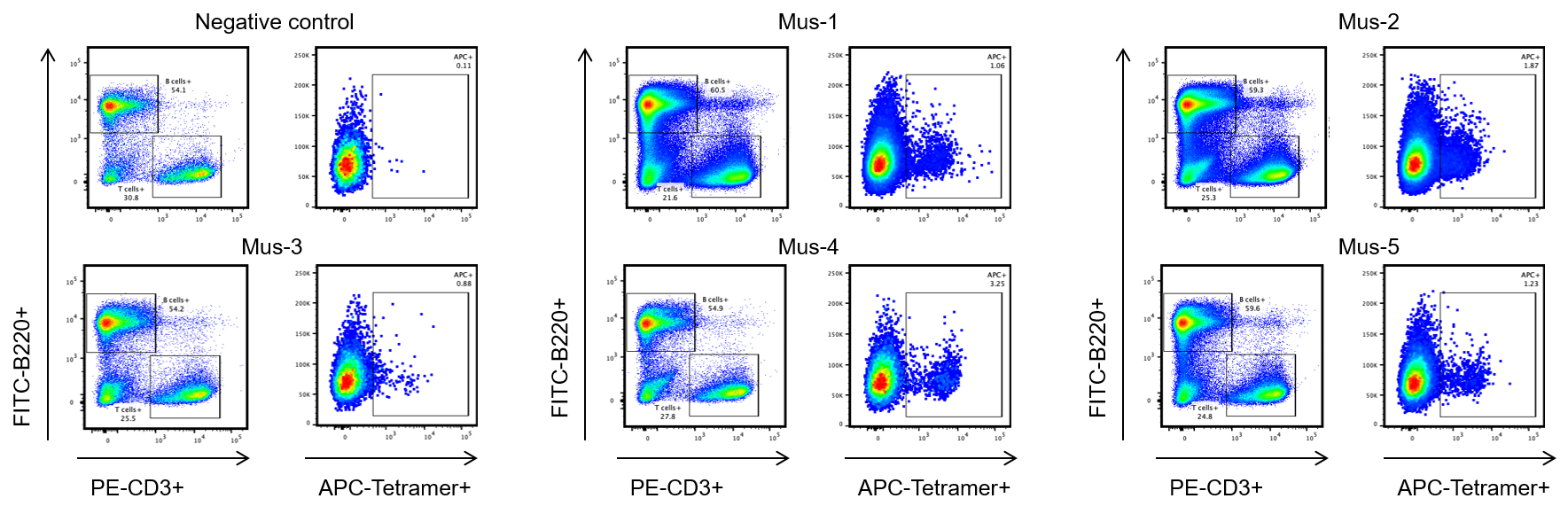
Single-cell isolation of target peptide-specific CD8⁺ T cells from HLA-A11.1 humanized mice. HLA-A11.1 humanized mice were subcutaneously immunized with the target peptide and the spleen cells from five mice displayed specific responses against the target peptide. Spleen cells from five mice (Mus-1, Mus-2, Mus-3, Mus-4 and Mus-5) that showed substantial target peptide specific responses were stained with target peptide/HLA-A*11:01 tetramer, and the tetramer positive CD8+ T cells were sorted by single-cell sorting with flow cytometry. Mice without target peptide immunization were enrolled as the negative control. These results demonstrated that the HLA-A11.1 humanized mice could be used for identifying target peptide specific T-cell receptors (TCRs) and investigating mechanisms of peptide presentation and TCR recognition of cancer targets epitopes in the context of HLA-A*11:01
Note: All results were provided by the client.

Establishment of a B-HLA-A11.1/hKRAS*G12V MC38 model and in vivo efficacy study of an anti-human NY-ESO-1 LNP mRNA in HLA-A11.1 humanized mice. B-HLA-A11.1/hKRAS*G12V MC38 cells were implanted subcutaneously into homozygous HLA-A11.1 humanized mice (female, 8-weeks-old, n=8).

Antitumor activity of KRAS*G12V mRNA vaccine against syngeneic tumors in HLA-A11.1 humanized mice. (A) Tumor growth curves. (B) Body weight changes during treatment. (C) Tumor cells growth of individual mice. These results demonstrate that HLA-A11.1 humanized mice provide a powerful preclinical model for in vivo evaluation of LNP-mRNA vaccines.
The overage of this tumor model is 40%.
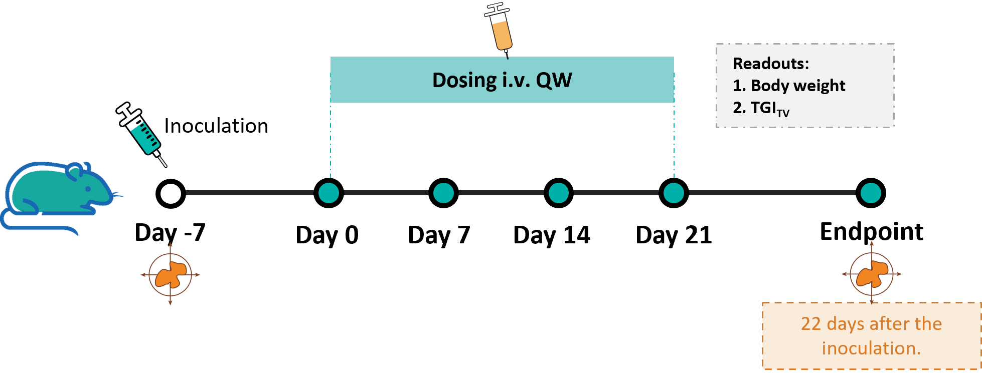
Establishment of a B-HLA-A11.1/hKRAS*G12V MC38 model and in vivo efficacy study of an anti-human KRAS*G12V LNP mRNA in HLA-A11.1 humanized mice. B-HLA-A11.1/hKRAS*G12V MC38 cells were implanted subcutaneously into homozygous HLA-A11.1 humanized mice (female, 8-weeks-old, n=8).
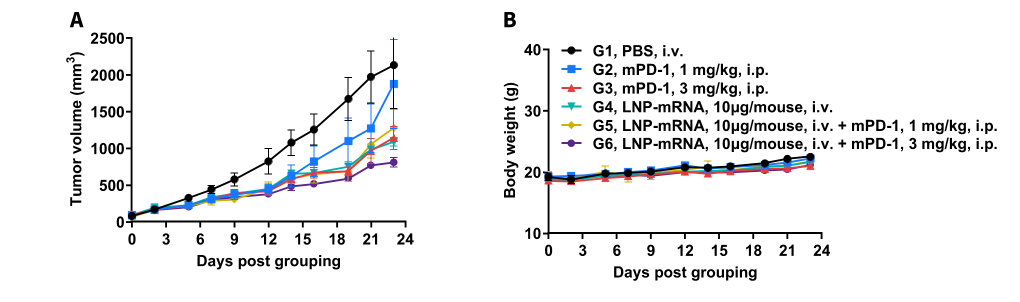
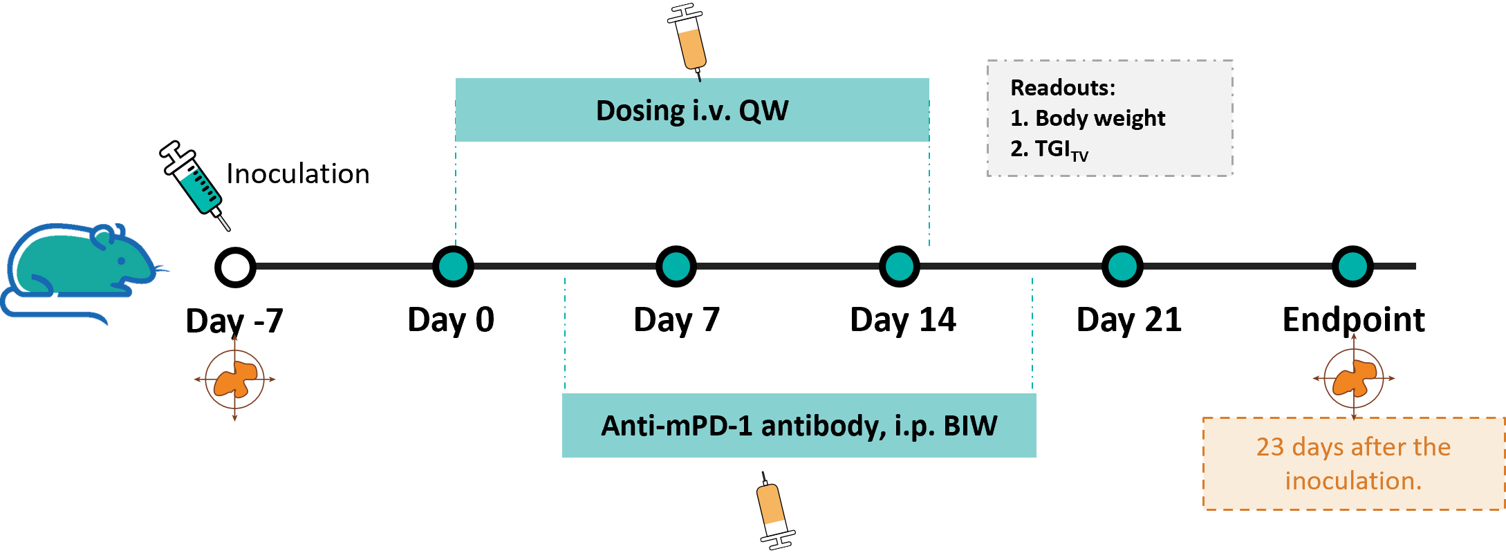
Antitumor activity of anti-mouse PD-1 antibody combined with KRAS*G12V mRNA vaccine against syngeneic tumors in HLA-A11.1 humanized mice. (A) Tumor growth curves. (B) Body weight changes during treatment. These results demonstrate that HLA-A11.1 humanized mice provide a powerful preclinical model for in vivo evaluation of LNP-mRNAvaccines. The overage of this tumor model is 40%.
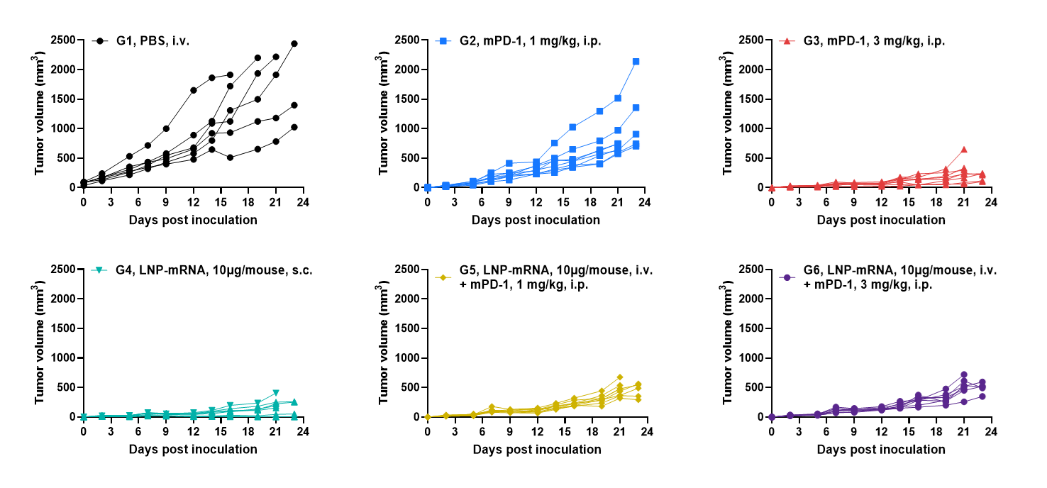
Antitumor activity of anti-mouse PD-1 antibody combined with KRAS*G12V mRNA vaccine against syngeneic tumors in HLA-A11.1 humanized mice. B-HLA-A11.1/hKRAS*G12V MC38 tumor cells growth of individual mice.
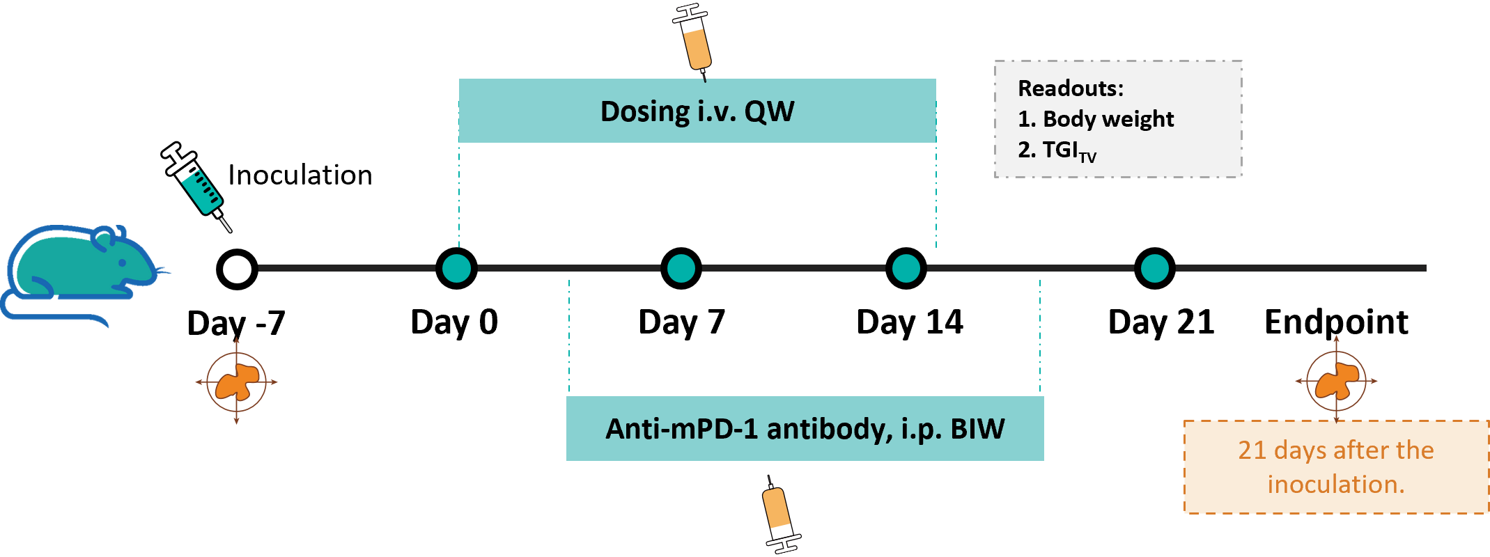
Establishment of a B-HLA-A11.1/hKRAS*G12V MC38 model and in vivo efficacy study of an anti-human KRAS*G12V LNP mRNA in HLA-A11.1 humanized mice. B-HLA-A11.1/hKRAS*G12V MC38 cells were implanted subcutaneously into homozygous HLA-A11.1 humanized mice (female, 8-weeks-old, n=8).
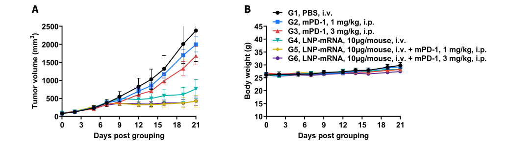
Antitumor activity of anti-mouse PD-1 antibody combined with KRAS*G12V mRNA vaccine against syngeneic tumors in HLA-A11.1 humanized mice. (A) Tumor growth curves. (B) Body weight changes during treatment. These results demonstrate that HLA-A11.1 humanized mice provide a powerful preclinical model for in vivo evaluation of lipid nanoparticle (LNP)-mRNA vaccines. The overage of this tumor model is 40%.
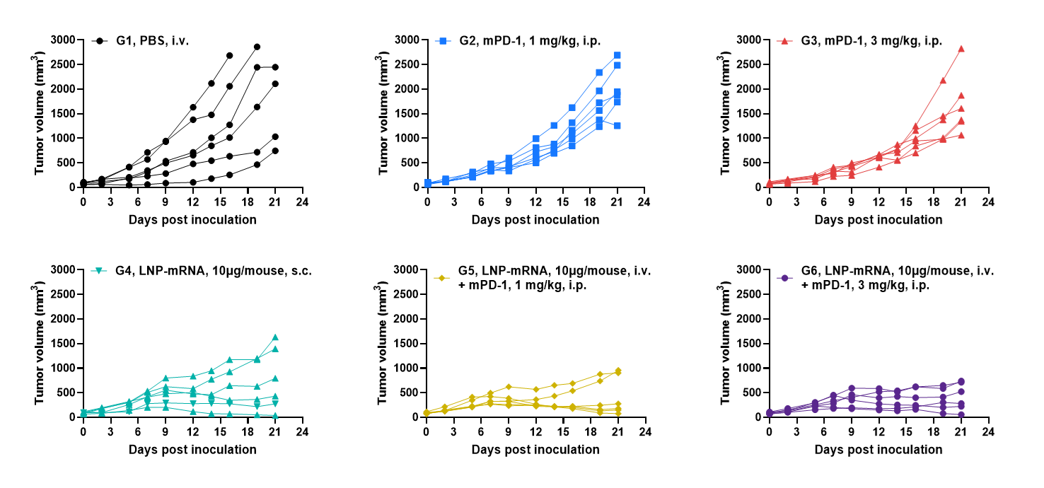
Antitumor activity of anti-mouse PD-1 antibody combined with KRAS*G12V mRNA vaccine against syngeneic tumors in HLA-A11.1 humanized mice. B-HLA-A11.1/hKRAS*G12V MC38 tumor cells growth of individual mice.
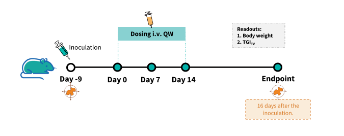
Establishment of a B-HLA-A11.1/hMAGEA3 MC38 model and in vivo efficacy study of an anti-human MAGEA3 LNP-mRNA in HLA-A11.1 humanized mice. B-HLA-A11.1/hKMAGEA3 MC38cells were implanted subcutaneously into homozygous HLA-A11.1 humanized mice (male, 8-weeks-old, n=5).

Antitumor activity of MAGEA3 mRNA vaccine against syngeneic tumors in HLA-A11.1 humanized mice. (A) Tumor growth curves. (B) Body weight changes during treatment. (C) Tumor cells growth of individual mice. These results demonstrate that HLA-A11.1 humanized mice provide a powerful preclinical model for in vivo evaluation of lipid nanoparticle (LNP)-mRNA vaccines.The overage of this tumor model is 40%.

Q1: What are B-HLA-A11.1 mice?
B-HLA-A11.1 mice are a humanized preclinical model. In this model, the mouse B2m gene (exons 1-3) is replaced by a chimeric construct containing the human B2M coding sequence and the α1-α2 domains of the human HLA-A*1101 allele, which are fused to the α3, transmembrane, and cytoplasmic domains of the murine H-2D gene. This enables the expression of the complete human B2M/HLA-A11.1 protein complex on the cell surface, replacing the endogenous mouse MHC I complex.
Q2: Why is the HLA-A11.1 humanized mouse model important for immunotherapy and vaccine research?
The HLA-A*1101 allele is one of the most prevalent HLA class I alleles in Asian populations. This model provides a critical in vivo platform to evaluate HLA-A11.1-restricted immune responses, and is essential for developing and testing personalized cancer vaccines, adoptive T cell therapies (like TCR-T), and other immunotherapies.
Q3: How was the human B2M and HLA-A*1101 gene knocked in?
The targeting strategy involved replacing exons 1-3 of the mouse B2m gene with a sequence containing the human B2M CDS followed by a chimeric gene: the leader sequence, α1, and α2 domains of human HLA-A*1101, linked to the α3, transmembrane, and cytoplasmic domains of the mouse H-2D gene. This design ensures proper folding, surface expression, and function of the humanized MHC I molecule in mouse cells.
Q4: Has the human HLA-A11.1 expression and function been validated in this model?
Yes. Human B2M and HLA-A11.1 protein expression is confirmed on immune cells (e.g., splenocytes) by species-specific flow cytometry. Functional validation is demonstrated through antigen presentation assays, in which immunization with HLA-A11.1–restricted peptides (e.g., a KRAS peptide) induces potent antigen-specific T cell responses.
Q5: What are the main applications of the B-HLA-A11.1 mouse model?
This model is primarily used for: