Programmed death-1 (PD-1) is primarily expressed on T cells and primary B cells, and its ligands PD-L1 and PD-L2 are broadly expressed on antigen-presenting cells (APCs). PD-1 signaling negatively regulates immune responses, and PD-L1 is frequently detected in human tumors, where it can support immune evasion; therefore, blocking PD-1/PD-L1 interaction is a key strategy in cancer immunotherapy.
Validation data confirm the successful generation of PD-1 humanized mice (B-hPD-1 mice plus), demonstrating their suitability as a robust preclinical platform for pharmacodynamic, efficacy, and safety evaluation of anti-human PD-1 therapeutics.
Key Advantages
Validation
Applications
PD-1 humanized mice (B-hPD-1 mice plus) provide an in vivo platform for evaluating anti-human PD-1 therapeutics—including target engagement (e.g., nivolumab/pembrolizumab binding) and antitumor efficacy studies.

PD-1
•A chimeric CDS including human PD-1 gene encoding the signal peptide and extracellular region, mouse PD-1 gene encoding the transmembrane and cytoplasmic region, a stop sequence WPRE were inserted after the initiation codon ATG of mouse PD-1 gene in PD-1 humanized mice (B-hPD-1 mice plus).
•The endogenous mouse promoter and 5′ UTR are retained. The chimeric PD-1 expression is driven by the endogenous mouse PD-1 promoter, while endogenous mouse PD-1 transcription and translation are disrupted.

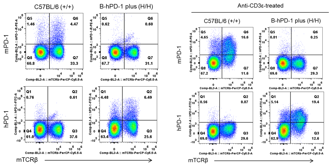
Strain-specific analysis of mouse and human PD-1 protein expression in wild-type C57BL/6 mice and PD-1 humanized mice plus by flow cytometry. Splenocytes were isolated from wild-type C57BL/6 mice (+/+) and homozygous PD-1 humanized mice plus (H/H) and stimulated with or without an anti-mouse CD3ε antibody. Mouse and human PD-1 protein expression on splenocytes was analyzed by flow cytometry using species-specific anti-mouse PD-1 (BioLegend, 109104) and anti-human PD-1 (BioLegend, 329904) antibodies.
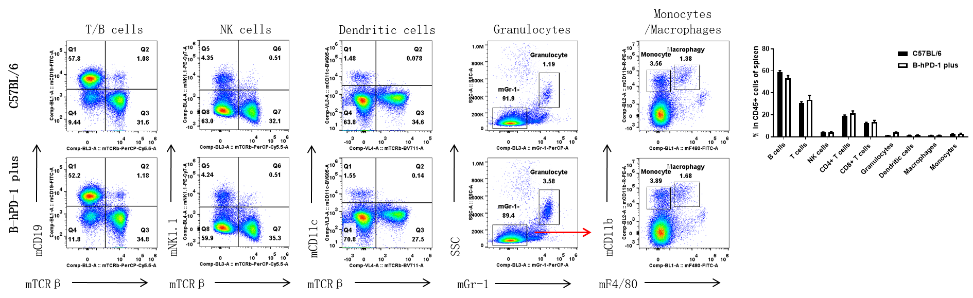
Flow cytometry analysis of splenic leukocyte subpopulations in wild-type C57BL/6 mice and PD-1 humanized mice plus. Splenocytes were collected from female wild-type C57BL/6 mice and PD-1 humanized mice plus (7 weeks old, n = 3). Leukocyte subpopulations were analyzed by flow cytometry. (A) Representative flow cytometry plots. Single live cells were gated on the CD45⁺ population for further analysis as indicated. (B) Quantification of leukocyte subpopulations by flow cytometry. The percentages of T cells, B cells, NK cells, dendritic cells (DCs), granulocytes, monocytes, and macrophages in homozygous PD-1 humanized mice plus were comparable to those in wild-type C57BL/6 controls, indicating that PD-1 humanization did not alter the overall development, differentiation, or distribution of these leukocyte populations in the spleen. Values are expressed as mean ± SEM.
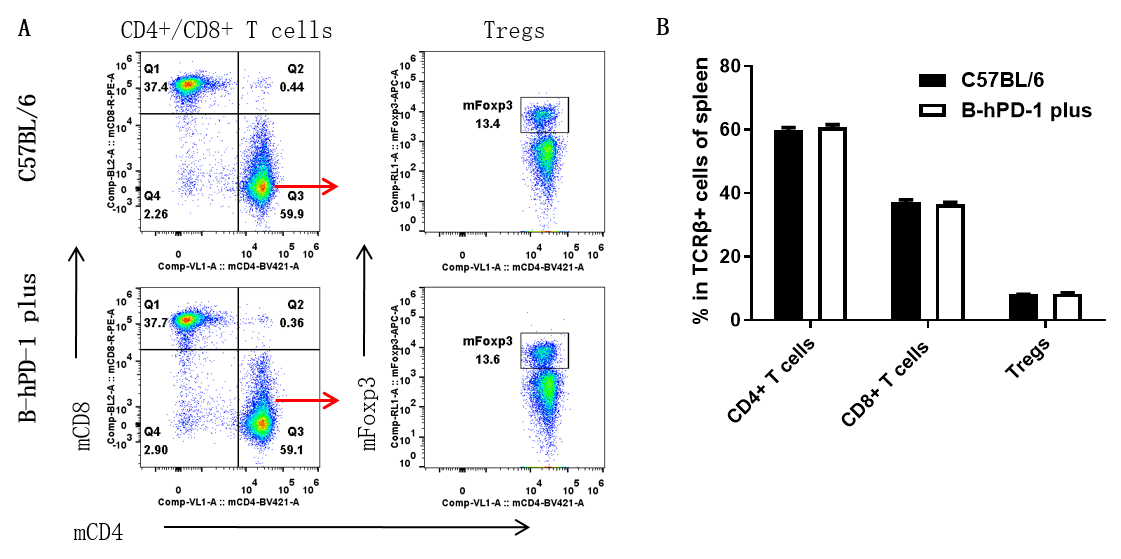
Flow cytometry analysis of splenic T-cell subpopulations in wild-type C57BL/6 mice and PD-1 humanized mice plus. Splenocytes were isolated from female wild-type C57BL/6 mice and PD-1 humanized mice plus (7 weeks old, n = 3). Single live CD45⁺ cells were gated on the TCRβ⁺ T-cell population for further flow cytometry analysis. The percentages of CD4⁺ T cells, CD8⁺ T cells, and regulatory T cells (Tregs) in homozygous PD-1 humanized mice plus were comparable to those in wild-type C57BL/6 controls, indicating that replacement of mouse PD-1 with human PD-1 did not alter the overall development, differentiation, or distribution of these T-cell subpopulations in the spleen. Values are expressed as mean ± SEM.
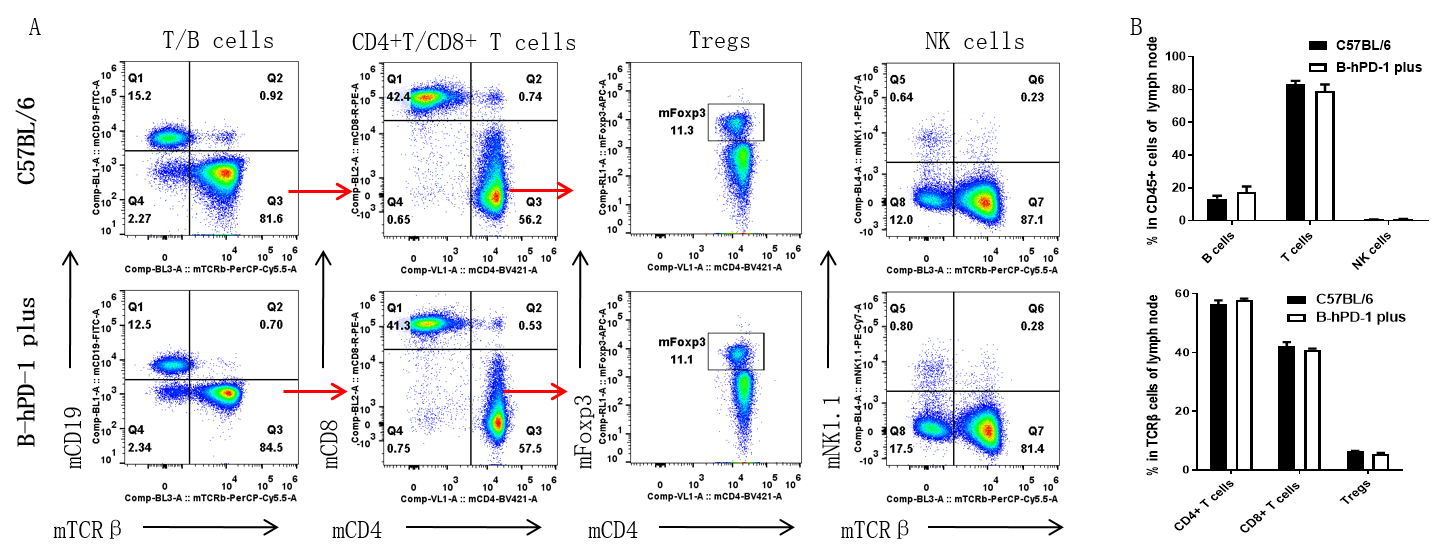
Flow cytometry analysis of leukocyte and T-cell subpopulations in lymph nodes of wild-type C57BL/6 mice and PD-1 humanized mice plus. Lymph nodes were collected from female wild-type C57BL/6 mice and PD-1 humanized mice plus (7 weeks old, n = 3). Single live cells were gated on the CD45⁺ population for further flow cytometry analysis. The percentages of T cells, B cells, CD4⁺ T cells, CD8⁺ T cells, regulatory T cells (Tregs), and NK cells in homozygous PD-1 humanized mice plus were comparable to those in wild-type C57BL/6 controls, indicating that PD-1 humanization did not alter the overall development, differentiation, or distribution of these immune cell populations in the lymph nodes. Values are expressed as mean ± SEM.

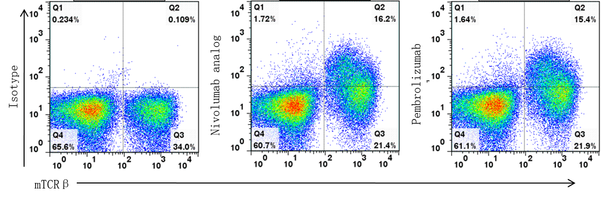
Flow cytometry analysis of human PD-1 expression and anti-human PD-1 antibody binding on T cells from PD-1 humanized mice plus. Splenocytes were isolated from female PD-1 humanized mice plus and analyzed for human PD-1 expression on T cells by flow cytometry. Single live cells were gated on the CD45⁺ population for further analysis. Human PD-1 expression was detected on T cells from PD-1 humanized mice plus, as demonstrated by the binding of in-house nivolumab and pembrolizumab analogs compared with the isotype control.
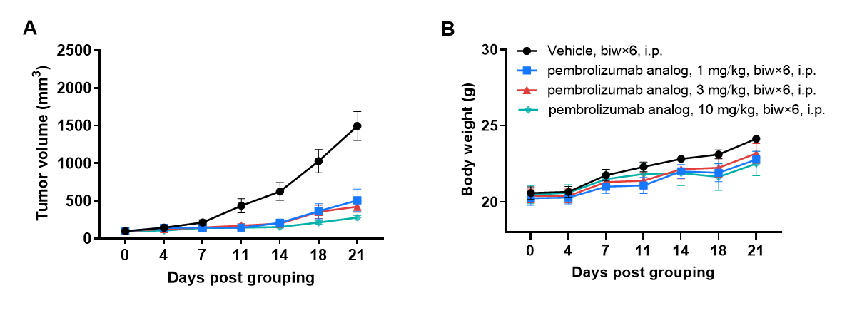
In vivo antitumor efficacy of an anti-human PD-1 antibody (pembrolizumab analog) in a subcutaneous MC38 tumor model using PD-1 humanized mice plus. (A) Tumor growth curves. Murine colon cancer MC38 cells (5 × 10⁵) were subcutaneously implanted into homozygous PD-1 humanized mice plus (female, 7 weeks old, n = 5). When tumor volumes reached approximately 150 mm³, mice were assigned to treatment groups and administered an in-house anti-human PD-1 antibody (pembrolizumab analog) according to the doses and schedules indicated in the panel. (B) Body weight changes during treatment. As shown in panel A, treatment with the pembrolizumab analog effectively inhibited MC38 tumor growth in PD-1 humanized mice plus, demonstrating the utility of this model for evaluating the in vivo antitumor efficacy of anti-human PD-1 antibodies. Values are expressed as mean ± SEM.
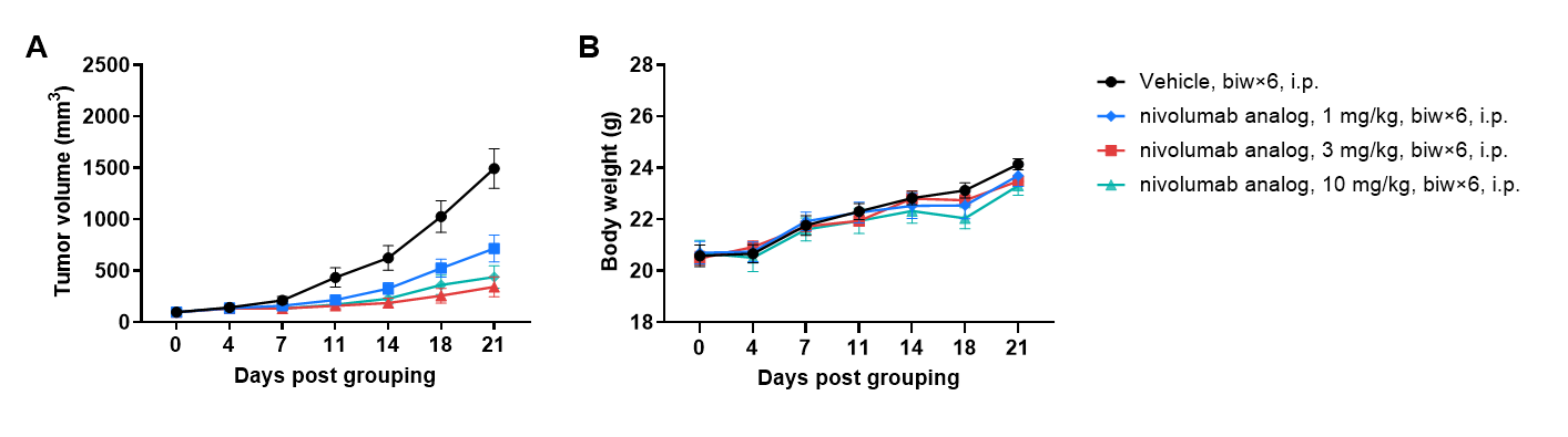
In vivo antitumor efficacy of an anti-human PD-1 antibody (nivolumab analog) in a subcutaneous MC38 tumor model using PD-1 humanized mice plus. (A) Tumor growth curves. Murine colon cancer MC38 cells (5 × 10⁵) were subcutaneously implanted into homozygous PD-1 humanized mice plus (female, 7 weeks old, n = 5). When tumor volumes reached approximately 150 mm³, mice were assigned to treatment groups and administered an in-house anti-human PD-1 antibody (nivolumab analog) according to the doses and schedules indicated in the panel. (B) Body weight changes during treatment. As shown in panel A, treatment with the nivolumab analog effectively inhibited MC38 tumor growth in PD-1 humanized mice plus, demonstrating the utility of this model for evaluating the in vivo antitumor efficacy of anti-human PD-1 antibodies. Values are expressed as mean ± SEM.