

SIRPA: A key immune checkpoints in macrophages phagocytosis and its therapeutic intervention
EGFR: A critical receptor tyrosine kinase in cancer signaling and its therapeutic intervention
Gene Information: The EGFR gene (located on chromosome 7p11.2) encodes the epidermal growth factor receptor (ErbB1/HER1), a 170-kDa transmembrane receptor tyrosine kinase that is frequently altered in human cancers via gene amplification, activating mutations (e.g., exon 19 deletions, L858R), or in-frame insertions.
Protein Expression: EGFR is physiologically localized to the plasma membrane of epithelial and stromal tissues, but it is pathologically overexpressed in a wide spectrum of human solid malignancies, including non-small cell lung cancer (NSCLC), colorectal cancer (CRC), head and neck squamous cell carcinoma (HNSCC), and glioblastoma.
Signaling Pathway: Phosphorylated EGFR activates downstream MAPK, PI3K-AKT, and STAT cascades to govern tumor cell proliferation, survival, and metastasis.
Therapeutic Inhibition: In this triple-humanized model, co-targeting EGFR alongside the CD47–SIRPA checkpoint dismantles tumor "don't eat me" signaling to unleash robust, macrophage-mediated phagocytosis (ADCP); moreover, it provides a cross-species-matched platform to evaluate EGFR × CD47 bispecific antibodies that selectively concentrate CD47 blockade at EGFR-rich tumor sites while minimizing on-target hematologic toxicity.

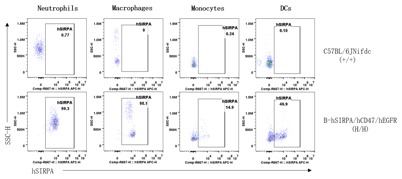
Strain specific SIRPA expression analysis in wild-type C57BL/6JNifdc mice and homozygous SIRPA/CD47/EGFR humanized mice by flow cytometry. Splenocytes were collected from wild-type C57BL/6JNifdc mice (+/+) and homozygous B-hSIRPA/hCD47/hEGFR mice (H/H) and analyzed by flow cytometry with anti-human SIRPA antibody (Biolegend, 323810).
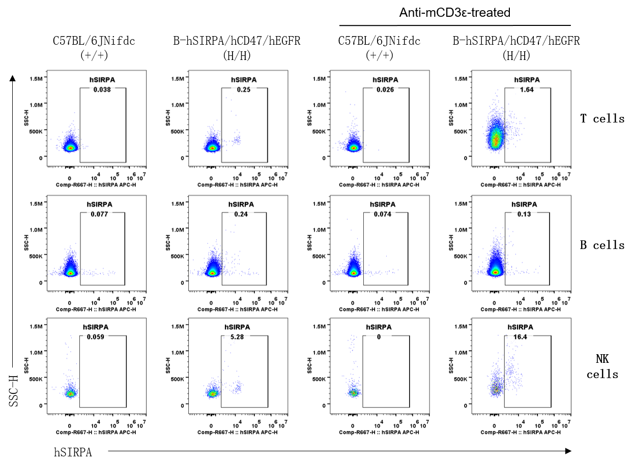
Strain specific SIRPA expression analysis in wild-type C57BL/6JNifdc mice and homozygous SIRPA/CD47/EGFR humanized mice by flow cytometry. Splenocytes were collected from wild-type C57BL/6JNifdc mice (+/+) (female, n=2, 6-week-old) and homozygous B-hSIRPA/hCD47/hEGFR mice (H/H) (female, 6-week-old, n=2,) unstimulated or stimulated with anti-mCD3ε antibody (Bioxcell, BE0001-1) in vivo (7.5 μg/mice, stimulation for 24 hours, i.p.). Protein expression was analyzed with anti-human SIRPA antibody (Biolegend, 323810) by flow cytometry.
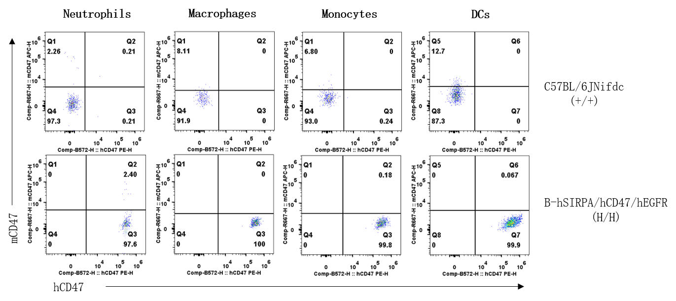
Strain specific CD47 expression analysis in wild-type C57BL/6JNifdc mice and homozygous SIRPA/CD47/EGFR humanized mice by flow cytometry. Splenocytes were collected from wild-type C57BL/6JNifdc mice (+/+) and homozygous B-hSIRPA/hCD47/hEGFR mice (H/H). Protein expression was analyzed with anti-mouse CD47 antibody (Biolegend, 127514) and anti-human CD47 antibody (Biolegend, 323108) by flow cytometry.
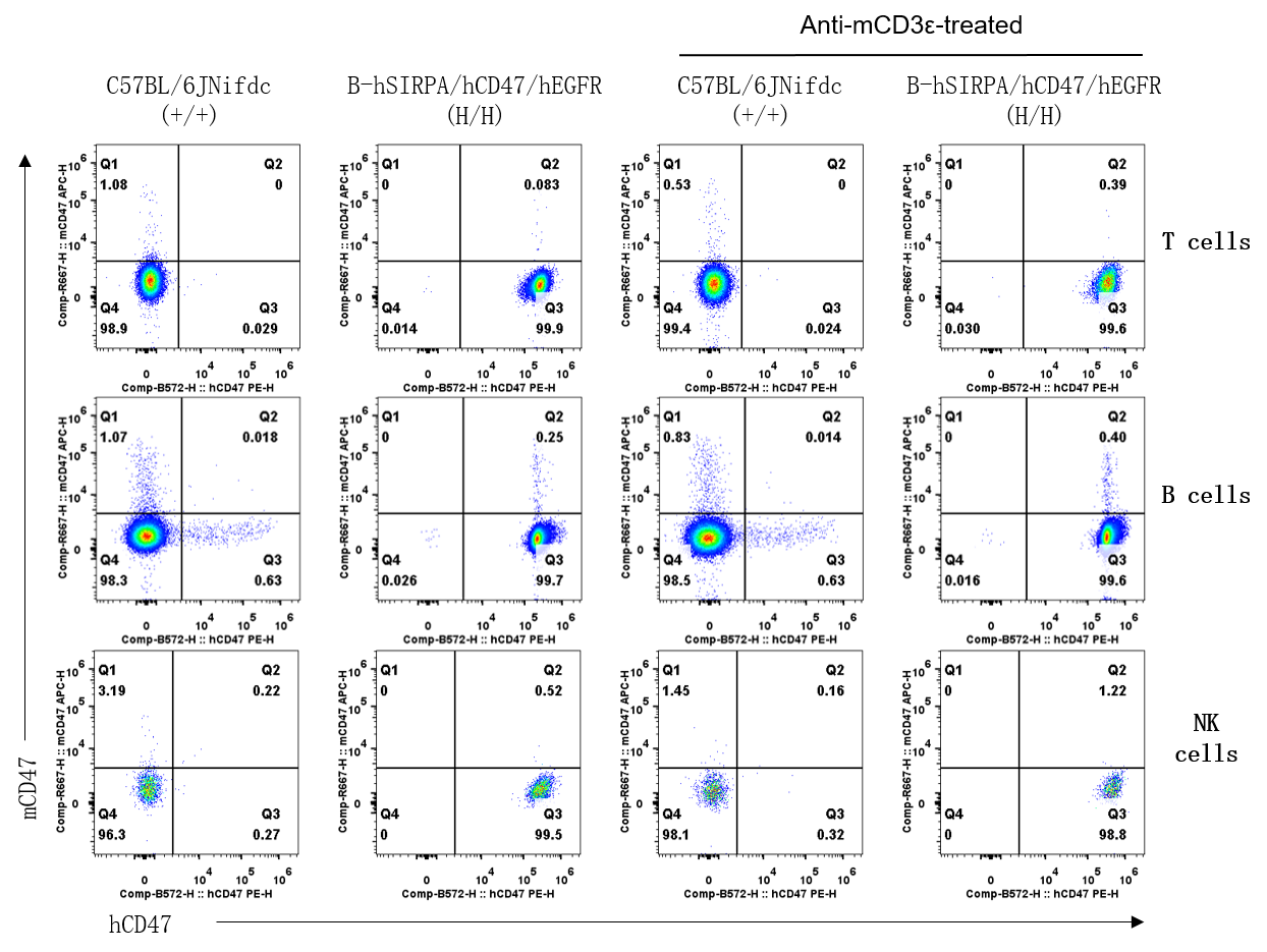
Strain specific CD47 expression analysis in wild-type C57BL/6JNifdc mice and homozygous SIRPA/CD47/EGFR humanized mice by flow cytometry. Splenocytes were collected from wild-type C57BL/6JNifdc mice (+/+) (female, 6-week-old, n=2) and homozygous B-hSIRPA/hCD47/hEGFR mice (H/H) (female, 6-week-old, n=2) unstimulated or stimulated with anti-mCD3ε antibody (Bioxcell, BE0001-1) in vivo (7.5 μg/mice, stimulation for 24 hours, i.p.). Protein expression was analyzed with anti-mouse CD47 antibody (Biolegend, 127514) and anti-human CD47 antibody (Biolegend, 323108) by flow cytometry.
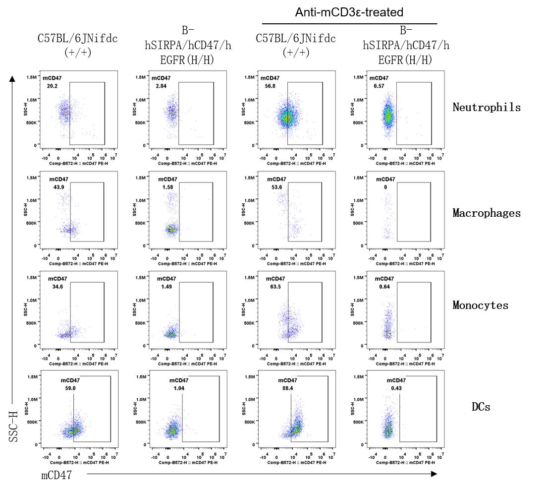
Strain specific CD47 expression analysis in wild-type C57BL/6JNifdc mice and homozygous SIRPA/CD47/EGFR humanized mice by flow cytometry. Splenocytes were collected from wild-type C57BL/6JNifdc mice (+/+) (female, 6-week-old, n=2) and homozygous B-hSIRPA/hCD47/hEGFR mice (H/H) (female, 6-week-old, n=2) unstimulated or stimulated with anti-mCD3ε antibody (Bioxcell, BE0001-1) in vivo (7.5 μg/mice, stimulation for 24 hours, i.p.). Protein expression was analyzed with anti-mouse CD47 antibody (Biolegend, 127508) by flow cytometry.
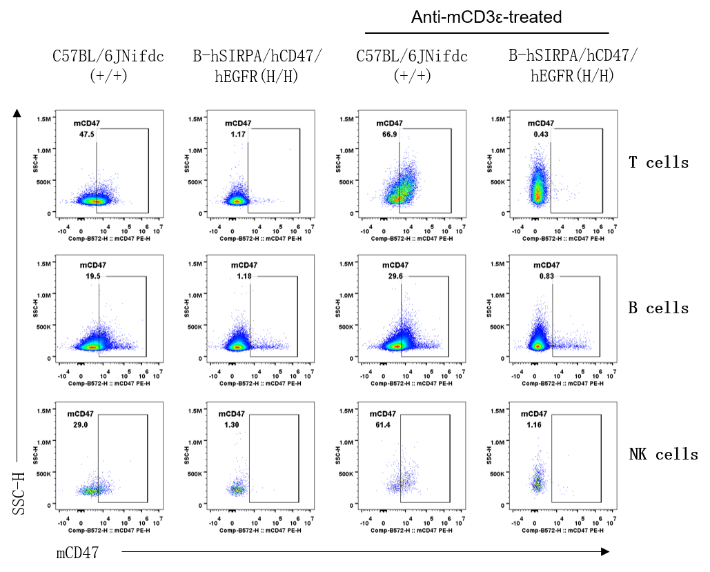
Strain specific CD47 expression analysis in wild-type C57BL/6JNifdc mice and homozygous SIRPA/CD47/EGFR humanized mice by flow cytometry. Splenocytes were collected from wild-type C57BL/6JNifdc mice (+/+) (female, 6-week-old, n=2) and homozygous B-hSIRPA/hCD47/hEGFR mice (H/H) (female, 6-week-old, n=2) unstimulated or stimulated with anti-mCD3ε antibody (Bioxcell, BE0001-1) in vivo (7.5 μg/mice, stimulation for 24 hours, i.p.). Protein expression was analyzed with anti-mouse CD47 antibody (Biolegend, 127508) by flow cytometry.
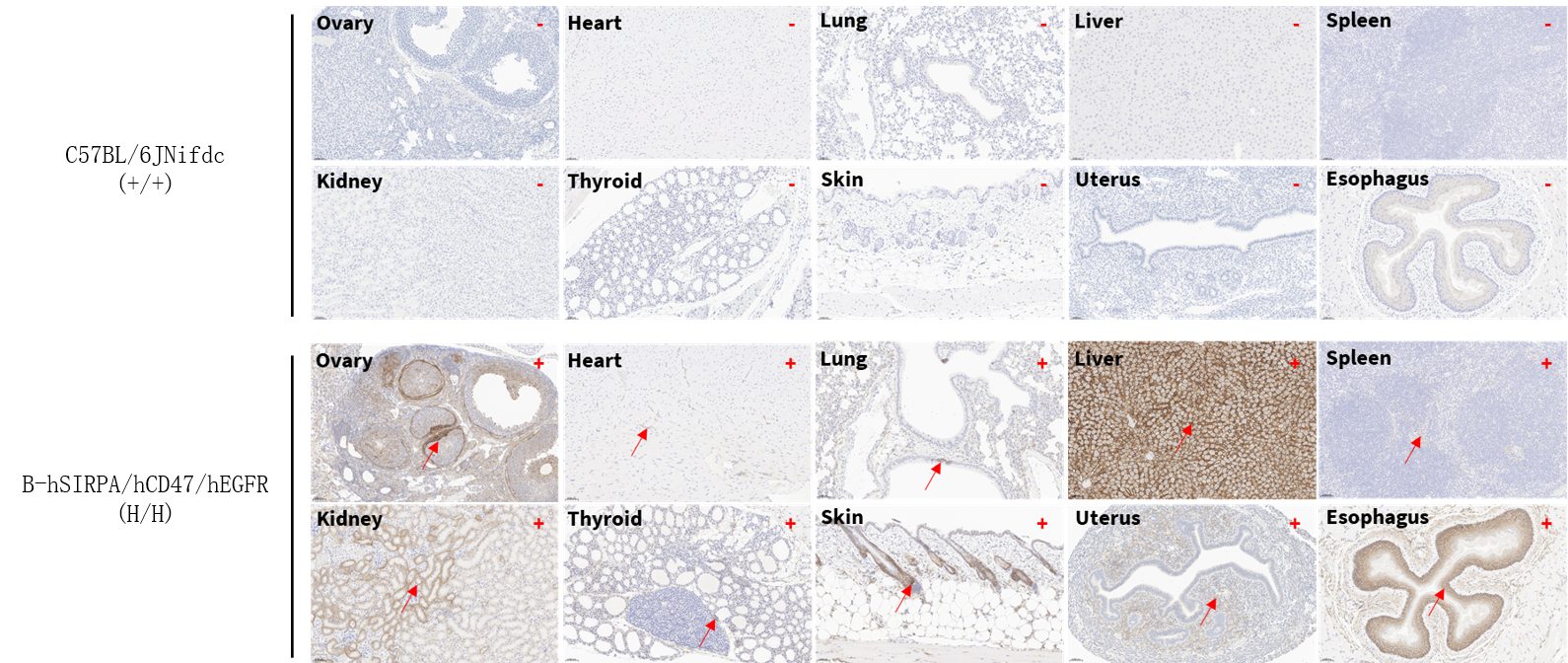
Immunohistochemical (IHC) analysis of EGFR protein expression in homozygous SIRPA/CD47/EGFR humanized mice. Various tissue were collected from wild-type C57BL/6JNifdc mice (+/+) and homozygous B-hSIRPA/hCD47/hEGFR mice (H/H), and then analyzed by IHC with anti-EGFR antibody (Invitrogen, MA5-49312). Red arrows indicate cells with positive EGFR staining (brown). '+' and '-' indicate positive and negative staining, respectively.