

Inhibitory LILRBs (LILRB1–4): Leukocyte immunoglobulin-like receptors and targets for tumor microenvironment reprogramming


Strain specific human LILRB1 expression analysis in LILRB2/LILRB3/LILRB1/LILRB4 humanized transgenic mice by flow cytometry. Blood cells were collected from wild-type C57BL/6 mice (+/+) and transgenic B-Tg(hLILRB2/hLILRB3/hLILRB1/hLILRB4) mice (Tg). Human LILRB1 expression was analyzed by flow cytometry using anti-human LILRB1 antibody (eBioscience™, 17-5129-41).

Strain specific hLILRB1 expression analysis in LILRB2/LILRB3/LILRB1/LILRB4 humanized transgenic mice by flow cytometry. Blood cells were collected from wild-type C57BL/6 mice (+/+) and transgenic B-Tg(hLILRB2/hLILRB3/hLILRB1/hLILRB4) mice (Tg). Human LILRB1 expression was analyzed by flow cytometry using anti-human LILRB1 antibody (eBioscience™, 17-5129-41).

Strain specific hLILRB2 expression analysis in LILRB2/LILRB3/LILRB1/LILRB4 humanized transgenic mice by flow cytometry. Blood cells were collected from wild-type C57BL/6 mice (+/+) and transgenic B-Tg(hLILRB2/hLILRB3/hLILRB1/hLILRB4) mice (Tg). Human LILRB2 expression was analyzed by flow cytometry using anti-human LILRB2 antibody (R&D, FAB2078P).

Strain specific hLILRB2 expression analysis in LILRB2/LILRB3/LILRB1/LILRB4 humanized transgenic mice by flow cytometry. Blood cells were collected from wild-type C57BL/6 mice (+/+) and transgenic B-Tg(hLILRB2/hLILRB3/hLILRB1/hLILRB4) mice (Tg). Human LILRB2 expression was analyzed by flow cytometry using anti-human LILRB2 antibody (R&D, FAB2078P).

Strain specific hLILRB3 expression analysis in LILRB2/LILRB3/LILRB1/LILRB4 humanized transgenic mice by flow cytometry. Blood cells were collected from wild-type C57BL/6 mice (+/+) and transgenic B-Tg(hLILRB2/hLILRB3/hLILRB1/hLILRB4) mice (Tg). Human LILRB3 expression was analyzed by flow cytometry using anti-human LILRB3 antibody (R&D, FAB1806P-100).

Strain specific hLILRB3 expression analysis in LILRB2/LILRB3/LILRB1/LILRB4 humanized transgenic mice by flow cytometry. Blood cells were collected from wild-type C57BL/6 mice (+/+) and transgenic B-Tg(hLILRB2/hLILRB3/hLILRB1/hLILRB4) mice (Tg). Human LILRB3 expression was analyzed by flow cytometry using anti-human LILRB3 antibody (R&D, FAB1806P-100).

Strain specific hLILRB4 expression analysis in LILRB2/LILRB3/LILRB1/LILRB4 humanized transgenic mice by flow cytometry. Blood cells were collected from wild-type C57BL/6 mice (+/+) and transgenic B-Tg(hLILRB2/hLILRB3/hLILRB1/hLILRB4) mice (Tg). Human LILRB4 expression was analyzed by flow cytometry using anti-human LILRB4 antibody (R&D, FAB24251P).

Strain specific hLILRB4 expression analysis in LILRB2/LILRB3/LILRB1/LILRB4 humanized transgenic mice by flow cytometry. Blood cells were collected from wild-type C57BL/6 mice (+/+) and transgenic B-Tg(hLILRB2/hLILRB3/hLILRB1/hLILRB4) mice (Tg). Human LILRB4 expression was analyzed by flow cytometry using anti-human LILRB4 antibody (R&D, FAB24251P).
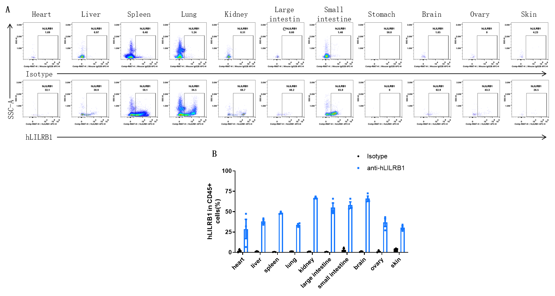
Strain specific hLILRB1 expression analysis in LILRB2/LILRB3/LILRB1/LILRB4 humanized transgenic mice by flow cytometry. Heart, liver, spleen, lung, kidney, large intestine, small intestine, stomach, brain, ovary, and skin were collected from transgenic B-Tg(hLILRB1/hLILRB2/hLILRB3/hLILRB4) mice (female, 8-week-old, n=3). Human LILRB1 expression was analyzed by flow cytometry using anti-human LILRB1 antibody (eBioscience™, 17-5129-41). (A). Representative FACS plots. (B). Results of FACS analysis. Values are expressed as mean ± SEM.
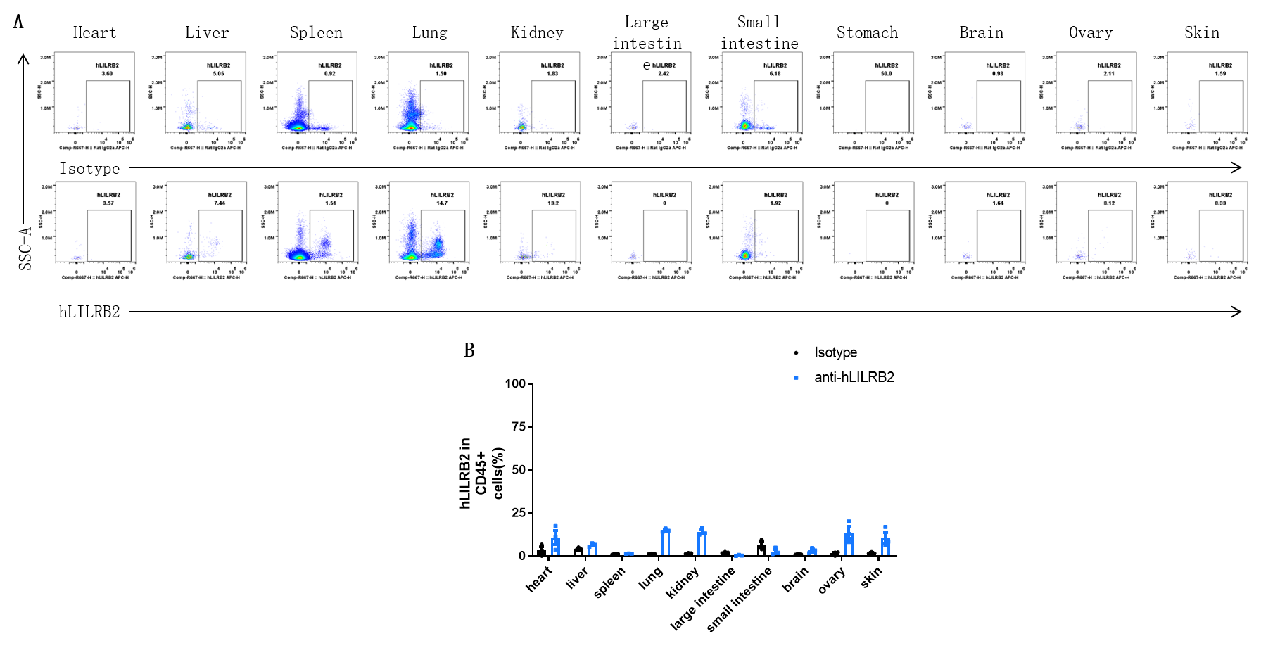
Strain specific hLILRB2 expression analysis in LILRB2/LILRB3/LILRB1/LILRB4 humanized transgenic mice by flow cytometry. Heart, liver, spleen, lung, kidney, large intestine, small intestine, stomach, brain, ovary, and skin were collected from transgenic B-Tg(hLILRB1/hLILRB2/hLILRB3/hLILRB4) mice (female, 8-week-old, n=3). Human LILRB2 expression was analyzed by flow cytometry using anti-human LILRB2 antibody (R&D, FAB2078P). (A). Representative FACS plots. (B). Results of FACS analysis. Values are expressed as mean ± SEM.
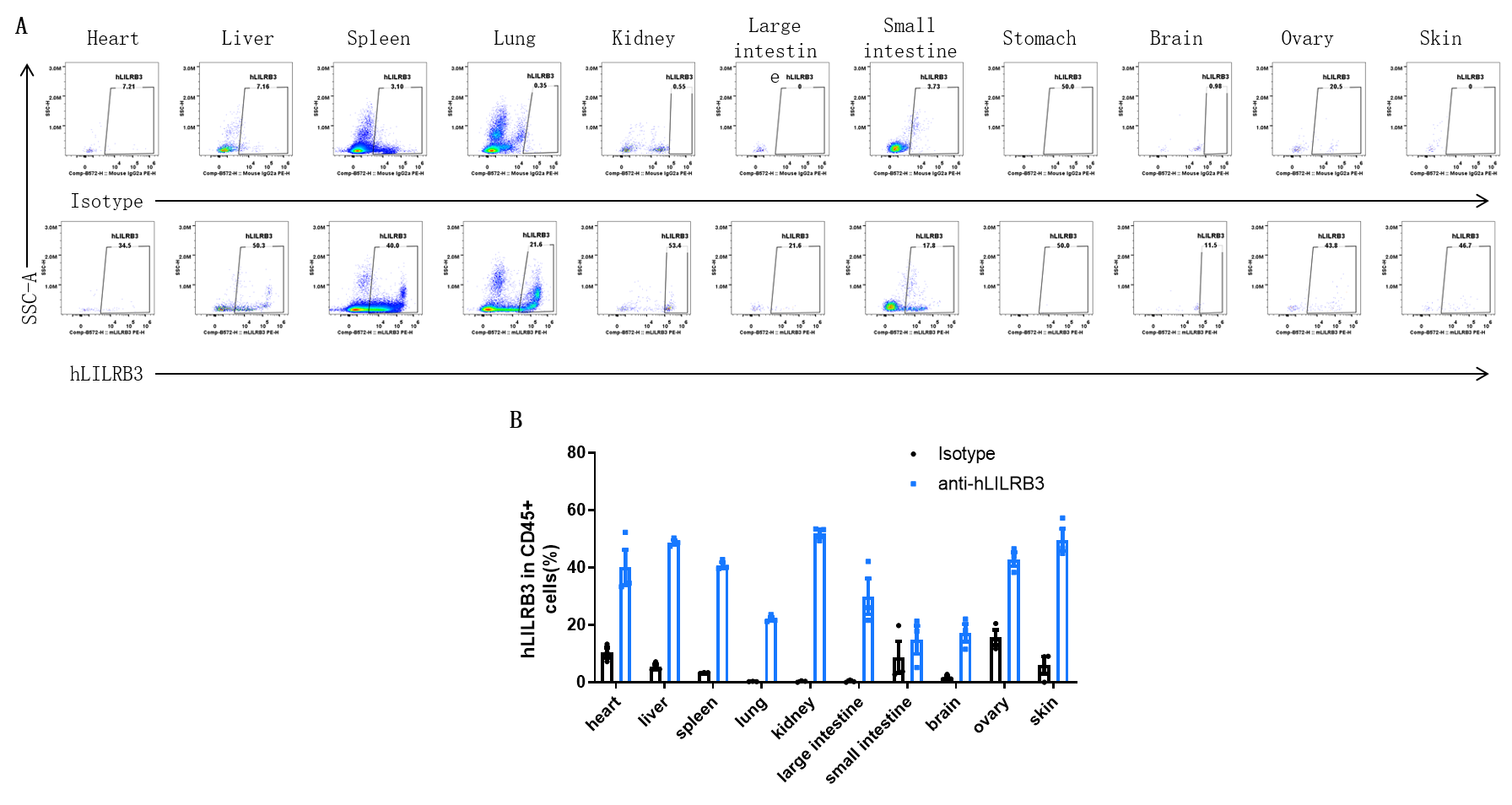
Strain specific hLILRB3 expression analysis in LILRB2/LILRB3/LILRB1/LILRB4 humanized transgenic mice by flow cytometry. Heart, liver, spleen, lung, kidney, large intestine, small intestine, stomach, brain, ovary, and skin were collected from transgenic B-Tg(hLILRB1/hLILRB2/hLILRB3/hLILRB4) mice. Human LILRB3 expression was analyzed by flow cytometry using anti-human LILRB3 antibody (R&D, FAB1806P-100). (A). Representative FACS plots. (B). Results of FACS analysis. Values are expressed as mean ± SEM.

Strain specific hLILRB4 expression analysis in LILRB2/LILRB3/LILRB1/LILRB4 humanized transgenic mice by flow cytometry. Heart, liver, spleen, lung, kidney, large intestine, small intestine, stomach, brain, ovary, and skin were collected from transgenic B-Tg(hLILRB1/hLILRB2/hLILRB3/hLILRB4) mice. Human LILRB4 expression was analyzed by flow cytometry using anti-human LILRB3 antibody (R&D, FAB24251P). (A). Representative FACS plots. (B). Results of FACS analysis. Values are expressed as mean ± SEM.
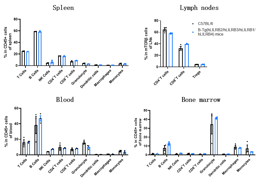
Analysis of leukocyte subpopulations by flow cytometry in immune organs and blood. Splenocytes, lymph nodes, peripheral blood, and bone marrow were isolated from wild-type C57BL/6 mice and B-Tg(hLILRB2/hLILRB3/hLILRB1/hLILRB4) mice (female, 8-week-old, n=3). Single live cells were gated on the CD45⁺ population and analyzed by flow cytometry as indicated. Values are expressed as mean ± SEM.
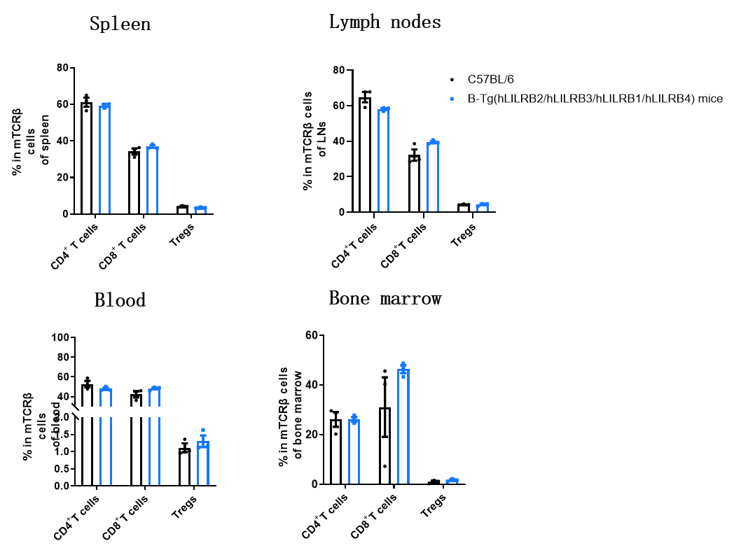
Analysis of T-cell subpopulations by flow cytometry in immune organs and blood. Splenocytes, lymph nodes, peripheral blood, and bone marrow were isolated from wild-type C57BL/6 mice and B-Tg(hLILRB2/hLILRB3/hLILRB1/hLILRB4) mice (female, 8-week-old, n=3). Single live cells were gated on the CD45⁺ CD3⁺ T cellspopulation and analyzed by flow cytometry as indicated. Values are expressed as mean ± SEM.

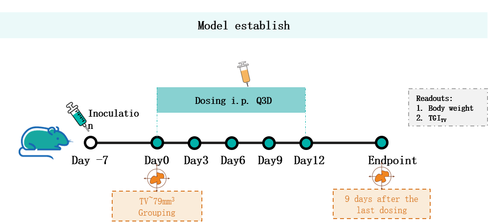
Establishment of a B-hB2M/HLA-G MC38 model and in vivo efficacy study of anti-HLA-G antibodies. B-hB2M/HLA-G MC38 cells were inoculated subcutaneously into transgenic B-Tg(hLILRB2/hLILRB3/hLILRB1/hLILRB4) mice (female, 6-8 weeks old, n = 4). When the average tumor volume reached approximately 79 mm³, mice were randomized and subsequently administered the anti-HLA-G antibodies via intraperitoneal injection.
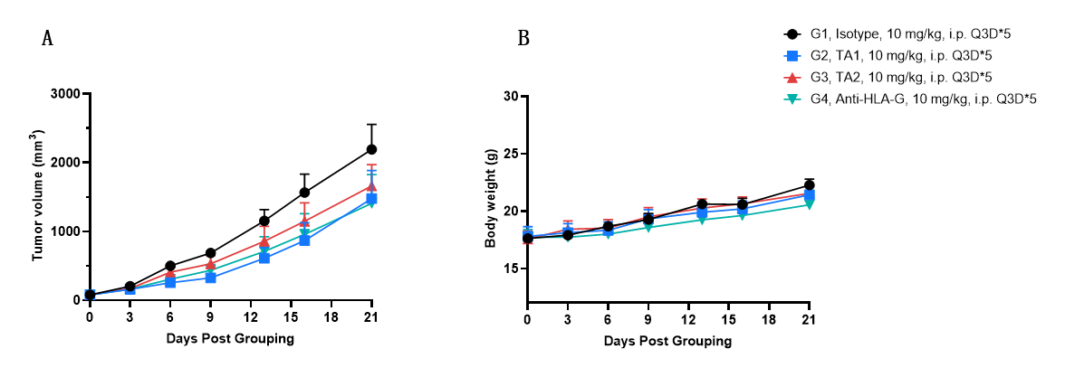
Antitumor activity of anti-HLA-G antibodies in LILRB2/LILRB3/LILRB1/LILRB4 humanized transgenic mice. (A) Anti-human HLA-G antibodies inhibited MC38 tumor growth in B-Tg(hLILRB2/hLILRB3/hLILRB1/hLILRB4) mice. Murine colon cancer B-hB2M/HLA-G MC38 cells (5 × 10⁵) were subcutaneously implanted into B-Tg(hLILRB2/hLILRB3/hLILRB1/hLILRB4) mice (female, 6-7 weeks old, n = 4). Tumor-bearing animals were randomly enrolled into four study groups when the mean tumor size reached 79 mm3, at which time they were intraperitoneally injected with anti-HLA-G antibodies (provided by a client) indicated in the panel. (B) Body weight changes during treatment. As shown in panel A, the anti-HLA-G antibodies inhibited tumor growth in B-Tg(hLILRB2/hLILRB3/hLILRB1/hLILRB4) mice. Values are expressed as mean ± SEM.

Q1: How were LILRB2/LILRB3/LILRB1/LILRB4 humanized transgenic mice generated/developed?
A1: The LILRB2/LILRB3/LILRB1/LILRB4 humanized transgenic mice was obtained by mating LILRB2/LILRB3 humanized transgenic mice and LILRB1/LILRB4 humanized transgenic mice.
Q2: What were the differences of LIRB1-4 protein expression between LILRB2/LILRB3/LILRB1/LILRB4 humanized transgenic mice and humans?
A2: Protein expression in different immune cells: LILRB1 leaks on neutrophils; LILRB2 expression profile is consistent with human; LILRB3 leaks on NK; LILRB4 leaks on B cells.