Description
- Gene Information: PD‑1 is encoded by the PDCD1 gene on chromosome 2q37.3, while PD‑L1 is encoded by CD274 on chromosome 9p24.1. Both are type I transmembrane proteins; PD‑1 belongs to the immunoglobulin superfamily and functions as an inhibitory receptor, whereas PD‑L1 is a member of the B7 family of co‑stimulatory molecules.
- Protein Expression: PD-1 is primarily expressed on activated T cells, B cells, and NK cells, whereas PD-L1 is expressed on antigen-presenting cells and many tumor cells, with expression often induced by IFN-γ.
- Signaling Pathway: Binding of PD-L1 to PD-1 recruits SHP2 phosphatase and suppresses TCR/CD28-mediated PI3K–AKT signaling, resulting in reduced T-cell proliferation, cytokine production, and cytotoxic activity.
- Therapeutic Inhibition: Anti-PD-1 and anti-PD-L1 antibodies block this inhibitory pathway, restoring T-cell function and enhancing anti-tumor immune responses.
VEGFA: a master regulator of angiogenesis and therapeutic intervention
- Gene Information: VEGFA is a protein‑coding gene located on chromosome 6p21.1. It encodes a potent angiogenic growth factor that belongs to the PDGF/VEGF family, with multiple isoforms generated by alternative splicing.
- Protein Expression: VEGFA is secreted by tumor cells, cancer‑associated fibroblasts, tumor‑infiltrating macrophages, and endothelial cells themselves. Its expression is strongly upregulated by hypoxia, oncogenic mutations, and inflammatory mediators such as IL‑6 and TGF‑β, creating a pro‑angiogenic gradient within the tumor microenvironment.
- Signaling Pathway: VEGFA binds with high affinity to receptor tyrosine kinases VEGFR‑1 and VEGFR‑2 on endothelial cells. This drives endothelial cell proliferation, migration, tube formation, vascular permeability, and survival. Additionally, VEGFA promotes the recruitment of immunosuppressive cells and upregulates PD‑L1 on endothelial and tumor cells, directly linking angiogenesis to immune evasion.
- Therapeutic Inhibition: Anti‑VEGFA monoclonal antibodies and small‑molecule multikinase inhibitors targeting VEGFRs block ligand‑receptor binding or downstream kinase activity.
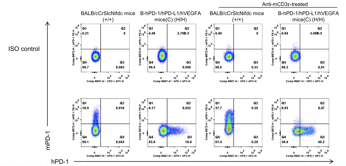
Human PD-1 Protein Expression Analysis in Spleen of PD-1/PD-L1/VEGFA Humanized Mice
- Mouse PD-1 was detectable in wild-type BALB/cCrSlcNifdc mice.
- Human PD-1 was detectable in homozygous PD-1/PD-L1/VEGFA humanized mice, but not in wild-type mice.
Mouse and human PD-1 expression analysis in splenocytes. Splenocytes were collected from wild-type BALB/cCrSlcNifdc mice (+/+) (female, n=2, 6-week-old) and homozygous B-hPD-1/hPD-L1/hVEGFA mice(C) (H/H) (female, n=2, 6-week-old) unstimulated or stimulated with anti-mCD3ε antibody (Bioxcell, BE0001-1) in vivo (7.5 μg/mice, stimulation for 24 hours, i.p.). Protein expression was analyzed with anti-mouse PD-1 antibody (Biolegend, 109104) and anti-human PD-1 antibody (Biolegend, 329908) by flow cytometry.
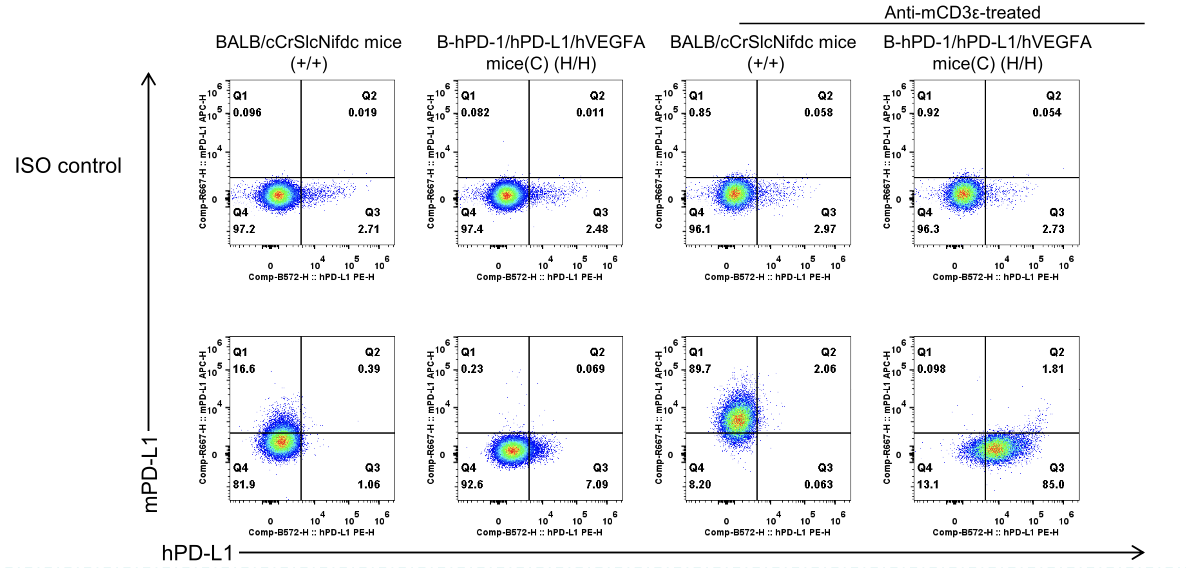
Human PD-L1 Protein Expression Analysis in Spleen of PD-1/PD-L1/VEGFA Humanized Mice
- Mouse PD-L1 was detectable in wild-type BALB/cCrSlcNifdc mice.
- Human PD-L1 was detectable in homozygous PD-1/PD-L1/VEGFA humanized mice, but not in wild-type mice.
Mouse and human PD-L1 expression analysis in splenocytes. Splenocytes were collected from wild-type BALB/cCrSlcNifdc mice (+/+) (female, n=2, 6-week-old) and homozygous B-hPD-1/hPD-L1/hVEGFA mice(C) (H/H) (female, n=2, 6-week-old) unstimulated or stimulated with anti-mCD3ε antibody (Bioxcell, BE0001-1) in vivo (7.5 μg/mice, stimulation for 24 hours, i.p.). Protein expression was analyzed with anti-mouse PD-L1 antibody (Biolegend, 124312) and anti-human PD-L1 antibody (Biolegend, 329706) by flow cytometry.
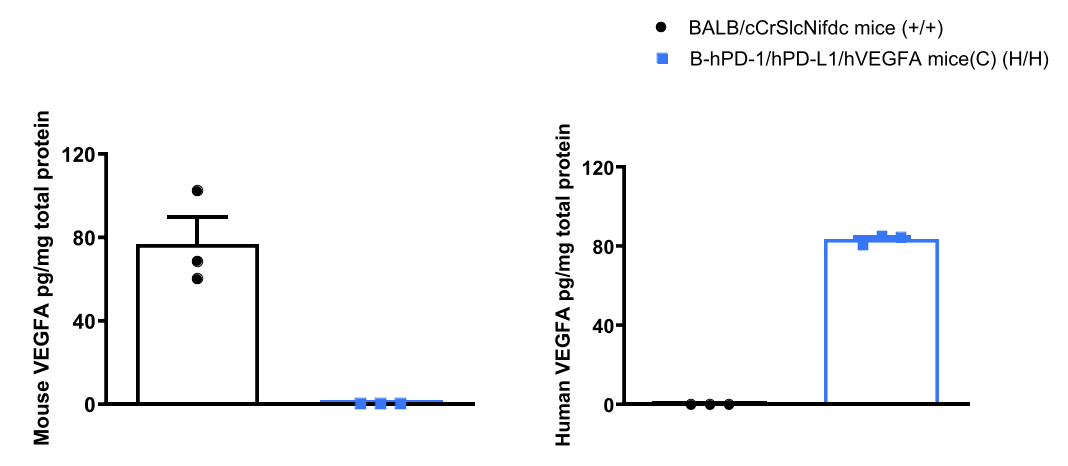
Human VEGFA Protein Expression Analysis in Lung of PD-1/PD-L1/VEGFA Humanized Mice
- Mouse VEGFA was detected exclusively in wild-type BALB/cCrSlcNifdc mice.
- Human VEGFA was detected in homozygous PD-1/PD-L1/VEGFA humanized mice, but not in wild-type mice.
Strain-specific VEGFA expression evaluated by ELISA in wild-type BALB/cCrSlcNifdc mice and homozygous PD-1/PD-L1/VEGFA humanized mice. Lung tissue homogenate was collected from BALB/cCrSlcNifdc mice (+/+) (female, n=3, 6-week-old) and homozygous B-hPD-1/hPD-L1/hVEGFA mice(C) (H/H) (female, n=3, 6-week-old). Expression level of mouse and human VEGFA were analyzed by ELISA (anti-mouse VEGFA ELISA kit: RD, MMV00-1; anti-human VEGFA ELISA kit: RD, DVE00).
* When publishing results obtained using this animal model, please acknowledge the source as follows: The animal model [B-hPD-1/hPD-L1/hVEGFA mice(C)] (Cat# 115055) was purchased from Biocytogen.