Gene Information: PD-1 (PDCD1) and PD-L1 (CD274) are key immune checkpoint molecules regulating T-cell activity, whereas PTK7 is a tumor-associated cell-surface glycoprotein. Together, these targets link tumor-targeted therapy with cancer immunotherapy.
Protein Expression: PD-1 is primarily expressed on activated T cells, while PD-L1 is expressed on tumor cells and various immune cells. PTK7 is frequently overexpressed in multiple solid tumors, including colorectal, breast, ovarian, and lung cancers, and is associated with tumor progression.
Signaling Pathway: PD-1 binding to PD-L1 inhibits T-cell activation and promotes tumor immune evasion. PTK7 primarily regulates non-canonical Wnt/planar cell polarity (PCP) signaling and interacts with Wnt pathway components to control cell polarity, migration, and invasion.
Model application: PD-1/PD-L1 blockade restores anti-tumor T-cell activity, while PTK7-targeted ADCs selectively eliminate PTK7-positive tumor cells. Their combination provides a promising strategy for enhancing anti-tumor efficacy and overcoming immune suppression.

PD-1

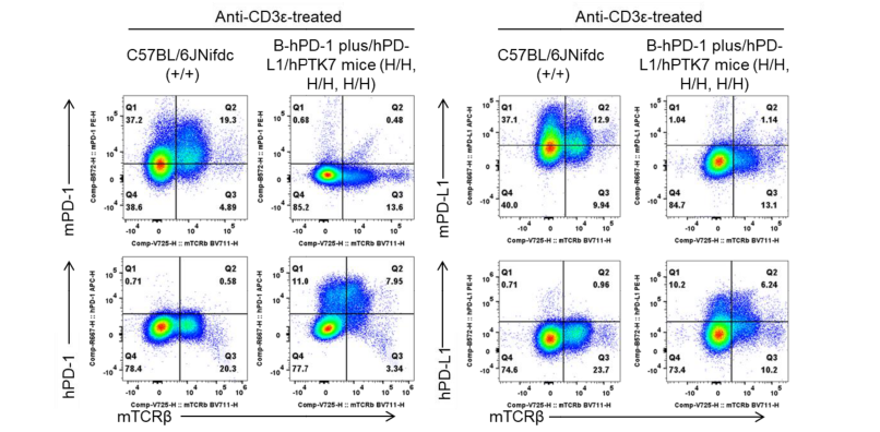
PD-1 and PD-L1 expression analysis in wild-type C57BL/6JNifdc mice and homozygous humanized B-hPD-1 plus/hPD-L1/hPTK7 mice by flow cytometry. Splenocytes were collected from wild-type C57BL/6JNifdc mice (+/+) and homozygous B-hPD-1 plus/hPD-L1/hPTK7 mice (H/H, H/H, H/H) (Female, 6-8-week-old, n=1) stimulated with anti-CD3ε in vivo (7.5 μg/mice, stimulation for 24 hours, i.p.). Protein expression was analyzed with anti-human PD-1 antibody (Biolegend, 329908), anti-mouse PD-1 antibody (Biolegend, 109104), anti-human PD-L1 antibody (Biolegend, 329706) and anti-mouse PD-L1 antibody (Biolegend, 124312) by flow cytometry.
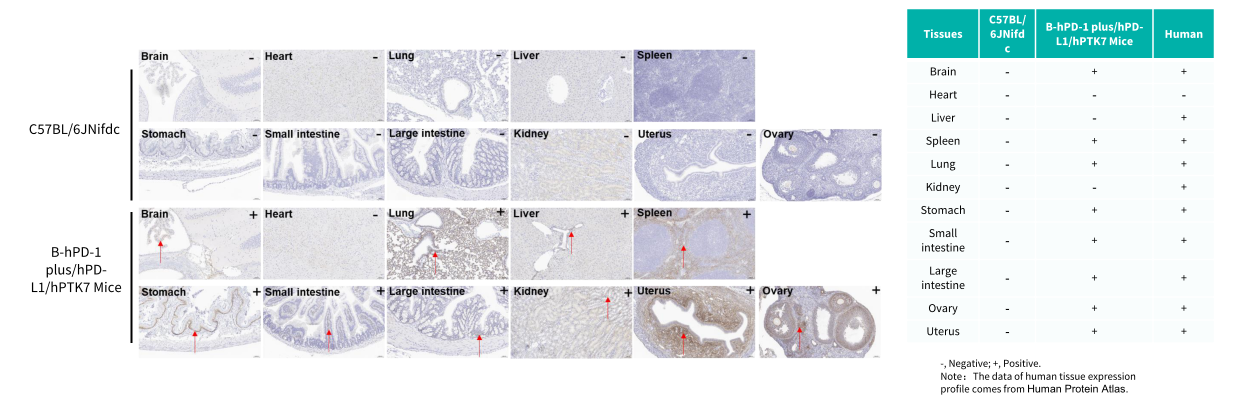
Immunohistochemical (IHC) analysis of PTK7 protein expression in wild-type C57BL/6JNifdc mice and B-hPD-1 plus/hPD-L1/hPTK7 mice. Major tissues were collected from wild-type C57BL/6JNifdc mice and homozygous B-hPD-1 plus/hPD-L1/hPTK7 mice and analyzed by IHC with anti-PTK7 antibody (Abcam, 325170).