

C57BL/6-Tfrctm1(TFRC)BcgenApptm1(APP*K670N*M671L*V717I )Bcgen/Bcgen • 114728

TFR1/APP: The combination of targeted antibody (TFR1) and therapeutic oligonucleotide (APP)

TFR1
The exons 4-19 of mouse Tfr1 gene that encode extracellular domain are replaced by human counterparts in B-hTFR1 mice. The genomic region of mouse Tfr1 gene that encodes cytoplasmic portion is retained.
The promoter, 5’UTR and 3’UTR region of the mouse gene are also retained. The chimeric TFR1 expression is driven by endogenous mouse Tfr1 promoter, while mouse Tfr1 gene transcription and translation will be disrupted.
APP
The partial 5’UTR and CDS that encodes the full-length human APP protein with Swedish (K670N, M671L) and London (V717I) mutation, followed by human 3’UTR-STOP is inserted right after mouse App 5’UTR to replace the exon 1 of mouse App gene.
The APP protein expression will be driven by endogenous mouse App promoter, while mouse App gene transcription and translation will be disrupted.

Human APP mRNA was exclusively detectable in the cortex and hippocampus of homozygous B-hTFR1/hAPP*K670N*M671L*V717I mice, but not in wild-type mice.
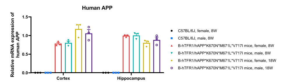
Species-specific analysis of APP gene expression in wild-type C57BL/6J mice and homozygous B-hTFR1/hAPP*K670N*M671L*V717I mice by RT-qPCR. Cortex and hippocampus were collected from wild-type C57BL/6J mice (8-week-old; 3 males and 3 females), homozygous B-hTFR1/hAPP*K670N*M671L*V717I mice (8-week-old, 18-week-old, 3 males and 3 females), then cDNA libraries were synthesized by reverse transcription, followed by PCR with human APP primers. Values are expressed as mean ± SEM.
Mouse App mRNA was exclusively detectable in the cortex and hippocampus of wild-type mice, but not in homozygous B-hTFR1/hAPP*K670N*M671L*V717I mice.
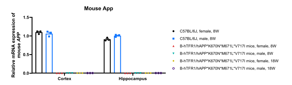
Species-specific analysis of APP gene expression in wild-type C57BL/6J mice and homozygous B-hTFR1/hAPP*K670N*M671L*V717I mice by RT-qPCR. Cortex and hippocampus were collected from wild-type C57BL/6J mice (8-week-old; 3 males and 3 females), homozygous B-hTFR1/hAPP*K670N*M671L*V717I mice (8-week-old, 18-week-old, 3 males and 3 females), then cDNA libraries were synthesized by reverse transcription, followed by PCR with mouse App primers. Values are expressed as mean ± SEM.
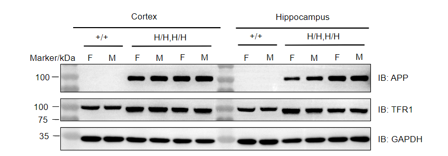
Protein expression analysis of APP and TFR1 in homozygous B-hTFR1/hAPP*K670N*M671L*V717I mice. Various tissue lysates were collected from wild-type C57BL/6JNifdc mice (+/+; 8-week-old), 8-week-old and 18-week-old B-hTFR1/hAPP*K670N*M671L*V717I mice (H/H,H/H), and then analyzed by western blot with anti-APP antibody (Abcam, ab133588) and anti-TFR1 antibody (Abcam, ab214039). 40 μg total proteins were loaded for western blotting analysis. F: female; M: male
