IL25: The alarmin IL-25 signals via the IL-17RA/IL-17RB receptor complex to trigger release of Th2 cytokines and promote inflammatory responses.
Gene Information: Interleukin-25 (IL25) is a protein-coding gene located on chromosome 14q11.2. It encodes a extracellular pro-inflammatory cytokine that is a member of the IL-17 family.
Protein Expression: IL25 (also referred to as IL-17E) is produced by eosinophils, Th2 cells, and epithelial cells, and belongs to the IL-17 family of cytokines. IL-17RB is highly expressed on Th2-associated immune cells including Th2 cells, eosinophils and mast cells, and its transcript abundance in Th2 cells correlates with serum IgE levels in allergic airway inflammation.
Signaling Pathway: IL-25 binds to its receptor complex of IL-17RA and IL-17RB to upregulate NF-κB, STAT6, GATA3 and NF-ATC1, activating and polarizing Th2 (but not Th17) cells and inducing Th2-cytokine expression and secretion.
Therapeutic Inhibition: The blockage of IL-17RB via SM17 or IL25 via XKH001 offers an attractive therapeutic target for Th2-mediated diseases, such as asthma.

IL17RA
Exons 2-11 of the mouse Il17ra gene encoding extracellular domain are replaced with the corresponding human IL7RB exons 2-11.
IL17RB
Exons 2-10 of the mouse Il7rb gene encoding the extracellular domain are replaced with the corresponding human IL7RB exons 2-10.
IL25
B-hIL17RA/hIL17RB/hIL25 mice were obtained by breeding B-hIL17RA/hIL17RB mice with B-hIL25 mice.


Experimental schedule for the induction of asthma model and in vivo efficacy of anti-human IL25 antibody in B-hIL17RA/hIL17RB/hIL25 mice. B-hIL17RA/hIL17RB/hIL25 mice(female, 7-week-old, n=5) were immunized with OVA etc. inducer to induce asthma. The anti-human IL25 antibody and anti-humanIL17RA antibody (in-house) was administered by intraperitoneal injection (n = 5).
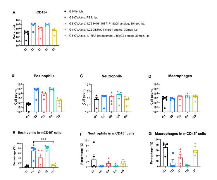
Analysis of immune cells in BALF by flow cytometry.
B-hIL17RA/hIL17RB/hIL25 mice (female, 7-week-old, n=5) were stimulated with inducer to induce asthma-like symptoms. Anti-human IL25 antibodies and anti-human IL17RA antibody (in-house) was intraperitoneally injected. Broncheoalveolar fluid (BALF) was collected at the end of the experiment to detect inflammatory cell infiltration in lung tissue. The results showed that the number and frequency of eosinophils induced in the untreated modeling group (G2) was significantly higher than that in the un-modeling group (G1), while the number and frequencies of these cells in the treated group (G5) decreased significantly when compared with the untreated modeling group (G2). Values are expressed as mean ± SEM. Significance was determined by one-way ANOVA test. *P < 0.05, P < 0.01, ***P < 0.001.
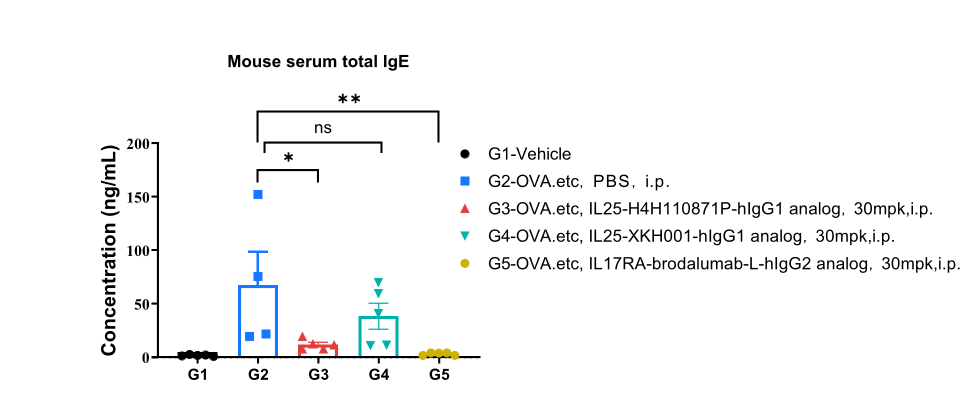
Total IgE in serum was significantly reduced in the mouse asthma model treated with anti-human IL25 antibodies and anti-human IL17RA antibody. Serum was collected at the study endpoint. IgE levels were analyzed by ELISA. The results showed that the levels of total IgE in mice treated with anti-human IL25 antibodies and anti-human IL17RA antibody (in-house) was lower than that in untreated mice. Values are expressed as mean ± SEM. *P < 0.05, **P < 0.01. ns: no Significance.
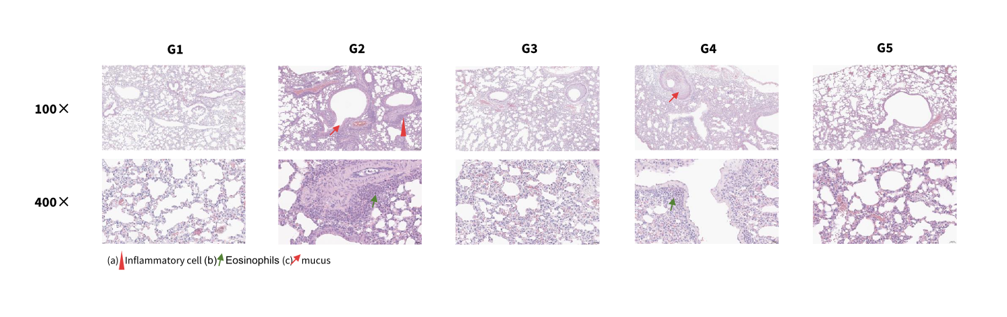
Hematoxylin and eosin (H&E) staining of an asthma model in B-hIL17RA/hIL17RB/hIL25 mice. Lung tissues were collected at the study endpoint and analyzed by H&E staining.
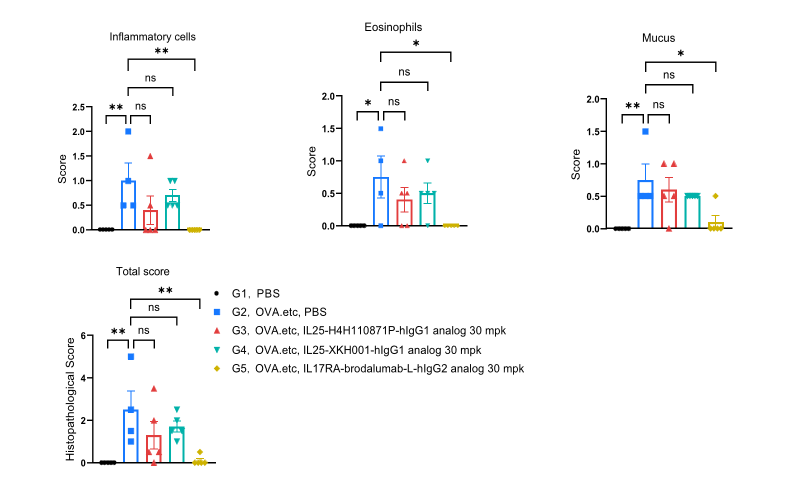
Hematoxylin and eosin (H&E) staining of an asthma model in B-hIL17RA/hIL17RB/hIL25 mice. Lung tissues were collected at the study endpoint and analyzed by H&E staining. Values are expressed as mean ± SEM. *P < 0.05, **P < 0.01, ***P < 0.001. ns: Not significant.