Key Advantages
Applications

TFR1 humanization (B-hTFR1 mice):
CD98HC humanization (B-hCD98HC mice):

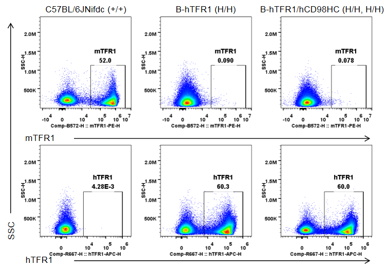
Strain-specific TFR1 expression was analyzed by flow cytometry in bone marrow erythrocytes from wild-type C57BL/6JNifdc mice (+/+), homozygous TFR1 humanized mice (H/H), and homozygous TFR1/CD98HC humanized mice (H/H, H/H). Bone marrow cells were analyzed with anti-mouse TFR1 antibody (BioLegend, 113808) and anti-human TFR1 antibody (BioLegend, 334108). Mouse TFR1 was detected only in erythrocytes of wild-type mice, whereas humanized TFR1 was exclusively detected in erythrocytes of homozygous TFR1 humanized mice and TFR1/CD98HC humanized mice, but not in wild-type mice.
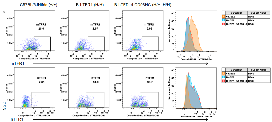
Brain cells were collected from wild-type C57BL/6JNifdc mice (+/+), homozygous TFR1 humanized mice (H/H), and homozygous TFR1/CD98HC humanized mice (H/H, H/H). Flow cytometry was performed using anti-mouse TFR1 antibody (BioLegend, 113808) and anti-human TFR1 antibody (BioLegend, 334108). Mouse TFR1 was detectable only in brain endothelial cells of wild-type mice, whereas humanized TFR1 was exclusively detected in brain endothelial cells of homozygous TFR1 humanized mice and TFR1/CD98HC humanized mice.
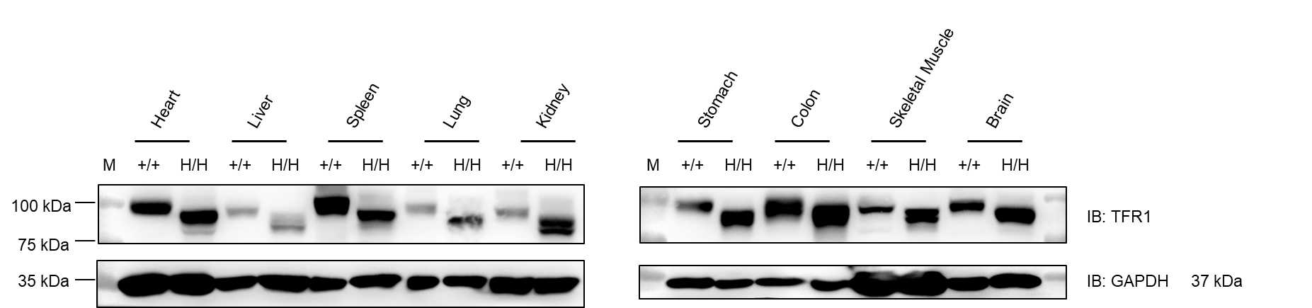
Western blot analysis was performed on tissue lysates from heart, liver, spleen, lung, kidney, stomach, colon, muscle, and brain of wild-type C57BL/6JNifdc mice (+/+) and homozygous TFR1 humanized mice (H/H). Samples were probed with anti-TFR1 antibody (Abcam, ab214039), which is cross-reactive between mouse and human TFR1. TFR1 protein was detectable in all examined tissues from both genotypes. GAPDH was used as a loading control. M: marker. Total protein loaded per lane: 40 μg.

Protein expression of TFR1 in the spinal cord, cerebral cortex, hippocampus, and cerebellum was analyzed by western blot in wild-type C57BL/6JNifdc mice (+/+) and homozygous TFR1/CD98HC humanized mice (H/H). Tissue lysates were probed with anti-transferrin receptor antibody (Abcam, ab214039), which is cross-reactive between species. TFR1 was detectable in CNS tissues from both wild-type and humanized mice. (A) Male. (B) Female. Total protein loaded per lane: 40 μg.
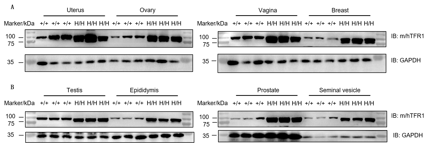
Western blot analysis of uterus, ovary, vagina, breast, testis, epididymis, prostate, and seminal vesicle was performed in wild-type C57BL/6JNifdc mice (+/+) and homozygous TFR1/CD98HC humanized mice (H/H) using anti-transferrin receptor antibody (Abcam, ab214039). TFR1 protein was detectable in all examined reproductive organs from both genotypes due to antibody cross-reactivity. Total protein loaded per lane: 30 μg.
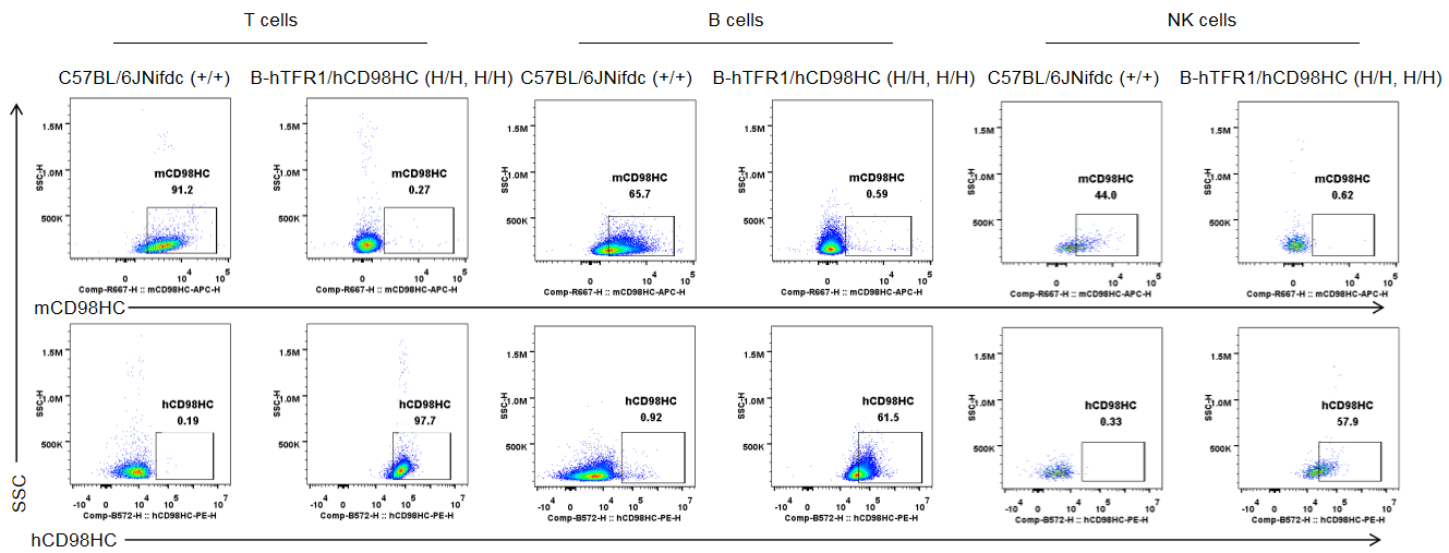
Strain-specific analysis of mouse and human CD98HC protein expression in wild-type C57BL/6JNifdc mice and TFR1/CD98HC humanized mice by flow cytometry. Blood cells were collected from wild-type C57BL/6JNifdc mice (+/+) and homozygous TFR1/CD98HC humanized mice (H/H; H/H). Mouse and human CD98HC protein expression was analyzed by flow cytometry using anti-mouse CD98HC (BioLegend, 128211) and anti-human CD98HC (231161-CD98BBBB-h1.L, produced in-house) antibodies.
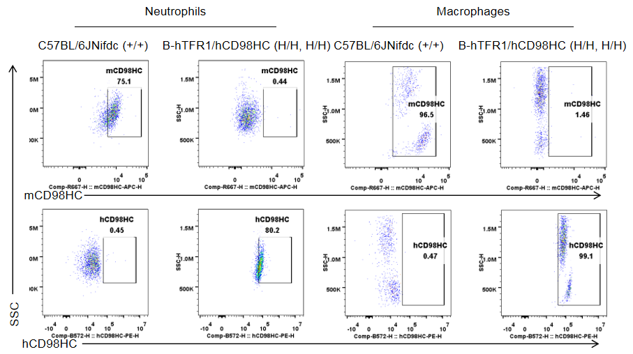
Strain-specific analysis of mouse and human CD98HC protein expression in wild-type C57BL/6JNifdc mice and TFR1/CD98HC humanized mice by flow cytometry. Blood cells were collected from wild-type C57BL/6JNifdc mice (+/+) and homozygous TFR1/CD98HC humanized mice (H/H; H/H). Mouse and human CD98HC protein expression was analyzed by flow cytometry using anti-mouse CD98HC (BioLegend, 128211) and anti-human CD98HC (231161-CD98BBBB-h1.L, produced in-house) antibodies.
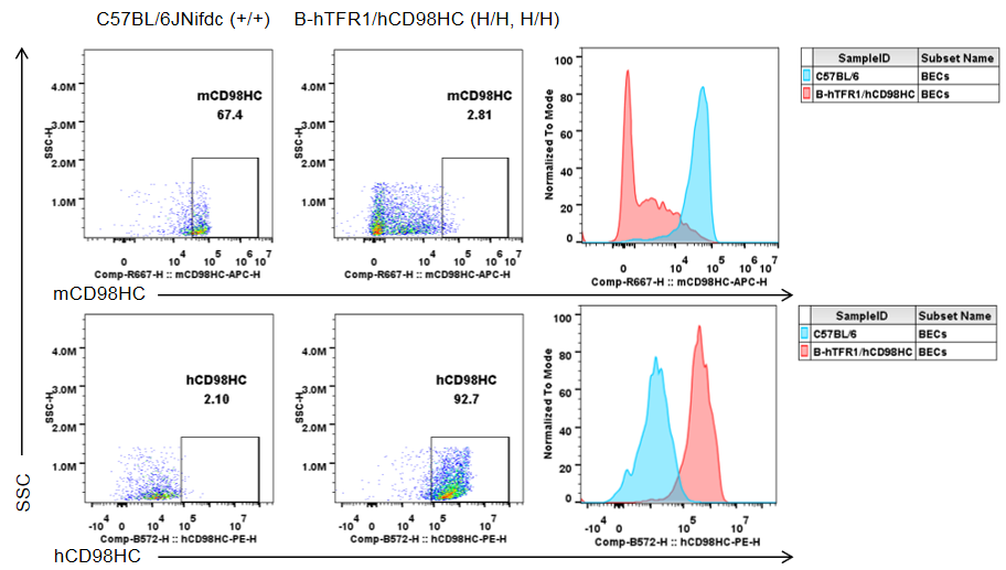
Strain-specific analysis of mouse and human CD98HC protein expression in brain endothelial cells of wild-type C57BL/6JNifdc mice and TFR1/CD98HC humanized mice by flow cytometry. Brain cells were collected from wild-type C57BL/6JNifdc mice (+/+) and homozygous TFR1/CD98HC humanized mice (H/H; H/H). Mouse and human CD98HC protein expression was analyzed by flow cytometry using anti-mouse CD98HC (BioLegend, 128211) and anti-human CD98HC (231161-CD98BBBB-h1.L, produced in-house) antibodies.
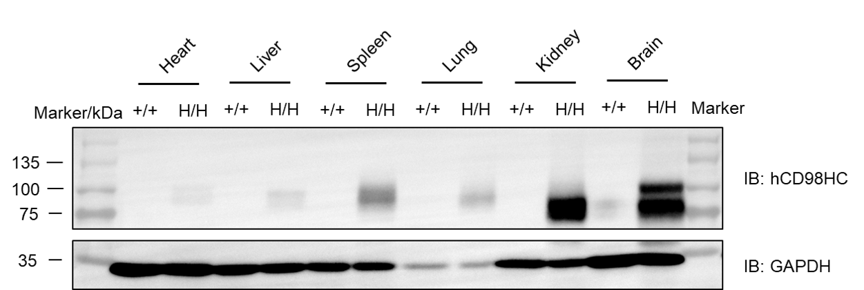
Western blot analysis of human CD98HC protein expression in multiple tissues of CD98HC humanized mice. Tissue lysates were collected from wild-type C57BL/6 mice and homozygous CD98HC humanized mice (H/H), including the spleen, lung, kidney, stomach, colon, spinal cord, skin, and brain. Human CD98HC protein expression was analyzed by Western blot using a species-specific anti-human CD98 antibody (Abcam, ab307587). A total of 50 μg of protein was loaded for Western blot analysis. M: protein marker.
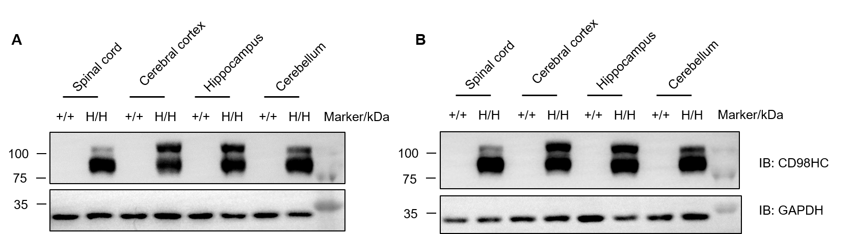
Western blot analysis of human CD98HC protein expression in central nervous system tissues of TFR1/CD98HC humanized mice. Tissue lysates were collected from the spinal cord, cerebral cortex, hippocampus, and cerebellum of wild-type C57BL/6JNifdc mice (+/+) and homozygous TFR1/CD98HC humanized mice (H/H). Human CD98HC protein expression was analyzed by Western blot using an anti-CD98 antibody (Abcam, ab307587). A total of 40 μg of protein was loaded for Western blot analysis. (A) Male mice. (B) Female mice.
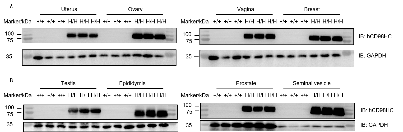
Western blot analysis of human CD98HC protein expression in reproductive tissues of TFR1/CD98HC humanized mice. Tissue lysates were collected from the uterus, ovary, vagina, mammary gland, testis, epididymis, prostate, and seminal vesicle of wild-type C57BL/6JNifdc mice (+/+) and homozygous TFR1/CD98HC humanized mice (H/H). Human CD98HC protein expression was analyzed by Western blot using an anti-CD98 antibody (Abcam, ab307587). A total of 30 μg of protein was loaded for Western blot analysis.
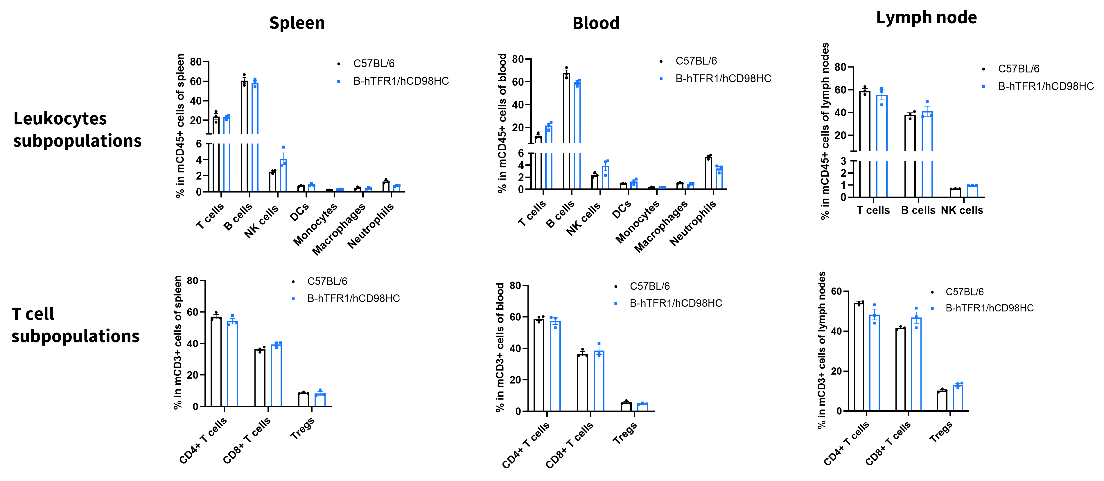
Values are expressed as mean ± SD.

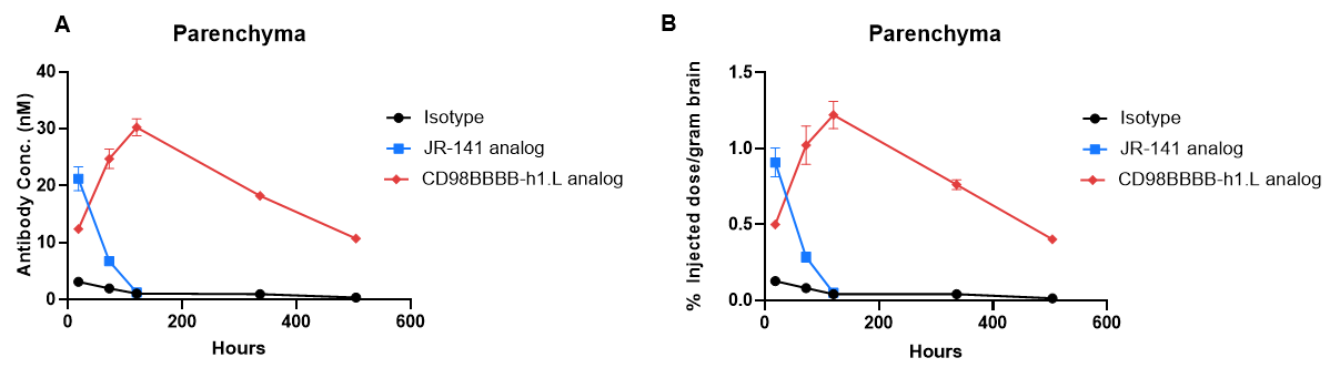
In vivo pharmacokinetic (PK) evaluation and comparison of brain exposure to anti-human TFR1 and anti-human CD98HC antibodies in TFR1/CD98HC humanized mice. Female TFR1/CD98HC humanized mice (8 weeks old, n = 2) received intravenous injections via the tail vein of control IgG (10 mg/kg), an anti-human TFR1 antibody (TFR1 Ab, JR-141 analog, monovalent, produced in-house, 12.56 mg/kg), or an anti-human CD98HC antibody (CD98HC Ab, CD98BBBB-h1.L analog, monovalent, produced in-house, 13.3 mg/kg). Brain tissues were collected at 18 hours and on days 3, 5, 14, and 21 after dosing for in vivo pharmacokinetic analysis. (A) Brain antibody concentrations. (B) Percentage of injected dose per gram of brain tissue (%ID/g). The anti-human TFR1 antibody exhibited higher brain exposure at the early time point (within 24 hours after dosing), whereas the anti-human CD98HC antibody exhibited higher brain exposure at 72 hours after dosing. These results demonstrate the utility of TFR1/CD98HC humanized mice for evaluating brain uptake and comparing the pharmacokinetic profiles of antibody shuttle molecules targeting human TFR1 and CD98HC. Data are presented as mean ± SEM.