B-hCD3EDG/hCD19 plus/hBCMA mice

C57BL/6-Cd3etm1(CD3E)Bcgen Cd3dtm1(CD3D)Bcgen Cd3gtm1(CD3G)Bcgen Cd19tm5(CD19)Bcgen Tnfrsf17tm2(TNFRSF17)Bcgen • 114828

B-hCD3EDG/hCD19 plus/hBCMA mice

Catalog Number: 114828
Strain Name: C57BL/6-Cd3etm1(CD3E)Bcgen Cd3dtm1(CD3D)Bcgen Cd3gtm1(CD3G)Bcgen Cd19tm5(CD19)Bcgen Tnfrsf17tm2(TNFRSF17)Bcgen
Strain Background: C57BL/6
NCBI gene ID: 12501,12500,12502,12478,21935 (Human)
Aliases: CD3; T3e; CD3epsilon; T3d; T3g; Ctg3; Ctg-3; BCM; BCMA; Tnfrsf13; Tnfrsf13a
---
라이선스 옵션 제공 가능
B-hCD3EDG/hCD19 plus/hBCMA mice

이 페이지에서

  • Description
  • Phenotypic analysis
  • Efficacy

포스터

모두 보기

    출판물

      Description

      Gene Information:

      • CD3: Encoded by CD3E, D, G; part of the Ig superfamily. It forms the essential signaling backbone of the T-cell receptor (TCR) complex. 
      • CD19: is a transmembrane glycoprotein. It belongs to the immunoglobulin superfamily and plays a critical role in B-cell development, activation, and maintenance throughout the immune system. 
      • BCMA: Encoded by the TNFRSF17 gene, a critical member of the tumor necrosis factor receptor (TNFR) superfamily.

      Protein Expression:

      • CD3: Constitutive and universal marker for all mature T cells (CD4+, CD8+). Always present on the cell surface. 
      • CD19: is expressed almost exclusively on B-lineage cells, beginning at the early B-cell stage and persisting through mature B cells. Its expression is typically absent or markedly reduced in plasma cells. 
      • BCMA: It is a B-cell maturation antigen, whose expression is highly in mature plasma cells and nearly all multiple myeloma cells.

      TCE Therapy in Hematologic Cancers

      • T-cell engagers (TCEs), mainly represented by bispecific antibodies (BsAbs), have become one of the most important immunotherapy platforms in hematologic malignancies, particularly multiple myeloma (MM) and B-cell lymphomas. By simultaneously binding a tumor-associated antigen and CD3 on T cells, TCEs redirect and activate the patient's own T cells to recognize and kill tumor cells.
      • Among these agents, CD19- and BCMA-targeting TCEs have shown remarkable clinical efficacy. CD19-directed TCEs are widely used in B-cell malignancies, while BCMA-directed TCEs have become important treatment options for relapsed or refractory MM. In addition, both CD19- and BCMA-targeting therapies are being actively explored in autoimmune diseases, where depletion of pathogenic B cells and plasma cells may help restore immune tolerance and achieve durable disease control.
      CD3E Protein Expression in Spleen
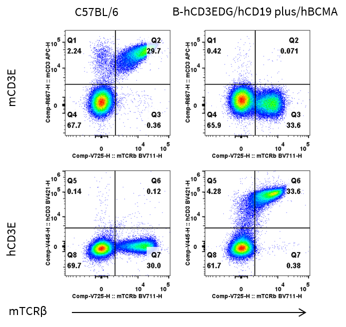
      • Mouse CD3E was detected on T cells populations in wild-type C57BL/6 mice, but not in B-hCD3EDG/hCD19 plus/hBCMA mice.
      • Human CD3E was detected on T cells populations in B-hCD3EDG/hCD19 plus/hBCMA mice, but not in wild-type C57BL/6 mice.

      Mouse and human CD3E expression analysis in splenocytes. Splenocytes were collected from wild-type C57BL/6 mice and homozygous B-hCD3EDG/hCD19 plus/hBCMA mice (female, 9-week-old, n = 1). CD3E expression on T cells was analyzed by flow cytometry using species-specific anti-CD3E antibodies (anti-human CD3E antibody, BD Horizon, 562426; anti-mouse CD3E antibody, Biolegend, 100312 ).
      CD3E Protein Expression in Blood
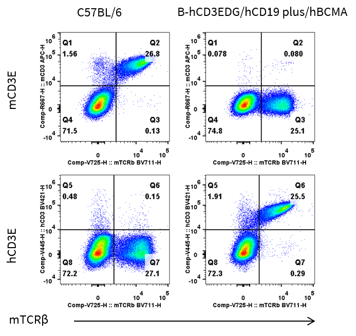
      • Mouse CD3E was detected on T cells populations in wild-type C57BL/6 mice, but not in B-hCD3EDG/hCD19 plus/hBCMA mice.
      • Human CD3E was detected on T cells populations in B-hCD3EDG/hCD19 plus/hBCMA mice, but not in wild-type C57BL/6 mice.

      Mouse and human CD3E expression analysis in blood. Blood cells were collected from wild-type C57BL/6 mice and homozygous B-hCD3EDG/hCD19 plus/hBCMA mice (female, 9-week-old, n = 1). CD3E expression on T cells was analyzed by flow cytometry using species-specific anti-CD3E antibodies (anti-human CD3E antibody, BD Horizon, 562426; anti-mouse CD3E antibody, Biolegend, 100312 ).
      CD19 Protein Expression in Spleen
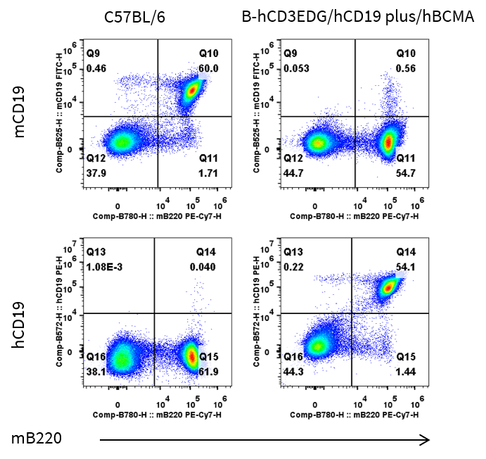
      • Mouse CD19 was detected on B cells populations in wild-type C57BL/6 mice, but not in B-hCD3EDG/hCD19 plus/hBCMA mice.
      • Human CD19 was detected on B cells populations in B-hCD3EDG/hCD19 plus/hBCMA mice, but not in wild-type C57BL/6 mice.

      Mouse and human CD19 expression analysis in splenocytes. Splenocytes were collected from wild-type C57BL/6 and homozygous B-hCD3EDG/hCD19 plus/hBCMA mice (female, 9-week-old, n = 1). CD19 expression on B cells was analyzed by flow cytometry using species-specific anti-CD19 antibodies (anti-human CD19 antibody, BD Horizon, 302208; anti-mouse CD19 antibody, Biolegend, 115538 ).
      CD19 Protein Expression in Blood
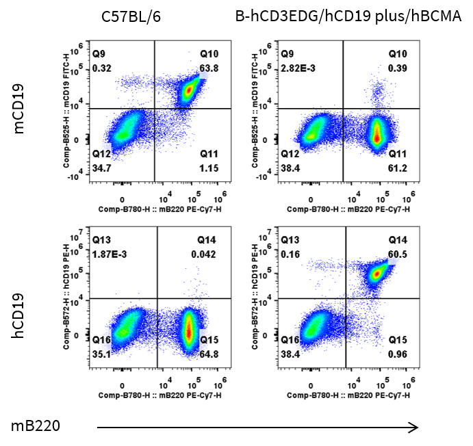
      • Mouse CD19 was detected on B cells populations in wild-type C57BL/6 mice, but not in B-hCD3EDG/hCD19 plus/hBCMA mice.
      • Human CD19 was detected on B cells populations in B-hCD3EDG/hCD19 plus/hBCMA mice, but not in wild-type C57BL/6 mice.

      Mouse and human CD19 expression analysis in blood. Blood cells were collected from wild-type C57BL/6 and homozygous B-hCD3EDG/hCD19 plus/hBCMA mice (female, 9-week-old, n = 1). CD19 expression on B cells was analyzed by flow cytometry using species-specific anti-CD19 antibodies (anti-human CD19 antibody, BD Horizon, 302208; anti-mouse CD19 antibody, Biolegend, 115538 ).
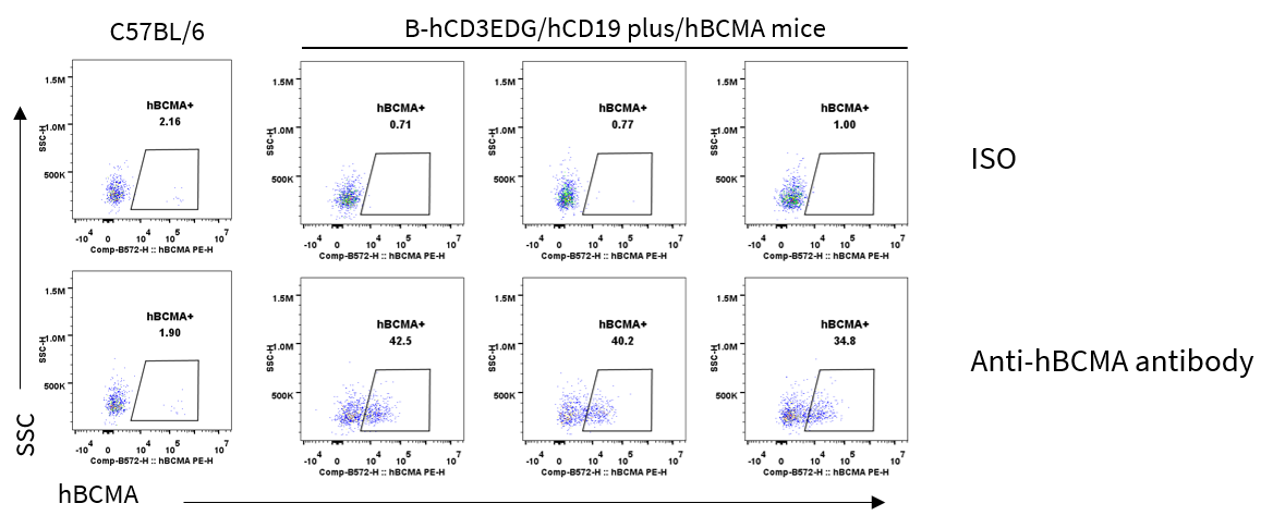
      BCMA Protein Expression in Spleen
      • Human BCMA was detectable in plasma cells of spleen from B-hCD3EDG/hCD19 plus/hBCMA mice but not in wild-type C57BL/6 mice.

      Human BCMA expression analysis in splenocytes. Spleen cells were collected from wild-type C57BL/6 mice and homozygous B-hCD3EDG/hCD19 plus/hBCMA mice. Human BCMA expression on plasma cells was analyzed by flow cytometry using a anti-human BCMA antibody (Biolegend, 357504). Mice were stimulated with LPS and treated with γ-secretase inhibitor to enhance membrane BCMA expression.
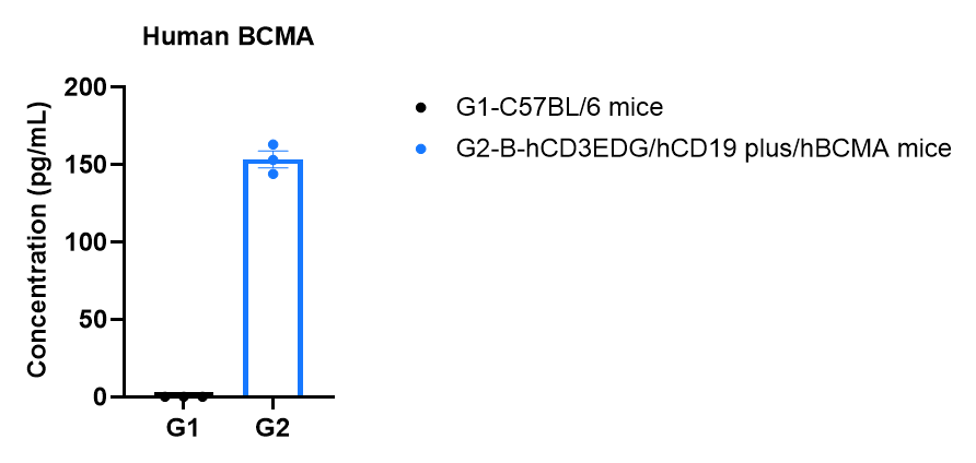
      sBCMA Protein Expression in Serum
      • Human soluble BCMA protein expression was detectable in serum of homozygous B-hCD3EDG/hCD19 plus/hBCMA mice, but not wild-type C57BL/6 mice.

      Strain specific soluble BCMA expression analysis in serum from homozygous of B-hCD3EDG/hCD19 plus/hBCMA mice by MSD. Serum were collected from wild-type C57BL/6 mice and homozygous B-hCD3EDG/hCD19 plus/hBCMA mice (8-week-old, n=3 per group). Values are expressed as mean ± SEM.
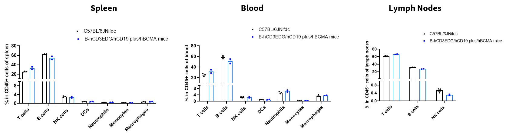
      Analysis of Leukocyte Subpopulations
      • The percentages of T cells, B cells, NK cells, DCs, neutrophils, monocytes, and macrophages in homozygous B-hCD3EDG/hCD19 plus/hBCMA mice were similar to those in C57BL/6 mice.
      • Humanization of CD3E, CD3D, CD3G, CD19, and BCMA does not affect normal immune cell development or splenic distribution.

      Analysis of leukocyte subpopulations by flow cytometry in immune organs and blood. Splenocytes, peripheral blood, and lymph nodes were isolated from female C57BL/6 and B-hCD3EDG/hCD19 plus/hBCMA mice (female, 14-week-old, n = 3). Single live cells were gated on the CD45⁺ population and analyzed by flow cytometry as indicated. Values are expressed as mean ± SEM.
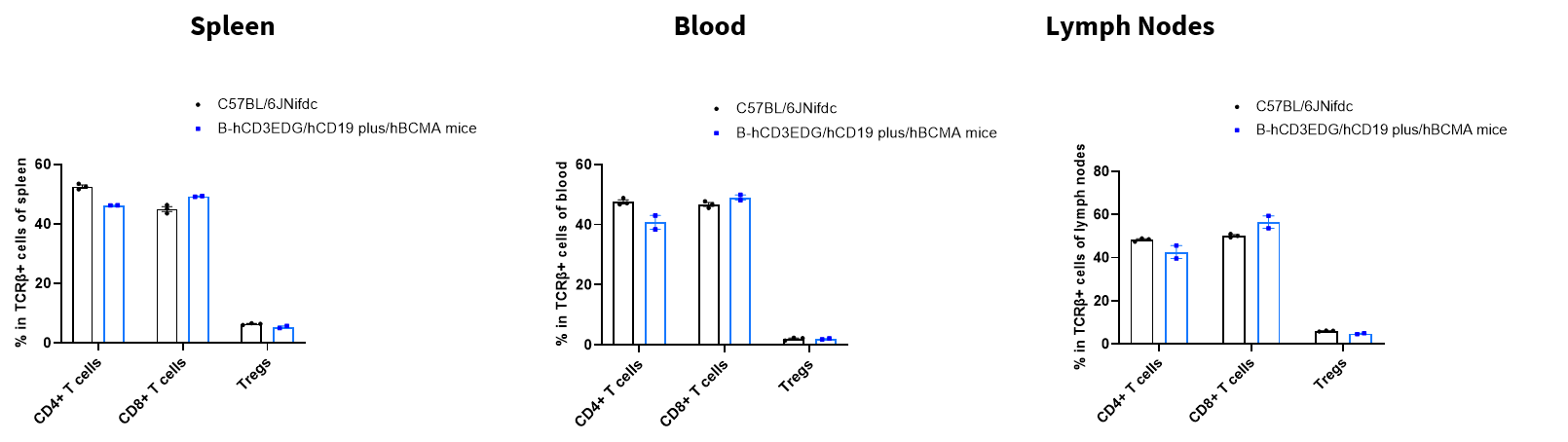
      Analysis of T Cell Subpopulations
      • The proportions of CD4⁺ T cells, CD8⁺ T cells, and Tregs in homozygous B-hCD3EDG/hCD19 plus/hBCMA mice were comparable to those in C57BL/6 mice.
      • Humanization of CD3E, CD3D, CD3G, CD19, and BCMA does not affect normal T cell development, differentiation, or splenic distribution.

      Analysis of T-cell subpopulations by flow cytometry in immune organs and blood. Splenocytes, peripheral blood, and lymph nodes were isolated from female C57BL/6 and B-hCD3EDG/hCD19 plus/hBCMA mice (female, 14-week-old, n = 3). Single live cells were gated on the TCRβ⁺ T-cell population and analyzed by flow cytometry as indicated. Values are expressed as mean ± SEM.

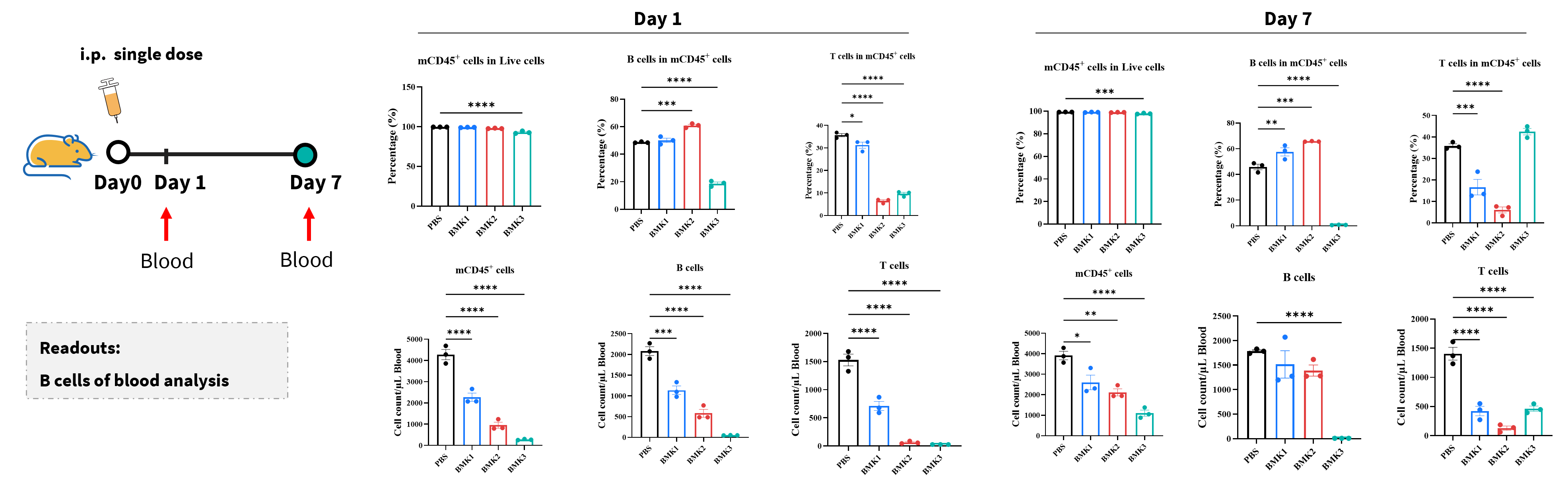
      In vivo B Cells and Plasma Cells Depletion

      In vivo B-cell depletion by anti-human CD3/BCMA or anti-human CD3/CD19 bispecific antibodies (BsAbs) in B-hCD3EDG/hCD19/hBCMA mice.​ Bispecific antibodies (BMK1 and BMK2: targeting BCMA/CD3; BMK3: targeting CD19/CD3) or PBS control were administered as a single dose to B-hCD3EDG/hCD19/hBCMA mice (n=3 per group). Blood cells were harvested on day 1 and day 7 post-treatment. The frequency and absolute numbers of mCD45⁺ cells, B cells, and T cells were quantified by flow cytometry. Data are presented as mean ± SEM and analyzed by one-way ANOVA followed by Dunnett’s multiple comparisons test versus the PBS control group (*p<0.05, **p<0.01, ***p<0.001,****p<0.0001). This study was conducted in collaboration with our partner.
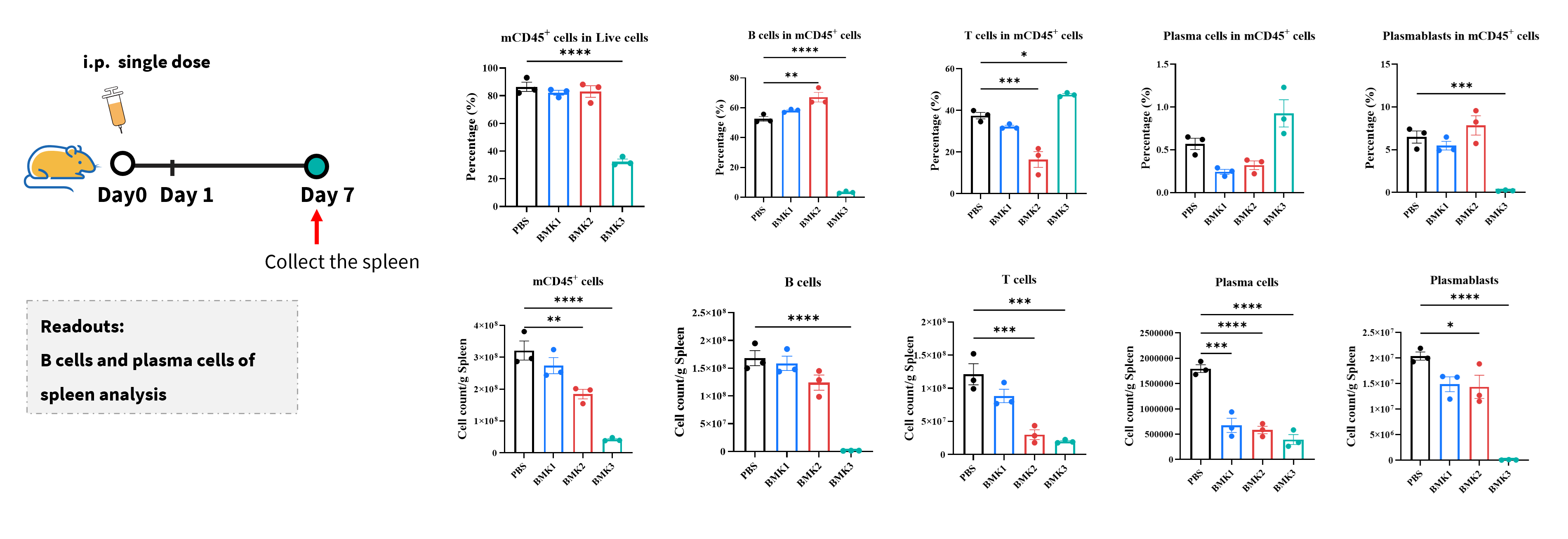
      In vivo B-cell depletion by anti-human CD3/BCMA or anti-human CD3/CD19 bispecific antibodies (BsAbs) in B-hCD3EDG/hCD19/hBCMA mice.​ Bispecific antibodies (BMK1 and BMK2: targeting BCMA/CD3; BMK3: targeting CD19/CD3) or PBS control were administered as a single dose to B-hCD3EDG/hCD19/hBCMA mice (n=3 per group). Spleens were harvested on day 7 post-treatment. The frequency and absolute numbers of mCD45⁺ cells, B cells, T cells, plasma cells, and plasmablasts were quantified by flow cytometry. Data are presented as mean ± SEM and analyzed by one-way ANOVA followed by Dunnett’s multiple comparisons test versus the PBS control group (*p<0.05, **p<0.01, ***p<0.001,****p<0.0001). This study was conducted in collaboration with our partner.

      * When publishing results obtained using this animal model, please acknowledge the source as follows: The animal model [B-hCD3EDG/hCD19 plus/hBCMA mice] (Cat# 114828) was purchased from Biocytogen.