

BALB/cCrSlcNifdc-Tfrctm3(TFRC)Bcgen/Bcgen • 114842


Gene targeting strategy for B-hTFR1 Mice Plus(C).

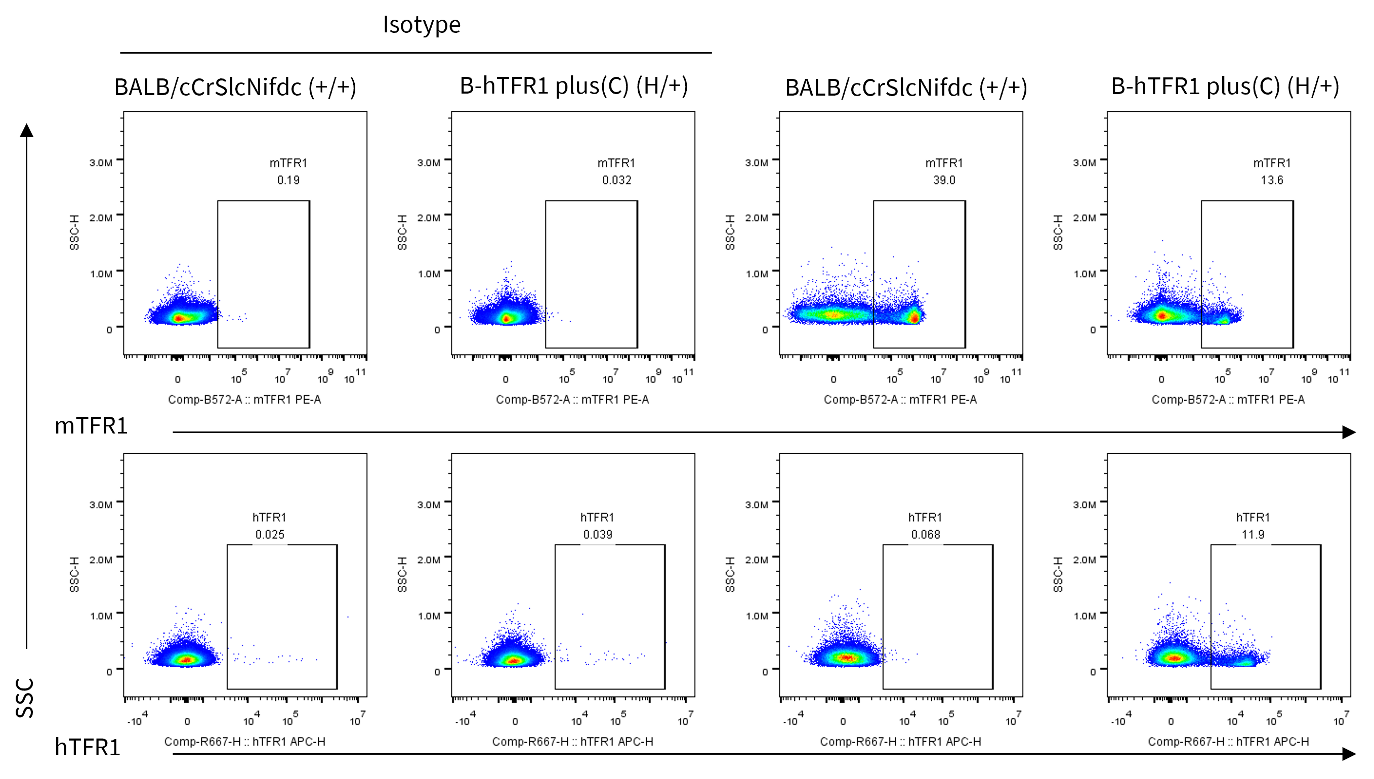
Strain specific TFR1 expression analysis in wild-type BALB/cCrSlcNifdc and heterozygous humanized B-hTFR1 mice plus(C) by flow cytometry. Bone marrow was collected from wild-type BALB/cCrSlcNifdc mice (+/+) and heterozygous B-hTFR1 mice plus(C) (H/+) and analyzed by flow cytometry. Protein expression was analyzed with anti-mouse CD71 antibody (Biolegend, 113808) and anti-human CD71 antibody (Biolegend, 334108) by flow cytometry. Mouse TFR1 was detectable in wild-type BALB/cCrSlcNifdc mice and heterozygous B-hTFR1 mice plus(C). Human TFR1 was exclusively detectable in heterozygous B-hTFR1 mice plus(C), but not in wild-type mice.
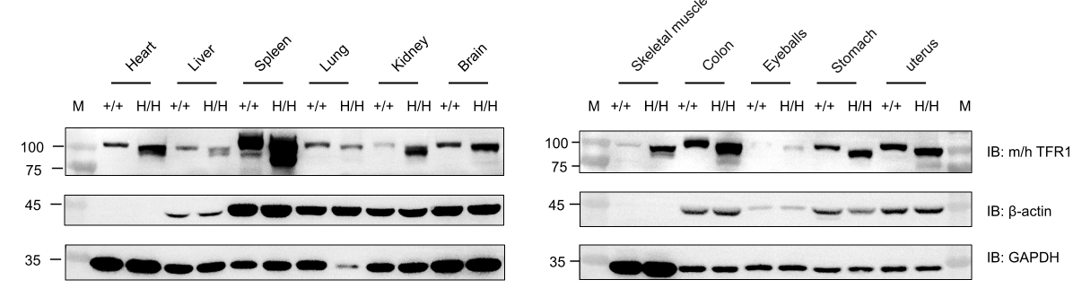
Western blot analysis of TFR1 protein expression in wild-type BALB/cCrSlcNifdc mice and homozygous B-hTFR1 plus (C) mice. Various tissues were collected from wild-type BALB/cCrSlcNifdc mice (+/+) and homozygous B-hTFR1 plus (C) mice (H/H) (female, 8-week-old) and then analyzed by western blot with an anti-TFR1 antibody (Abcam, ab214039). 40 μg total proteins were loaded for western blot analysis. GAPDH and β-actin were detected as internal controls. M, marker.
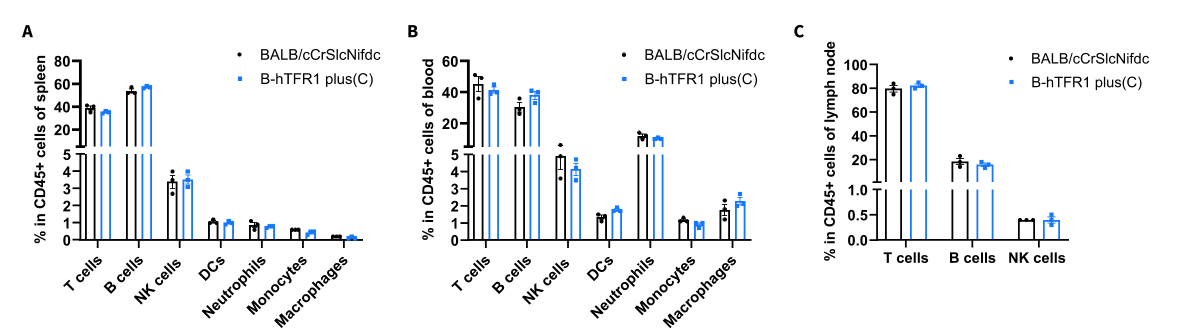
Frequency of leukocyte subpopulations by flow cytometry. Splenocytes (A), blood (B), and lymph node cells (C) were isolated from wild-type BALB/cCrSlcNifdc mice (female, n=3, 8-week-old) and homozygous B-hTFR1 plus (C) mice (female, n=3, 8-week-old). Flow cytometry analysis was performed to assess the frequency of leukocyte subpopulations. The frequencies of T cells, B cells, NK cells, DCs, neutrophils, monocytes, and macrophages in homozygous B-hTFR1 plus (C) mice were similar to those in wild-type BALB/cCrSlcNifdc mice. Humanization of TFR1 did not change the overall frequency or distribution of immune cell types in the spleen, blood, and lymph nodes. Values are expressed as mean ± SEM. Significance was determined by two-way ANOVA.
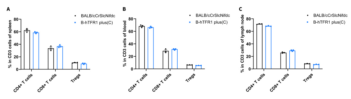
Frequency of T cell subpopulations by flow cytometry. Splenocytes (A), blood (B), and lymph node cells (C) were isolated from wild-type BALB/cCrSlcNifdc mice (female, n=3, 8-week-old) and homozygous B-hTFR1 plus (C) mice (female, n=3, 8-week-old). Flow cytometry analysis was performed to assess the frequency of T cell subpopulations. The frequencies of CD4+ T cells, CD8+ T cells, and Tregs in homozygous B-hTFR1 plus (C) mice were similar to those in wild-type BALB/cCrSlcNifdc mice. Humanization of TFR1 did not change the overall frequency or distribution of T cell types in the spleen, blood, and lymph nodes. Values are expressed as mean ± SEM. Significance was determined by two-way ANOVA.
